Click image to see more details
-

-
-
-
-
+3
Product Info Summary
| SKU: | A00231 |
|---|---|
| Size: | 80 µl |
| Reactive Species: | Human, Mouse |
| Host: | Rabbit |
| Application: | Flow Cytometry, IF, IHC-P, WB |
Customers Who Bought This Also Bought

Product info
Product Name
Anti-FGFR2 Antibody (N-term)
SKU/Catalog Number
A00231
Size
80 µl
Form
Liquid
Description
Boster Bio Anti-FGFR2 Antibody (N-term) (Catalog # A00231). Tested in IHC-P, WB, Flow Cytometry, IF application(s). This antibody reacts with Human, Mouse.
Storage & Handling
Maintain refrigerated at 2-8°C for up to 2 weeks. For long-term storage, store at -20°C in small aliquots to prevent freeze-thaw cycles.
Cite This Product
Anti-FGFR2 Antibody (N-term) (Boster Biological Technology, Pleasanton CA, USA, Catalog # A00231)
Host
Rabbit
Contents
Purified polyclonal antibody supplied in PBS with 0.09% (W/V) sodium azide.
Clonality
Polyclonal
Isotype
Rabbit IgG
Immunogen
This FGFR2 antibody is generated from rabbits immunized with a KLH conjugated synthetic peptide between 22-51 amino acids from the N-terminal region of human FGFR2.
Cross-reactivity
No cross reactivity with other proteins.
Reactive Species
A00231 is reactive to FGFR2 in Human, Mouse
Calculated molecular weight
92.0 kDa
Background of FGFR2
FGFR2 is a member of the fibroblast growth factor receptor family, where amino acid sequence is highly conserved between members and throughout evolution. FGFR family members differ from one another in their ligand affinities and tissue distribution. A full-length representative protein consists of an extracellular region, composed of three immunoglobulin-like domains, a single hydrophobic membrane-spanning segment and a cytoplasmic tyrosine kinase domain. The extracellular portion of the protein interacts with fibroblast growth factors, setting in motion a cascade of downstream signals, ultimately influencing mitogenesis and differentiation. This particular family member is a high-affinity receptor for acidic, basic and/or keratinocyte growth factor, depending on the isoform. Mutations in the gene are associated with many craniosynostotic syndromes and bone malformations. The genomic organization of the gene encompasses 20 exons. Alternative splicing in multiple exons, including those encoding the Ig-like domains, the transmembrane region and the carboxyl terminus, results in varied isoforms which differ in structure and specificity. Isoform 1 has equal affinity for aFGF and bFGF but does not bind KGF.
Antibody Validation
Boster validates all antibodies on WB, IHC, ICC, Immunofluorescence, and ELISA with known positive control and negative samples to ensure specificity and high affinity, including thorough antibody incubations.
Application & Images
Applications
A00231 is guaranteed for Flow Cytometry, IF, IHC-P, WB Boster Guarantee
Recommend Dilution
IF: 1:25
WB: 1:1000-1:2000
IHC-P: 1:50-1:100
FC: 1:25
Validation Images & Assay Conditions
Click image to see more details
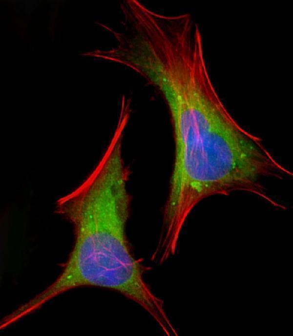
Immunofluorescent analysis of 4% paraformaldehyde-fixed, 0. 1% Triton X-100 permeabilized HeLa (human cervical epithelial adenocarcinoma cell line) cells labeling FGFR2 with A00231 at 1/25 dilution, followed by Dylight® 488-conjugated goat anti-rabbit IgG secondary antibody at 1/200 dilution (green). Immunofluorescence image showing cytoplasm and weak nucleus staining on HeLa cell line. Cytoplasmic actin is detected with Dylight® 554 Phalloidin at 1/100 dilution (red). The nuclear counter stain is DAPI (blue).

Click image to see more details
FGFR2 Antibody (N-term) (Cat. #A00231) western blot analysis in mouse NIH-3T3 cell line lysates (35ug/lane). This demonstrates the FGFR2 antibody detected the FGFR2 protein (arrow).

Click image to see more details
All lanes : Anti-FGFR2 Antibody (N-term) at 1:1000-1:2000 dilution
Lane 1: DU145 whole cell lysate
Lane 2: A549 whole cell lysate
Lane 3: T47D whole cell lysate
Lane 4: Hela whole cell lysate
Lane 5: K562 whole cell lysate
Lane 6: M. brain whole lysate
Lysates/proteins at 20 µg per lane.
Secondary
Goat Anti-Rabbit IgG, (H+L), Peroxidase conjugated at 1/10000 dilution.
Predicted band size : 92 kDa
Blocking/Dilution buffer: 5% NFDM/TBST.

Click image to see more details
Formalin-fixed and paraffin-embedded human cancer tissue reacted with the primary antibody, which was peroxidase-conjugated to the secondary antibody, followed by AEC staining. This data demonstrates the use of this antibody for immunohistochemistry; clinical relevance has not been evaluated. BC = breast carcinoma; HC = hepatocarcinoma.

Click image to see more details
Overlay histogram showing Hela cells stained with A00231 (green line). The cells were fixed with 2% paraformaldehyde (10 min) and then permeabilized with 90% methanol for 10 min. The cells were then icubated in 2% bovine serum albumin to block non-specific protein-protein interactions followed by the antibody (A00231, 1:25 dilution) for 60 min at 37ºC. The secondary antibody used was Goat-Anti-Rabbit IgG, DyLight® 488 Conjugated Highly Cross-Adsorbed at 1/200 dilution for 40 min at 37ºC. Isotype control antibody (blue line) was rabbit IgG1 (1μg/1x10^6 cells) used under the same conditions. Acquisition of >10, 000 events was performed.
Specific Publications For Anti-FGFR2 Antibody (N-term) (A00231)
Loading publications
Recommended Resources
Here are featured tools and databases that you might find useful.
- Boster's Pathways Library
- Protein Databases
- Bioscience Research Protocol Resources
- Data Processing & Analysis Software
- Photo Editing Software
- Scientific Literature Resources
- Research Paper Management Tools
- Molecular Biology Software
- Primer Design Tools
- Bioinformatics Tools
- Phylogenetic Tree Analysis
Customer Reviews
Have you used Anti-FGFR2 Antibody (N-term)?
Share your experimental results or join a short interview to earn up to $1,000 in product credits or other rewards.
0 Reviews For Anti-FGFR2 Antibody (N-term)
Customer Q&As
Have a question?
Find answers in Q&As, reviews.
Can't find your answer?
Submit your question
