This website uses cookies to ensure you get the best experience on our website.


- Table of Contents
Plan chromogenic FHIT IHC on paraffin sections using the catalog antibody at 0.5–1 µg/mL (datasheet PB9181). Select comparison tissues and score cytoplasmic staining by cell type using the reported tissue pattern (HPA tissue IHC).

Expected localisation, validated protocols, controls and antibodies — the at-a-glance facts below, then the full design guide.
| Expected localisation | General cytoplasmic tissue staining (HPA tissue IHC) | |
| Staining pattern | Cytoplasmic staining across multiple cell types (HPA tissue IHC) | |
| Antigen retrieval | EDTA pH 8.0 HIER, heat-mediated (datasheet PB9181) | |
| Positive control | Cerebellum+4 more · see all | |
| Negative control | Vagina |
| Fixation | Keep fixation consistent across sections (standard IHC practice; not target-specific) | |
| Caveat | Vaginal squamous cells may show no signal (HPA tissue IHC) | |
| Regulation | FHIT levels vary by cell type (HPA tissue IHC) | |
| Isoform / epitope | One chain, residues 1–147; no isoforms reported (UniProt) |
The catalog antibody’s IHC-P protocol (datasheet: PB9181) is accompanied by four published FHIT protocols (PMC2409597; PMC2376126; PMC2394260; PMC2762784).
| Sample | Paraffin-embedded human mammary cancer tissue; fixative not specified (datasheet PB9181) |
| Fixation | Image fixative and duration unreported (datasheet PB9181); verify before use. |
| Sectioning | 4–5 µm sections on charged slides (standard) |
| Deparaffinisation | Xylene, graded ethanol series to water (standard) |
| Antigen retrieval | Heat retrieval: EDTA pH 8.0 (datasheet PB9181); 20 min, 95–100 °C (standard) |
| Peroxidase block | 3% H2O2, 10 min, room temperature (standard) |
| Blocking | 10% goat serum (datasheet PB9181) |
| Primary antibody | Rabbit anti-FHIT, 0.5-1μg/ml (datasheet PB9181) |
| Primary incubation | Overnight at 4 °C (datasheet PB9181) |
| Detection | Streptavidin-biotin complex (SABC), DAB chromogen (datasheet PB9181) |
| Counterstain | Hematoxylin, blue, dehydrate and mount (standard) |
| Expected result | FHIT-positive staining in gLUC cells - nucleus of cerebellum (HPA tissue IHC: High). HPA tissue profile: General cytoplasmic expression. No signal in the no-primary control. |
In paraffin sections, expect chiefly cytoplasmic FHIT staining (HPA: general cytoplasmic IHC expression). Kidney tubular cells, cerebral cortical neurons, and thyroid and parathyroid glandular cells are useful high staining examples (HPA: High in each). Nuclear staining can occur in cerebellar GLUC cells (HPA: High nuclear staining). FHIT has no transmembrane segment, although UniProt also lists nuclear and mitochondrial locations (UniProt P49789: topology and subcellular location). HPA rates its tissue IHC evidence Enhanced, with medium agreement between staining and RNA data (HPA: reliability).
| Distinct cytoplasmic staining in kidney tubules or thyroid glandular cells, with recognizable cell boundaries. | This fits the reported tissue pattern and offers a practical positive reference (HPA: High in kidney tubular and thyroid glandular cells). Judge the named cells separately from surrounding tissue; a whole-section impression can hide a cell-specific result (general IHC practice). |
| Nuclear staining in cerebellar GLUC cells, or nuclear signal alongside cytoplasmic signal elsewhere. | Cerebellar GLUC nuclear staining is reported as High (HPA: cerebellum). UniProt also lists the nucleus (UniProt P49789: subcellular location). Nuclear signal alone in an unlisted cell type needs control-based review; the cerebellar observation does not establish that every tissue should stain in nuclei. |
| Signal confined to extracellular deposits, luminal material, or a crisp membrane rim in tissue IHC. | Treat this as a possible artefact and compare it with positive-cell morphology and controls (general IHC practice). FHIT lacks a transmembrane segment (UniProt P49789: topology), while HPA reports a plasma membrane location in ICC-IF, so a membrane rim alone cannot prove either specificity or error (HPA: subcellular ICC-IF). |
| Strong color in cells outside the expected positive population, including across a control section. | Consider cross-reactivity or endogenous detection activity before calling FHIT positive (general IHC practice). HPA's levels refer to specified cell types, such as High in rectal endocrine cells and not detected in vaginal squamous epithelial cells (HPA: tissue IHC); they do not assign one intensity to every cell in those tissues. |
| Diffuse haze throughout the section, or no signal in kidney tubular cells used as a positive reference. | Diffuse haze obscures compartment and cell-type assessment and suggests background (general IHC practice). An absent kidney tubular signal conflicts with a reported High population (HPA: kidney); check the run and antibody conditions before interpreting other negative fields. |
| Tissue and cell choice | Kidney tubular cells and cerebral cortical neurons are reported High, while vaginal squamous epithelial cells are not detected (HPA: tissue IHC). Use the named populations when comparing sections; a negative cell population is a contextual reference, not proof that every unstained field is technically valid (general IHC practice). |
| Antibody validation | HPA018840 and HPA018909 have Enhanced IHC status; CAB002684 has Supported IHC status (HPA: antibodies). HPA describes overall tissue IHC reliability as Enhanced with medium staining/RNA consistency (HPA: reliability). Record which antibody produced the image when comparing patterns across runs (general IHC practice). |
| Subcellular context | HPA summarizes tissue IHC as generally cytoplasmic but reports approved nucleoli fibrillar center and plasma membrane locations in ICC-IF (HPA: tissue IHC; HPA: subcellular ICC-IF). These are observations from different assays; do not require an ICC-IF membrane pattern in paraffin IHC. |
| Phosphorylation and antibody specificity | UniProt lists phosphotyrosines at residues 114 and 145 and reports tissue-dependent phospho-FHIT observations (UniProt P49789: modified residues and tissue specificity). These facts do not show whether an unspecified IHC antibody distinguishes phosphorylated FHIT; interpret its staining as total FHIT only if its own specificity documentation supports that call. |
| IF/ICC Q&A: Should the paraffin IHC pattern be copied into IF/ICC? | No. HPA reports nucleoli fibrillar center and plasma membrane as approved ICC-IF locations, with images from HEK293, U-251MG, and U2OS (HPA: subcellular ICC-IF). Use the separate IF/ICC guide for that application; the tissue IHC profile alone cannot predict the compartment pattern in those cells. |
| Situation | Likely cause | Next action |
|---|---|---|
| No color in kidney tubular cells and no convincing signal elsewhere. | The run may have failed or the chosen antibody conditions may be unsuitable (general IHC practice); kidney tubular cells are reported High (HPA: kidney). | Check a known-positive section, reagent order, retrieval step, and detection controls within the antibody's IHC-P instructions (general IHC practice). Do not score a test section as FHIT-negative until the positive reference stains. |
| Diffuse color covers both cells and open tissue spaces. | Nonspecific background or endogenous detection activity can obscure a cell-localized result (general IHC practice). | Compare a no-primary control, review blocking and washes, and inspect whether signal follows recognizable cells (general IHC practice). Reassess the slide after the background source is identified; haze does not match HPA's general cytoplasmic profile (HPA: tissue IHC). |
| Only a nuclear signal appears in an unlisted tissue or cell population. | FHIT can localize to the nucleus (UniProt P49789: subcellular location), but HPA's explicit High nuclear tissue example is cerebellar GLUC cells (HPA: cerebellum). | Check morphology and controls, then compare the same antibody in a reported positive population (general IHC practice; HPA: tissue IHC). Avoid declaring the result false solely because it is nuclear. |
| Bright staining appears in vaginal squamous epithelial cells. | HPA reports FHIT as not detected in that named cell population (HPA: vagina); cross-reactivity or detection background is possible (general IHC practice). | Review a no-primary control and compare the antibody in a reported positive tissue (general IHC practice; HPA: tissue IHC). Treat disagreement as a validation question, not proof of a new FHIT distribution. |
| A membrane rim dominates the paraffin section. | HPA's tissue summary is generally cytoplasmic (HPA: tissue IHC), although plasma membrane localization is approved in ICC-IF (HPA: subcellular ICC-IF). The two observations alone cannot resolve this slide. | Inspect whether cytoplasmic signal accompanies the rim and whether controls show the same outline (general IHC practice). Report the compartment observed rather than assigning an ICC-IF localization to tissue IHC. |
| Signal differs sharply between two antibodies on adjacent sections. | Antibodies have distinct HPA IHC validation statuses: HPA018840 and HPA018909 are Enhanced, while CAB002684 is Supported (HPA: antibodies). That status does not identify the cause of a specific discordant slide. | Document antibody identity, compare the named positive cell populations and controls, and review each antibody's IHC-P instructions (general IHC practice; HPA: tissue IHC). Keep the discrepant result unresolved until the comparison supports an interpretation. |


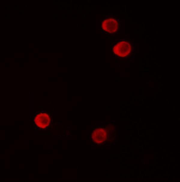
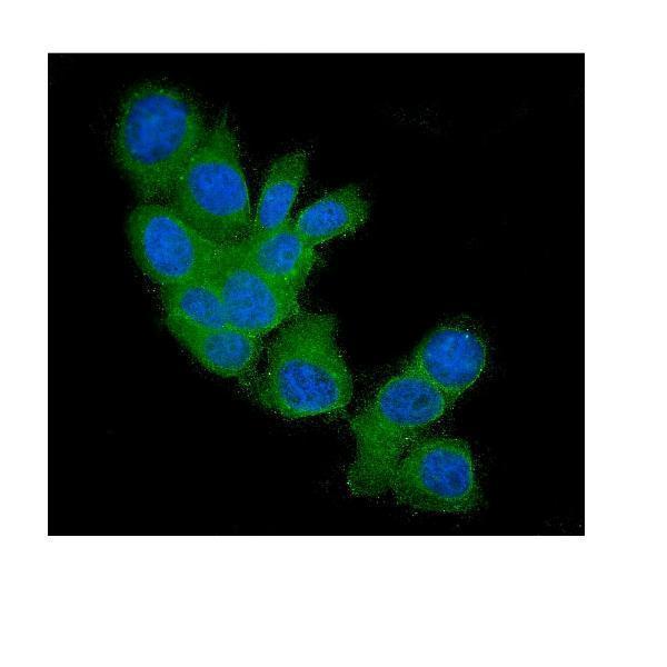


Comprehensive Human Protein Atlas IHC scoring per tissue (reliability: Enhanced — Medium consistency between antibody staining and RNA expression data.). Rows are taken directly from the HPA tissue chart — click any row's HPA link to view the source.
| Tissue | Cell type | Level | Evidence | Source |
|---|---|---|---|---|
| Cerebellum | GLUC cells - nucleus | High | Protein (IHC) | HPA → |
| Cerebral cortex | Neuronal cells | High | Protein (IHC) | HPA → |
| Kidney | Cells in tubules | High | Protein (IHC) | HPA → |
| Parathyroid gland | Glandular cells | High | Protein (IHC) | HPA → |
| Rectum | Endocrine cells | High | Protein (IHC) | HPA → |
| Tissue | Cell type | Level | Evidence | Source |
|---|---|---|---|---|
| Vagina | Squamous epithelial cells | Not detected | Protein (IHC) | HPA → |
Troubleshoot FHIT staining in paraffin sections by checking retrieval, compartment, cell type, controls and scoring before interpreting weak or absent signal.
Anti-FHIT antibodies have IHC data from human mammary cancer paraffin sections (PB9181 image caption) and IF data from human cell samples (A01200, M01200 image captions). Rat and mouse reactivity are catalog-listed (catalog: PB9181, A01200).
PB9181 has IHC data from a human mammary cancer paraffin section and is listed for IHC and IF (PB9181 image caption; catalog: PB9181 applications). A01200 has IF data from HeLa cells, while M01200 has IF data from MCF7 cells and is listed for IF/ICC (A01200 and M01200 image captions; catalog: M01200 applications).
Which to pick: Choose PB9181 for paraffin-section IHC: its IHC caption documents heat retrieval in EDTA at pH 8.0 and primary antibody at 1 μg/ml; the fixative is unreported (PB9181 image caption). For IF/ICC, M01200 is a mouse monoclonal listed for both applications, while A01200 is a rabbit antibody listed for IF (catalog: M01200 clone, host and applications; catalog: A01200 host and applications). For cross-species planning, PB9181 lists human and rat reactivity, and A01200 lists human and mouse reactivity; the cited IHC and IF figures show human samples (catalog: PB9181 and A01200 reactivity; PB9181 and A01200 image captions).