This website uses cookies to ensure you get the best experience on our website.
- Table of Contents
2 Citations 7 Q&As
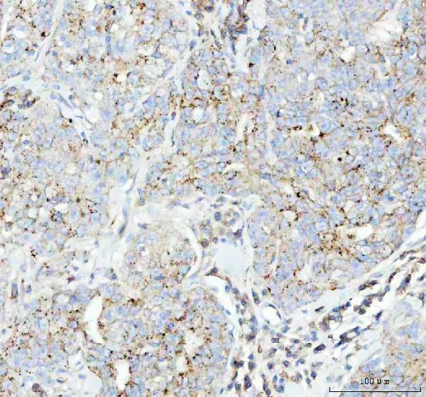
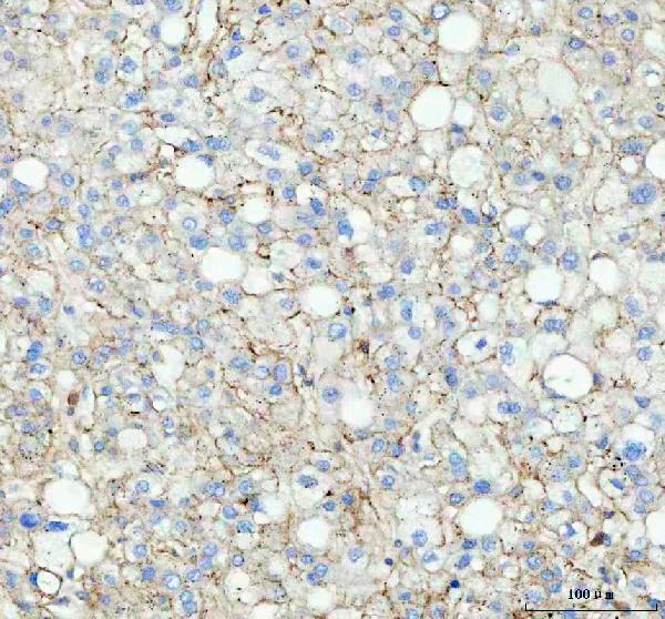
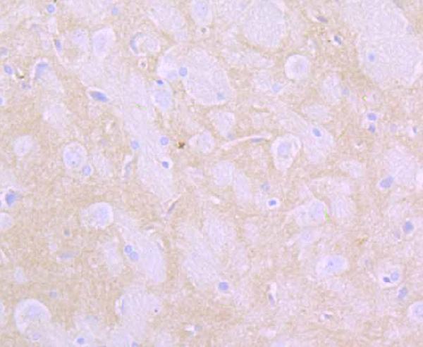
Facts about Flotillin-1.

| Human | |
|---|---|
| Gene Name: | FLOT1 |
| Uniprot: | O75955 |
| Entrez: | 10211 |

| Belongs to: |
|---|
| band 7/mec-2 family |

flotillin 1; flotillin-1; integral membrane component of caveolae
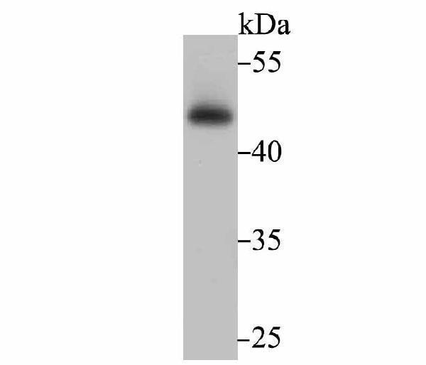
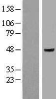
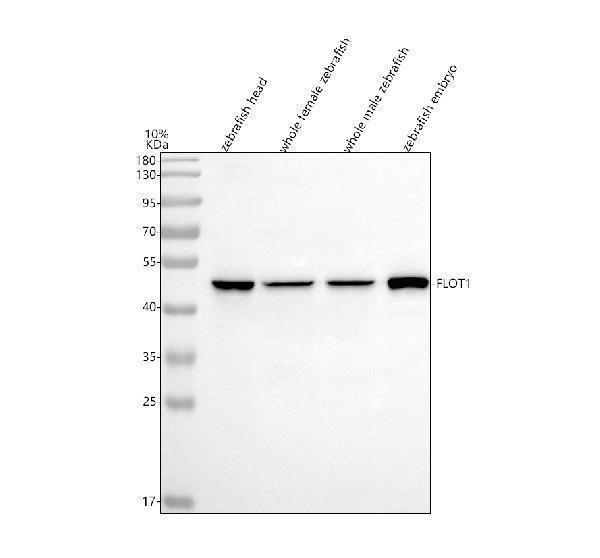
Mass (kDA):
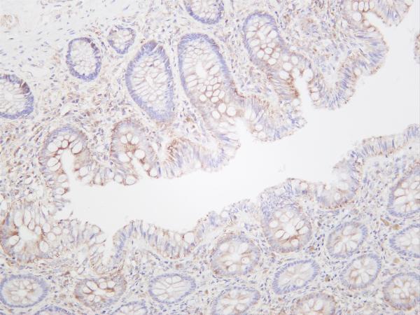
47.355 kDA

| Human | |
|---|---|
| Location: | 6p21.33 |
| Sequence: | 6; NC_000006.12 (30727709..30742851, complement) |
Cell membrane; Peripheral membrane protein. Endosome. Membrane, caveola; Peripheral membrane protein. Melanosome. Membrane raft. Identified by mass spectrometry in melanosome fractions from stage I to stage IV (PubMed:17081065). Membrane-associated protein of caveola (By similarity).


PMID: 11167132 by Edgar A.J., et al. Flotillin-1: gene structure: cDNA cloning from human lung and the identification of alternative polyadenylation signals.
PMID: 20682791 by Gorbea C., et al. A protein interaction network for Ecm29 links the 26 S proteasome to molecular motors and endosomal components.
*More publications can be found for each product on its corresponding product page