Click image to see more details
-

-
-
-
-
+6
Product Info Summary
| SKU: | PA2033 |
|---|---|
| Size: | 100 μg/vial |
| Reactive Species: | Human, Mouse, Rat |
| Host: | Rabbit |
| Application: | IP, IF, IHC, WB |
Customers Who Bought This Also Bought

Product info
Product Name
Anti-Flotillin 1/FLOT1 Antibody Picoband®
SKU/Catalog Number
PA2033
BA2904-2 is an alternative SKU for this antibody, used in previous lots.
Size
100 μg/vial
Form
Lyophilized
Description
Boster Bio Anti-Flotillin 1/FLOT1 Antibody catalog # PA2033. Tested in IF, IP, IHC, WB applications. This antibody reacts with Human, Mouse, Rat. The brand Picoband indicates this is a premium antibody that guarantees superior quality, high affinity, and strong signals with minimal background in Western blot applications. Only our best-performing antibodies are designated as Picoband, ensuring unmatched performance.
Storage & Handling
Store at -20˚C for one year from date of receipt. After reconstitution, at 4˚C for one month. It can also be aliquotted and stored frozen at -20˚C for six months. Avoid repeated freeze-thaw cycles.
Cite This Product
Anti-Flotillin 1/FLOT1 Antibody Picoband® (Boster Biological Technology, Pleasanton CA, USA, Catalog # PA2033)
Host
Rabbit
Contents
Each vial contains 4 mg Trehalose, 0.9 mg NaCl and 0.2 mg Na2HPO4.
Clonality
Polyclonal
Isotype
Rabbit IgG
Immunogen
A synthetic peptide corresponding to a sequence in the middle region of human Flotillin 1, different from the related rat and mouse sequences by one amino acid.
Cross-reactivity
No cross-reactivity with other proteins
Reactive Species
PA2033 is reactive to FLOT1 in Human, Mouse, Rat
Observed Molecular Weight
47 kDa
Calculated molecular weight
47.4 kDa
Background of FLOT1
FLOT1 (Flotillin 1), is a protein that in humans is encoded by the FLOT1 gene. The International Radiation Hybrid Mapping Consortium mapped the FLOT1 gene to chromosome 6. Bickel et al. (1997) found that mouse Flot1 behaves as a resident integral membrane protein of caveolae. It consistently copurified with Flot2 and with caveolin-1 in the purification of caveolin-rich membranes. Hazarika et al. (1999) found that stable transfection of Flot1, which they called ESA/flotillin-2, in COS-1 cells induced filopodia formation and changed the epithelial morphology to that of neuronal cells. Santamaria et al. (2005) found that prostate tumor overexpressed gene-1 interacted with flotillin-1 in detergent-insoluble membrane fractions. Flotillin-1 colocalized with PTOV1 at the plasma membrane and in the nucleus, and it entered the nucleus concomitant with PTOV1 shortly before initiation of S phase.
Antibody Validation
Boster validates all antibodies on WB, IHC, ICC, Immunofluorescence, and ELISA with known positive control and negative samples to ensure specificity and high affinity, including thorough antibody incubations.
Application & Images
Applications
PA2033 is guaranteed for IP, IF, IHC, WB Boster Guarantee
Assay Dilutions Recommendation
The recommendations below provide a starting point for assay optimization. The actual working concentration varies and should be decided by the user.
Western blot, 0.1-0.5μg/ml, Human, Mouse, Rat
Immunohistochemistry(Paraffin-embedded Section), 2-5 μg/ml, Human
Immunofluorescence, 5 μg/ml, Human
Immunoprecipitation, 0.5-2 μg/ml, Human
Positive Control
WB: human A431 whole cell lysates, human U251 whole cell lysates, human PC-3 whole cell lysates, human Hacat whole cell lysates, rat brain tissue lysates, rat skin tissue lysates, mouse brain tissue lysates, mouse skin tissue lysates
IHC: human stomach camcer, human liver cancer tissue, human thyroid cancer tissue, mouse brain, rat brain
IP: A431 cell
IF: human non-small cell lung cancer tissue
Validation Images & Assay Conditions
Click image to see more details
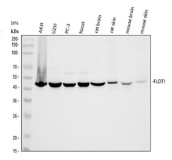
Western blot analysis of FLOT1 using anti-FLOT1 antibody (PA2033).
Electrophoresis was performed on a 5-20% SDS-PAGE gel at 70V (Stacking gel) / 90V (Resolving gel) for 2-3 hours. The sample well of each lane was loaded with 30 ug of sample under reducing conditions.
Lane 1: human A431 whole cell lysates,
Lane 2: human U251 whole cell lysates,
Lane 3: human PC-3 whole cell lysates,
Lane 4: human Hacat whole cell lysates,
Lane 5: rat brain tissue lysates,
Lane 6: rat skin tissue lysates,
Lane 7: mouse brain tissue lysates,
Lane 8: mouse skin tissue lysates.
After electrophoresis, proteins were transferred to a nitrocellulose membrane at 150 mA for 50-90 minutes. Blocked the membrane with 5% non-fat milk/TBS for 1.5 hour at RT. The membrane was incubated with rabbit anti-FLOT1 antigen affinity purified polyclonal antibody (Catalog # PA2033) at 0.5 μg/mL overnight at 4°C, then washed with TBS-0.1%Tween 3 times with 5 minutes each and probed with a goat anti-rabbit IgG-HRP secondary antibody at a dilution of 1:5000 for 1.5 hour at RT. The signal is developed using an Enhanced Chemiluminescent detection (ECL) kit (Catalog # EK1002) with Tanon 5200 system. A specific band was detected for FLOT1 at approximately 47 kDa. The expected band size for FLOT1 is at 47 kDa.

Click image to see more details
IHC analysis of FLOT1 using anti-FLOT1 antibody (PA2033).
FLOT1 was detected in a paraffin-embedded section of human stomach camcer tissue. Heat mediated antigen retrieval was performed in EDTA buffer (pH 8.0, epitope retrieval solution). The tissue section was blocked with 10% goat serum. The tissue section was then incubated with 2 μg/ml rabbit anti-FLOT1 Antibody (PA2033) overnight at 4°C. Peroxidase Conjugated Goat Anti-rabbit IgG was used as secondary antibody and incubated for 30 minutes at 37°C. The tissue section was developed using HRP Conjugated Rabbit IgG Super Vision Assay Kit (Catalog # SV0002) with DAB as the chromogen.

Click image to see more details
IHC analysis of FLOT1 using anti-FLOT1 antibody (PA2033).
FLOT1 was detected in a paraffin-embedded section of mouse brain tissue. Heat mediated antigen retrieval was performed in EDTA buffer (pH 8.0, epitope retrieval solution). The tissue section was blocked with 10% goat serum. The tissue section was then incubated with 2 μg/ml rabbit anti-FLOT1 Antibody (PA2033) overnight at 4°C. Peroxidase Conjugated Goat Anti-rabbit IgG was used as secondary antibody and incubated for 30 minutes at 37°C. The tissue section was developed using HRP Conjugated Rabbit IgG Super Vision Assay Kit (Catalog # SV0002) with DAB as the chromogen.

Click image to see more details
IHC analysis of FLOT1 using anti-FLOT1 antibody (PA2033).
FLOT1 was detected in a paraffin-embedded section of rat brain tissue. Heat mediated antigen retrieval was performed in EDTA buffer (pH 8.0, epitope retrieval solution). The tissue section was blocked with 10% goat serum. The tissue section was then incubated with 2 μg/ml rabbit anti-FLOT1 Antibody (PA2033) overnight at 4°C. Peroxidase Conjugated Goat Anti-rabbit IgG was used as secondary antibody and incubated for 30 minutes at 37°C. The tissue section was developed using HRP Conjugated Rabbit IgG Super Vision Assay Kit (Catalog # SV0002) with DAB as the chromogen.

Click image to see more details
IHC analysis of FLOT1 using anti-FLOT1 antibody (PA2033).
FLOT1 was detected in a paraffin-embedded section of human liver cancer tissue. Heat mediated antigen retrieval was performed in EDTA buffer (pH 8.0, epitope retrieval solution). The tissue section was blocked with 10% goat serum. The tissue section was then incubated with 2 μg/ml rabbit anti-FLOT1 Antibody (PA2033) overnight at 4°C. Peroxidase Conjugated Goat Anti-rabbit IgG was used as secondary antibody and incubated for 30 minutes at 37°C. The tissue section was developed using HRP Conjugated Rabbit IgG Super Vision Assay Kit (Catalog # SV0002) with DAB as the chromogen.

Click image to see more details
IHC analysis of FLOT1 using anti-FLOT1 antibody (PA2033).
FLOT1 was detected in a paraffin-embedded section of human thyroid cancer tissue. Heat mediated antigen retrieval was performed in EDTA buffer (pH 8.0, epitope retrieval solution). The tissue section was blocked with 10% goat serum. The tissue section was then incubated with 2 μg/ml rabbit anti-FLOT1 Antibody (PA2033) overnight at 4°C. Peroxidase Conjugated Goat Anti-rabbit IgG was used as secondary antibody and incubated for 30 minutes at 37°C. The tissue section was developed using HRP Conjugated Rabbit IgG Super Vision Assay Kit (Catalog # SV0002) with DAB as the chromogen.

Click image to see more details
IF analysis of FLOT1 using anti-FLOT1 antibody (PA2033).
FLOT1 was detected in a paraffin-embedded section of human non-small cell lung cancer tissue. Heat mediated antigen retrieval was performed in EDTA buffer (pH 8.0, epitope retrieval solution). The tissue section was blocked with 10% goat serum. The tissue section was then incubated with 5 μg/mL rabbit anti-FLOT1 Antibody (PA2033) overnight at 4°C. Cy3 Conjugated Goat Anti-Rabbit IgG (BA1032) was used as secondary antibody at 1:500 dilution and incubated for 30 minutes at 37°C. The section was counterstained with DAPI. Visualize using a fluorescence microscope and filter sets appropriate for the label used.

Click image to see more details
Immunoprecipitating FLOT1 in A431 whole cell lysate.
Western blot analysis of FLOT1 using anti-FLOT1 antibody (PA2033).
Lane 1: A431 whole cell lysates (30ug),
Lane 2: Rabbit control IgG instead of anti-FLOT1 antibody in A431 whole cell lysate,
Lane 3: anti-FLOT1 antibody (2μg) + A431 whole cell lysate (500μg).
After electrophoresis, proteins were transferred to a membrane. Then the membrane was incubated with rabbit anti-FLOT1 antigen affinity purified polyclonal antibody (PA2033) at a dilution of 0.5 μg/mL and probed with a mouse anti-rabbit IgG-HRP secondary antibody (Light Chain). The signal is developed using ECL Plus Western Blotting Substrate (Catalog # AR1196-200). A specific band was detected for FLOT1 at approximately 47 kDa. The expected band size for FLOT1 is at 47 kDa.
Specific Publications For Anti-Flotillin 1/FLOT1 Antibody Picoband® (PA2033)
Loading publications
Recommended Resources
Here are featured tools and databases that you might find useful.
- Boster's Pathways Library
- Protein Databases
- Bioscience Research Protocol Resources
- Data Processing & Analysis Software
- Photo Editing Software
- Scientific Literature Resources
- Research Paper Management Tools
- Molecular Biology Software
- Primer Design Tools
- Bioinformatics Tools
- Phylogenetic Tree Analysis
Customer Reviews
Have you used Anti-Flotillin 1/FLOT1 Antibody Picoband®?
Share your experimental results or join a short interview to earn up to $1,000 in product credits or other rewards.
0 Reviews For Anti-Flotillin 1/FLOT1 Antibody Picoband®
Customer Q&As
Have a question?
Find answers in Q&As, reviews.
Can't find your answer?
Submit your question
7 Customer Q&As for Anti-Flotillin 1/FLOT1 Antibody Picoband®
Question
Does PA2033 anti-Flotillin 1/FLOT1 antibody work on parafin embedded sections? If so, which fixation method do you recommend we use (PFA, paraformaldehyde, other)?
Verified Customer
Verified customer
Asked: 2019-12-12
Answer
As indicated on the product datasheet, PA2033 anti-Flotillin 1/FLOT1 antibody as been validated on WB. It is best to use PFA for fixation because it has better tissue penetration ability. PFA needs to be prepared fresh before use. Long term stored PFA turns into formalin, as the PFA molecules congregate and become formalin.
Boster Scientific Support
Answered: 2019-12-12
Question
I am interested in to test anti-Flotillin 1/FLOT1 antibody PA2033 on rat stomach for research purposes, then I may be interested in using anti-Flotillin 1/FLOT1 antibody PA2033 for diagnostic purposes as well. Is the antibody suitable for diagnostic purposes?
Verified Customer
Verified customer
Asked: 2019-11-13
Answer
The products we sell, including anti-Flotillin 1/FLOT1 antibody PA2033, are only intended for research use. They would not be suitable for use in diagnostic work. If you have the means to develop a product into diagnostic use, and are interested in collaborating with us and develop our product into an IVD product, please contact us for more discussions.
Boster Scientific Support
Answered: 2019-11-13
Question
Is this PA2033 anti-Flotillin 1/FLOT1 antibody reactive to the isotypes of FLOT1?
Verified Customer
Verified customer
Asked: 2019-10-09
Answer
The immunogen of PA2033 anti-Flotillin 1/FLOT1 antibody is A synthetic peptide corresponding to a sequence in the middle region of human Flotillin 1(219-234aa KKAAYDIEVNTRRAQA), different from the related rat and mouse sequences by one amino acid. Could you tell me which isotype you are interested in so I can help see if the immunogen is part of this isotype?
Boster Scientific Support
Answered: 2019-10-09
Question
Thank you for helping with my inquiry over the phone. Here are the WB image, lot number and protocol we used for stomach using anti-Flotillin 1/FLOT1 antibody PA2033. Let me know if you need anything else.
Verified Customer
Verified customer
Asked: 2019-06-27
Answer
Thanks for the data. You have provided everything we needed. Our lab team are working to resolve your inquiry as quickly as possible, and we appreciate your patience and understanding! Please let me know if there is anything you need in the meantime.
Boster Scientific Support
Answered: 2019-06-27
Question
I have attached the WB image, lot number and protocol we used for stomach using anti-Flotillin 1/FLOT1 antibody PA2033. Please let me know if you require anything else.
Verified Customer
Verified customer
Asked: 2019-06-21
Answer
Thank you very much for the data. Our lab team are working to resolve this as quickly as possible, and we appreciate your patience and understanding! You have provided everything we needed. Please let me know if there is anything you need in the meantime.
Boster Scientific Support
Answered: 2019-06-21
Question
We are currently using anti-Flotillin 1/FLOT1 antibody PA2033 for mouse tissue, and we are happy with the WB results. The species of reactivity given in the datasheet says human, mouse, rat. Is it likely that the antibody can work on dog tissues as well?
Verified Customer
Verified customer
Asked: 2018-05-04
Answer
The anti-Flotillin 1/FLOT1 antibody (PA2033) has not been validated for cross reactivity specifically with dog tissues, though there is a good chance of cross reactivity. We have an innovator award program that if you test this antibody and show it works in dog you can get your next antibody for free. Please contact me if I can help you with anything.
Boster Scientific Support
Answered: 2018-05-04
Question
Is a blocking peptide available for product anti-Flotillin 1/FLOT1 antibody (PA2033)?
M. Johnson
Verified customer
Asked: 2013-08-14
Answer
We do provide the blocking peptide for product anti-Flotillin 1/FLOT1 antibody (PA2033). If you would like to place an order for it please contact support@bosterbio.com and make a special request.
Boster Scientific Support
Answered: 2013-08-14
