FRMD6 / FERM domain-containing protein 6 · IHC design guide

Design Immunohistochemistry for FRMD6

Plan FRMD6 staining in paraffin sections using the catalog antibody's IHC protocol (datasheet A08826-2). Compare cytoplasmic staining across tissues while accounting for the uncertain reliability of the tissue profile (HPA tissue IHC).

Evidence assembled Oct 2026 · For research use; verify linked source records and product datasheet before use
Immunohistochemistry protocol sheet for FRMD6 (IHC for FRMD6): expected localisation Cytoplasmic in tissue sections (HPA tissue IHC), antibody A08826-2, validated IHC image, and IHC protocol steps
Printable FRMD6 IHC protocol sheet — expected localisation Cytoplasmic in tissue sections (HPA tissue IHC), antibody A08826-2, controls and protocol steps. Open the full FRMD6 IHC guide →

FRMD6 Immunohistochemistry Experimental Design Guide

Expected localisation, validated protocols, controls and antibodies — the at-a-glance facts below, then the full design guide.

Must know before staining
Expected localisation Cytoplasmic in tissue sections (HPA tissue IHC)
Staining pattern Cytoplasmic staining across several tissue types (HPA tissue IHC)
Antigen retrieval EDTA pH 8.0 HIER, heat-mediated (datasheet A08826-2)
Positive control ⓘ Bone marrow+4 more · see all
Negative control ⓘ Adipose tissue+4 more · see all
Important caveats
Reasons your staining may differ from the expected pattern.
Fixation Keep fixation consistent across sections. (standard IHC practice; not target-specific)
Caveat Tissue staining has uncertain reliability (HPA tissue IHC)
Regulation Expression regulation is unannotated (UniProt)
Isoform / epitope 3 isoforms; epitope coverage needs verification (UniProt)
Section 1

Recommended FRMD6 IHC & IF Protocols

The catalog antibody protocol is followed by published FRMD6 IHC methods for colorectal tissue, mouse neural cryosections, and colon carcinoma tissue microarrays (PMC6382106; PMC7002571; PMC8073668).

Recommended immunohistochemistry (IHC-P) protocol parameters
SampleParaffin-embedded human rectum cancer tissue; fixative not specified (datasheet A08826-2)
FixationImage fixative and duration unreported (datasheet A08826-2); verify before use.
Sectioning4–5 µm sections on charged slides (standard)
DeparaffinisationXylene, graded ethanol series to water (standard)
Antigen retrievalHeat retrieval: EDTA pH 8.0 (datasheet A08826-2); 20 min, 95–100 °C (standard)
Peroxidase block3% H2O2, 10 min, room temperature (standard)
Blocking10% goat serum (datasheet A08826-2)
Primary antibodyRabbit anti-FRMD6, 0.5-1μg/ml (datasheet A08826-2)
Primary incubationOvernight at 4 °C (datasheet A08826-2)
DetectionStreptavidin-biotin complex (SABC), DAB chromogen (datasheet A08826-2)
CounterstainHematoxylin, blue, dehydrate and mount (standard)
Expected resultFRMD6-positive staining in hematopoietic cells of bone marrow (HPA tissue IHC: Medium). HPA tissue profile: Cytoplasmic expression in several different tissue types. No signal in the no-primary control.
💡Decision noteStart with heat-mediated EDTA at pH 8.0 for the catalog antibody (datasheet: A08826-2). The colorectal tissue study used Tris-EDTA at pH 9.0 (PMC6382106).
Section 2

What Is the Expected FRMD6 Staining Pattern?

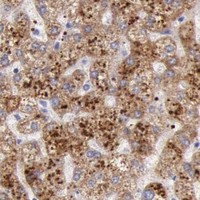
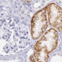
In paraffin tissue sections, expect mainly cytoplasmic FRMD6 staining in selected cells, including colon glandular cells, kidney tubule cells and hepatocytes, each reported at medium intensity (HPA: tissue IHC). UniProt also places FRMD6 at the cell membrane and reports no transmembrane segment (UniProt Q96NE9: localization and topology). Treat the tissue pattern as provisional: the available IHC antibody is rated Uncertain because staining and RNA expression show low consistency (HPA: IHC reliability).

What am I looking at on my slide?
Cytoplasmic signal appears in colon glandular cells, kidney tubule cells or hepatocytes.These cell types have medium staining in the HPA tissue panel (HPA: tissue IHC). Score the relevant cells and compartment together; medium is the reported category, not a required intensity for every section (HPA: tissue IHC; standard IHC practice).
Signal appears only in nuclei, or as a sharply restricted organelle pattern on the chromogenic slide.That pattern does not match the broad cytoplasmic tissue profile (HPA: tissue IHC). Check controls and antibody specificity before assigning it to FRMD6; UniProt also lists cell membrane localization (UniProt Q96NE9: localization).
Strong staining appears in adipocytes or salivary glandular cells while the expected cell population is weak.Both named populations are reported as not detected (HPA: tissue IHC). Consider antibody cross-reactivity or endogenous chromogenic activity, then compare a negative reagent control and a separately stained positive reference (standard IHC practice).
Brown signal covers stroma, lumina and several cell types without a clear cellular boundary.This is difficult to score as FRMD6 because the reported tissue pattern is cellular and cytoplasmic (HPA: tissue IHC). Diffuse signal can arise from incomplete blocking, excess detection reagent or nonspecific antibody binding (standard IHC practice).
No staining appears in a colon, kidney or liver section selected as a positive reference.Those named cell populations showed medium staining in the HPA panel, so an empty slide warrants a workflow check (HPA: tissue IHC). Because IHC reliability is Uncertain, one negative section alone cannot establish that FRMD6 is absent (HPA: IHC reliability).
💡Expected FRMD6 appearanceCall a result consistent with the reported pattern when cytoplasmic staining is discernible in colon glandular cells, kidney tubule cells or hepatocytes, with medium intensity as the HPA reference; isolated nuclear signal or staining confined to HPA-negative cell populations is suspect (HPA: tissue IHC; UniProt Q96NE9: localization).
How each factor affects the staining
Tissue and cell selectionHPA reports medium staining in several epithelial and hematopoietic populations, but not detection in several other listed populations (HPA: tissue IHC). Compare the exact cell type, since whole-tissue labels can conceal a mismatch (standard IHC practice).
Localization across applicationsWhy can IF images look different? HPA reports approved mitochondrial localization in ICC-IF images, while its tissue IHC profile is cytoplasmic and Uncertain (HPA: subcellular ICC-IF; HPA: tissue IHC). Judge each application against its own evidence.
Protein topologyFRMD6 has no transmembrane segment, despite its UniProt cell membrane annotation, and can colocalize with actin (UniProt Q96NE9: topology and localization). Membrane-adjacent cytoplasmic signal can therefore merit review; topology alone does not define an IHC staining boundary.
Antibody validationThe listed antibody HPA001297 is rated Uncertain for IHC and Approved for ICC (HPA: antibody validation). Its ICC status does not resolve the low consistency between tissue staining and RNA expression that underlies the IHC rating (HPA: IHC reliability).
Isoforms and epitope coverageUniProt lists 3 FRMD6 isoforms and a FERM domain at residues 16–328 (UniProt Q96NE9: isoforms and domains). No antibody epitope is supplied here, so isoform-specific staining or antigen retrieval requirements cannot be predicted from this record.
Why is my staining missing, weak or wrong?
SituationLikely causeNext action
Expected cytoplasmic cells are blank, including the selected reference section.The detection run may have failed, or the chosen section may not reproduce HPA's medium-staining examples (standard IHC practice; HPA: tissue IHC).Inspect the positive reference and reagent controls; review the run's retrieval, antibody incubation and chromogenic detection steps (standard IHC practice). Keep HPA's Uncertain IHC rating in view (HPA: IHC reliability).
The entire slide has a light brown haze.Background may reflect incomplete blocking, excessive primary antibody or prolonged chromogen development (standard IHC practice).Check the negative reagent control, then adjust blocking, antibody concentration or development time one variable at a time (standard IHC practice). Avoid scoring diffuse haze as cytoplasmic FRMD6 (HPA: tissue IHC).
Brown deposits persist in the negative reagent control.Endogenous peroxidase activity or nonspecific detection reagents may contribute to a chromogenic signal (standard IHC practice).Review the peroxidase-blocking and secondary detection steps; compare deposits with the control before interpreting any cell-specific staining (standard IHC practice).
Only nuclei stain in otherwise interpretable tissue.Nuclear-only staining conflicts with the reported cytoplasmic tissue pattern and UniProt localization (HPA: tissue IHC; UniProt Q96NE9: localization).Check counterstain and negative controls, then reassess antibody-dependent signal with a suitable positive reference (standard IHC practice). Do not call nuclear-only signal a confirmed FRMD6 pattern.
Adipocytes or salivary glandular cells stain more strongly than the chosen positive reference.These populations were not detected in the HPA panel, so cross-reactivity or background is plausible; the IHC antibody rating is Uncertain (HPA: tissue IHC; HPA: antibody validation).Compare matched negative reagent and positive reference sections, then review blocking and detection conditions (standard IHC practice). Record the discordance rather than assigning a new FRMD6 expression pattern.
ICC-IF images suggest mitochondria, but the tissue slide looks broadly cytoplasmic.The two HPA application summaries differ: mitochondrial localization is Approved in ICC-IF, whereas cytoplasmic tissue IHC is Uncertain (HPA: subcellular ICC-IF; HPA: tissue IHC).Interpret the paraffin chromogenic slide against the tissue IHC cell-type pattern and its reliability rating; report the cross-application difference without using ICC-IF localization as an IHC requirement (HPA: tissue IHC; HPA: subcellular ICC-IF).

Sample controls for FRMD6 IHC & IF

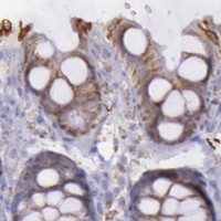
🧪Run colon first and look for staining in glandular cells, where HPA reports medium FRMD6 expression (HPA: colon glandular cells, Medium). Use adipose tissue adipocytes as the negative tissue (HPA: adipocytes, Not detected); on the colon slide, treat cells outside the reported glandular compartment as internal background comparators, without assuming HPA has established them as FRMD6-negative (HPA: colon glandular cells, Medium).
Positive control tissue: Bone marrow (Hematopoietic cells, HPA Medium)
Negative control tissue: Adipose tissue (HPA Not detected)
ICC-IF cell lines (HPA subcellular resource): HPA ICC-IF images show FRMD6 in A-431, U-251MG, U2OS, with annotated localisation: Mitochondria (approved) (HPA subcellular).
Technical controls: Include a no-primary, secondary-only control and a matched rabbit IgG isotype control for the rabbit primary, plus a matched FRMD6-knockout specimen if available (selected-SKU caption: rabbit anti-FRMD6; standard IHC practice). Block endogenous peroxidase and assess endogenous biotin background because the caption uses a streptavidin–biotin complex with DAB (selected-SKU caption: SABC/DAB; standard IHC practice).
⚠️Feasibility: No target-specific fixation window or fixation effect is reported, and the selected-SKU tissue-IHC caption leaves the fixative unreported (selected-SKU caption: fixative not stated). Heat retrieval in EDTA at pH 8.0 is a documented starting condition for paraffin sections, but the caption does not establish that retrieval is required or that frozen sections or IF are easier (selected-SKU caption: heat retrieval in EDTA, pH 8.0). In colon, distinguish glandular-cell staining from material in glandular lumens when scoring, using the no-primary slide to assess background (HPA: colon glandular cells, Medium; standard IHC practice).

HPA tissue IHC evidence for FRMD6

Comprehensive Human Protein Atlas IHC scoring per tissue (reliability: Uncertain — Low consistency between antibody staining and RNA expression data.). Rows are taken directly from the HPA tissue chart — click any row's HPA link to view the source.

Positive expression · recommended positive controls

TissueCell typeLevelEvidenceSource
Bone marrow Hematopoietic cells Medium Protein (IHC) HPA →
Bronchus Respiratory epithelial cells Medium Protein (IHC) HPA →
Colon Glandular cells Medium Protein (IHC) HPA →
Duodenum Glandular cells Medium Protein (IHC) HPA →
Epididymis Glandular cells Medium Protein (IHC) HPA →

Undetected expression · recommended negative controls

TissueCell typeLevelEvidenceSource
Adipose tissue Adipocytes Not detected Protein (IHC) HPA →
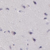
Caudate Glial cells Not detected Protein (IHC) HPA →
Cerebellum Cells in granular layer Not detected Protein (IHC) HPA →
Hippocampus Glial cells Not detected Protein (IHC) HPA →
Ovary Ovarian stroma cells Not detected Protein (IHC) HPA →
Section 3

Advanced FRMD6 IHC Tips

Troubleshoot FRMD6 staining in paraffin sections by checking retrieval, compartment, cell type and controls before interpreting chromogenic signal.

What retrieval should I try first if FRMD6 staining is weak?
Start with heat-mediated retrieval in EDTA pH 8.0 for paraffin sections (datasheet A08826-2). The selected tissue image used this retrieval before 1 μg/mL primary antibody overnight at 4°C, but its caption does not specify heating time or fixative (datasheet A08826-2). Keep the detection sequence comparable while checking whether staining improves in an expected positive cell population, such as colon glandular cells (datasheet A08826-2; HPA: Medium in colon glandular cells). If staining remains weak, compare a separately controlled retrieval condition and record tissue preservation alongside signal, since excessive heating can damage section morphology (standard IHC practice).
Could fixation explain weak or uneven FRMD6 staining?
FRMD6-specific sensitivity to fixation is unknown: the selected paraffin-section caption does not report a fixative, and the available record provides no target-specific fixation comparison (datasheet A08826-2; UniProt Q96NE9). Record each specimen’s fixative and fixation duration, then compare sections processed together under the stated EDTA pH 8.0 retrieval condition (datasheet A08826-2; standard IHC practice). Inspect morphology and staining across the section before changing antibody concentration, because processing differences can produce uneven antigen access (standard IHC practice). Use a matched positive-control section in each run; a negative result alone cannot establish absent FRMD6 when fixation history is uncertain (standard IHC practice).
Which FRMD6 staining pattern should I accept in tissue sections?
Assess cytoplasmic staining first: tissue IHC reports cytoplasmic expression across several tissue types, although its antibody-to-RNA consistency is rated uncertain (HPA: tissue IHC). Peripheral staining can be plausible because FRMD6 is annotated in the cytoplasm and at the cell membrane, with possible actin colocalisation and no transmembrane segment (UniProt Q96NE9). An approved mitochondrial location comes from subcellular ICC/IF data and should prompt comparison with tissue-cell morphology rather than automatically redefine a chromogenic pattern (HPA: subcellular; standard IHC practice). Compare the same compartment in expected positive and lower-signal cells, such as colon glandular cells and adipocytes, while checking that the counterstain preserves cell boundaries (HPA: tissue IHC; standard IHC practice).
How can isoforms or epitope placement affect my FRMD6 result?
FRMD6 has 3 annotated isoforms and a FERM domain at residues 16–328, but the supplied antibody evidence does not map its epitope to a shared region (UniProt Q96NE9; datasheet A08826-2). Therefore, a negative chromogenic result cannot identify which isoform is absent, and an unexpected compartment cannot establish isoform-specific trafficking (UniProt Q96NE9; standard IHC interpretation). Document the antibody’s epitope information if it becomes available, and compare staining with an independently validated reagent recognizing a distinct region (standard IHC practice). Phosphorylation sites include residues 522–544; their presence in the sequence does not establish that this antibody detects phosphorylation-dependent staining (UniProt Q96NE9; datasheet A08826-2).
How should I compare an IF pattern with this chromogenic FRMD6 result?
Use IF as a separate comparison with the chromogenic tissue result, since the selected antibody caption documents paraffin-section IHC and supplies no IF validation for that antibody (datasheet A08826-2). Multiplex FRMD6 with a marker that identifies the cell population under study, then check whether the two signals occupy the same cells while keeping compartment assignment separate (standard IF practice). Choose a fluorophore channel with low tissue autofluorescence and include single-label controls to detect bleed-through before comparing IF intensity with DAB staining (standard IF practice). Because FRMD6 has no transmembrane segment and is annotated in the cytoplasm and at the cell membrane, assess whether permeabilisation exposes the intracellular epitope being tested (UniProt Q96NE9; standard IF practice).
How do I reduce diffuse DAB background without losing FRMD6 signal?
First compare the stained section with a no-primary control and inspect whether deposits follow cells or collect at folds, edges and damaged areas (standard IHC practice). The selected tissue image used 10% goat serum blocking, 1 μg/mL primary overnight at 4°C, and a biotinylated secondary followed by SABC and DAB (datasheet A08826-2). Apply a peroxidase block for chromogenic detection, and check endogenous biotin background when using the stated biotin-based detection sequence (standard IHC practice; datasheet A08826-2). If background persists, adjust blocking, washing or primary concentration one variable at a time while retaining an expected positive tissue control (standard IHC practice).
What should I measure when comparing FRMD6 across sections? ⚠ ANSWER MARKED FOR VERIFICATION
Define the cell population and cytoplasmic scoring compartment before examining group assignments, since tissue IHC describes cytoplasmic FRMD6 expression but rates antibody-to-RNA consistency uncertain (HPA: tissue IHC; standard IHC practice). For a cell-based result, report the percentage of positive target cells and an H-score calculated from intensity grades 0–3, giving a possible range of 0–300 (standard IHC scoring practice). If counting positive cells per mm², normalise to the viable tissue area actually assessed and report the number of evaluable target cells (standard IHC quantification practice). Keep retrieval, DAB development, counterstain and image-analysis thresholds consistent across compared sections, and exclude folds or necrotic regions by predefined rules (standard IHC practice).
When is apparent FRMD6 positivity more likely to be artefact?
Treat reproducible staining in identifiable cytoplasmic cell regions as more credible when it occurs in an expected population, such as colon glandular cells or kidney tubule cells (HPA: Medium in both populations; standard IHC interpretation). Strong nuclear-only deposits warrant scrutiny because the supplied localisation annotations identify cytoplasm, cell membrane and, in ICC/IF, mitochondria (UniProt Q96NE9; HPA: subcellular). Check whether signal instead tracks section edges, necrosis or the no-primary control, and exclude endogenous peroxidase deposits before assigning DAB positivity (standard IHC practice). Even a convincing pattern needs independent support: HPA rates tissue staining reliability uncertain because antibody staining and RNA expression show low consistency (HPA: tissue IHC).
Boster reagents

Best FRMD6 / FERM domain-containing protein 6 IHC Antibodies

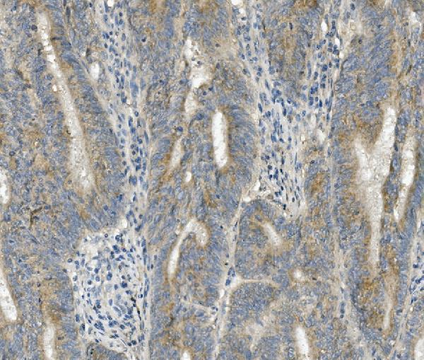
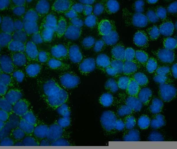
Anti-FRMD6 antibody A08826-2 has IHC data from paraffin-embedded human rectum cancer tissue and IF data from A431 cells (catalog image captions); its listed reactivity is human, mouse, and rat (catalog reactivity).

Real IHC data IHC analysis of FRMD6 using anti-FRMD6 antibody (A08826-2). FRMD6 was detected in paraffin-embedded section of human rectum cancer tissue. Heat mediated antigen retrieval was performed in EDTA buffer (pH8.0, epitope retrieval solution). The tissue section was blocked with 10% goat serum. The tissue section was then incubated with 1μg/ml rabbit anti-FRMD6 Antibody (A08826-2) overnight at 4°C. Biotinylated goat anti-rabbit IgG was used as secondary antibody and incubated for 30 minutes at 37°C. The tissue section was developed using Strepavidin-Biotin-Complex (SABC) (Catalog # SA1022) with DAB as the chromogen.
Anti-FRMD6/Willin Antibody ®
Cat # A08826-2

A08826-2 is listed for IHC and has an IHC image from a paraffin-embedded human rectum cancer section (catalog applications; IHC image caption). The same SKU is listed for IF/ICC and has an IF image from A431 cells (catalog applications; IF image caption).

Which to pick: Choose A08826-2 for paraffin-section IHC: its image caption reports EDTA retrieval at pH 8.0 and a primary concentration of 1 μg/ml in human rectum cancer tissue; the fixative is unreported (IHC image caption). For IF/ICC, the same SKU has an A431-cell image at 2 μg/ml (IF image caption). Human, mouse, and rat are listed as reactive species, but the supplied IHC image shows human tissue only; clonality is unreported (catalog reactivity; IHC image caption; catalog payload).

Each figure is that product's own IHC / IF validation image from its datasheet.

References

  1. UniProt Consortium. UniProt entry Q96NE9 (FRMD6_HUMAN, FERM domain-containing protein 6).
  2. Human Protein Atlas. FRMD6 tissue IHC expression (reliability: Uncertain).
  3. Human Protein Atlas. FRMD6 subcellular location (ICC-IF): Localized to the mitochondria..
  4. Human Protein Atlas. FRMD6 antibody validation summary (1 antibodies).
  5. Early-stage serrated adenocarcinomas are divided into several molecularly distinct subtypes. PloS one 2019 — PMC6382106.
  6. Expression and regulation of FRMD6 in mouse DRG neurons and spinal cord after nerve injury. Scientific reports 2020 — PMC7002571.
  7. PD-L1 as a Prognostic Factor in Early-Stage Colon Carcinoma within the Immunohistochemical Molecular Subtype Classification. Cancers 2021 — PMC8073668.
  8. FRMD6 inhibits human glioblastoma growth and progression by negatively regulating activity of receptor tyrosine kinases. Oncotarget 2016 — PMC5342536.
  9. PubMed PMID:14702039 — UniProt-cited evidence.
  10. PubMed PMID:17974005 — UniProt-cited evidence.
  11. PubMed PMID:12508121 — UniProt-cited evidence.