FYN / Tyrosine-protein kinase Fyn · IHC design guide

Design Immunohistochemistry for FYN

FYN shows cytoplasmic expression in several tissues, including lymphoid organs (HPA tissue IHC). Use tonsil non-germinal center cells as a positive reference (HPA tissue IHC) and start the catalog antibody at 1:25 for IHC-P (datasheet: 1:25).

Evidence assembled Oct 2026 · For research use; verify linked source records and product datasheet before use
Immunohistochemistry protocol sheet for FYN (IHC for FYN): expected localisation Cytoplasmic staining in several tissues (HPA tissue IHC), antibody M00684-2, validated IHC image, and IHC protocol steps
Printable FYN IHC protocol sheet — expected localisation Cytoplasmic staining in several tissues (HPA tissue IHC), antibody M00684-2, controls and protocol steps. Open the full FYN IHC guide →

FYN Immunohistochemistry Experimental Design Guide

Expected localisation, validated protocols, controls and antibodies — the at-a-glance facts below, then the full design guide.

Must know before staining
Expected localisation Cytoplasmic staining in several tissues (HPA tissue IHC)
Staining pattern Cytoplasmic staining in lymphoid organs (HPA tissue IHC)
Antigen retrieval Tris-EDTA pH 9.0 HIER, 95–98 °C, 20 min (rule: nuclear antigen)
Positive control ⓘ Fallopian tube+4 more · see all
Negative control ⓘ Adipose tissue+4 more · see all
Important caveats
Reasons your staining may differ from the expected pattern.
Fixation Keep fixation consistent across sections. (standard IHC practice; not target-specific)
Caveat Staining and RNA show low consistency (HPA tissue IHC)
Regulation T-cell isoform 2 expression (UniProt)
Isoform / epitope 3 isoforms; verify epitope coverage (UniProt)
Section 1

Recommended FYN IHC & IF Protocols

The catalog antibody IHC-P protocol is followed by four published FYN IHC methods, including three paraffin-section studies and one frozen-section study (PMC11391251; PMC2741693; PMC5358011; PMC9468808).

Recommended immunohistochemistry (IHC-P) protocol parameters
SampleParaffin-embedded H. tonsil tissue; fixative not specified (datasheet M00684-2)
FixationImage fixative and duration unreported (datasheet M00684-2); verify before use.
Sectioning4–5 µm sections on charged slides (standard)
DeparaffinisationXylene, graded ethanol series to water (standard)
Antigen retrievalHeat-induced epitope retrieval in Tris-EDTA buffer, pH 9.0, 20 min at 95–98 °C (standard rule: nuclear antigen)
Peroxidase block3% H2O2, 10 min, room temperature (standard)
Blocking10% normal serum of the secondary host, 30 min, room temperature (standard)
Primary antibodyMouse monoclonal (clone 1302CT390.118.237) anti-FYN, 1:25 (datasheet M00684-2)
Primary incubationOvernight at 4 °C (standard)
DetectionHRP-polymer secondary, DAB chromogen 5–10 min (standard)
CounterstainHematoxylin, blue, dehydrate and mount (standard)
Expected resultFYN-positive staining in ciliated cells (tip of cilia) of fallopian tube (HPA tissue IHC: High). HPA tissue profile: Cytoplasmic expression in several tissues, including lymphoid organs. No signal in the no-primary control.
💡Decision noteStart with Tris-EDTA pH 9.0 HIER at 95–98 °C for 20 min (page retrieval); two paraffin-section studies used pH 9.0 autoclave retrieval (PMC11391251; PMC9468808).
Section 2

What Is the Expected FYN Staining Pattern?

FYN is a non-transmembrane kinase found at the cell membrane and in the cytoplasm; UniProt also lists nuclear and perikaryal localization (UniProt P06241). In tissue IHC, expect cytoplasmic staining in several tissues, including lymphoid organs, with stronger staining in lymph-node and tonsil non-germinal-center cells (HPA tissue IHC). Treat this as a reference pattern, since HPA rates the IHC evidence Approved but reports low consistency with RNA expression (HPA tissue IHC).

What am I looking at on my slide?
Cytoplasmic staining in lymph-node or tonsil non-germinal-center cells, with some membrane-associated signal.This fits HPA's High staining in those cell populations and its broader cytoplasmic tissue profile (HPA tissue IHC). Membrane association is biologically plausible, but the supported plasma-membrane assignment comes from ICC-IF rather than paraffin IHC (HPA subcellular; UniProt P06241).
Predominantly nuclear staining with little expected cytoplasmic staining.Investigate staining artefact or antibody specificity before scoring it as the expected IHC pattern (HPA tissue IHC; standard IHC practice). Nuclear localization is listed by UniProt, so a nuclear signal alone does not prove cross-reactivity (UniProt P06241).
Strong staining in a cell population listed as Not detected, such as adipocytes in adipose tissue.Check cell identity and controls; cross-reactivity or endogenous detection activity may explain the signal (HPA tissue IHC; standard IHC practice). HPA's Not detected designation describes its observed pattern, not a universal absence of FYN (HPA tissue IHC).
Uniform colour across cells and extracellular areas, obscuring cell boundaries.This is diffuse background rather than an interpretable cell-specific pattern (standard IHC practice). Examine blocking, washes, primary-antibody concentration and detection controls before assigning FYN positivity (standard IHC practice).
No staining in lymph-node or tonsil non-germinal-center cells.A negative result in these HPA High reference populations warrants a control and workflow check (HPA tissue IHC). Confirm tissue integrity, retrieval and detection performance before interpreting an experimental section as FYN-negative (standard IHC practice).
💡Expected FYN appearanceCall a section positive when identifiable lymph-node or tonsil non-germinal-center cells show convincing cytoplasmic FYN staining, potentially with membrane-associated signal; HPA rates these cells High, while uniform haze or strong staining in an HPA Not detected population needs investigation (HPA tissue IHC; HPA subcellular; standard IHC practice).
How each factor affects the staining
Which compartment should guide IHC scoring?Use the cytoplasmic tissue pattern as the IHC reference (HPA tissue IHC). FYN has no transmembrane segment, yet UniProt lists cell-membrane localization and lipid-raft activity; membrane-associated staining is plausible without requiring a continuous membrane rim (UniProt P06241).
Which cell populations provide useful comparisons?Lymph-node and tonsil non-germinal-center cells are High, whereas bone-marrow hematopoietic cells and cerebral-cortex neuropil are Medium (HPA tissue IHC). Adipocytes in adipose tissue are listed as Not detected; compare the specified cells, not an entire tissue indiscriminately (HPA tissue IHC).
Can isoforms alter interpretation?UniProt lists three FYN isoforms: isoform 1 is highly expressed in brain, and isoform 2 occurs in hemopoietic lineages, especially T-lymphocytes (UniProt P06241). The supplied evidence does not establish which isoforms the IHC antibodies detect, so tissue staining cannot be assigned to one isoform here (UniProt P06241; HPA antibodies).
How strong is the tissue-pattern evidence?HPA calls the tissue IHC result Approved while reporting low consistency between staining and RNA expression (HPA tissue IHC). Its listed IHC antibodies have Approved status, but the supplied record does not establish an IHC Enhanced pattern; use cell-level morphology and controls when interpreting an unexpected result (HPA antibodies; standard IHC practice).
What should IF/ICC show?HPA reports mainly plasma-membrane FYN with additional cytosolic localization, both supported in ICC-IF (HPA subcellular). This answers the localization question only; ICC-IF images do not establish a paraffin-section staining intensity or an IHC protocol (HPA subcellular; standard IHC practice).
Why is my staining missing, weak or wrong?
SituationLikely causeNext action
Expected lymphoid cells are unstained.Target-specific fixation effects are not established by the supplied assay evidence. Verify with a matched IHC source before attributing a result to fixation.Check a concurrently processed positive section and detection controls, then optimize retrieval and antibody concentration using the validated IHC instructions for the chosen antibody (standard IHC practice).
Slide-wide brown haze obscures cells.Excess primary antibody, incomplete washing or nonspecific detection can elevate background (standard IHC practice).Compare a primary-omission control, improve washes and blocking, and adjust antibody concentration within its validated IHC range (standard IHC practice).
Signal appears in an HPA Not detected cell population.Cross-reactivity, mistaken cell identification or endogenous detection activity are possibilities; HPA's entry alone cannot identify the cause (HPA tissue IHC; standard IHC practice).Verify morphology and compare primary-omission and appropriate endogenous-activity controls before calling the cells FYN-positive (standard IHC practice).
Only nuclei stain strongly.This diverges from HPA's mainly cytoplasmic tissue profile, although UniProt lists the nucleus as a possible FYN location (HPA tissue IHC; UniProt P06241).Check counterstain and detection controls, then assess whether cytoplasmic signal is present in HPA High reference cells before interpreting nuclear staining (standard IHC practice; HPA tissue IHC).
Membrane staining is weak or discontinuous.Paraffin tissue IHC is described as cytoplasmic by HPA; its supported plasma-membrane assignment comes from ICC-IF (HPA tissue IHC; HPA subcellular).Score the specified cells and cytoplasmic pattern first; avoid requiring a complete membrane outline to accept an otherwise convincing IHC result (HPA tissue IHC; standard IHC practice).
Results differ between antibodies or sections.HPA reports low staining-to-RNA consistency, and antibody validation status does not guarantee identical staining in every section (HPA tissue IHC; HPA antibodies).Compare matched tissue areas, cell identities and controls; document the antibody and scoring criteria, and review discordant staining before drawing a biological conclusion (standard IHC practice).

Sample controls for FYN IHC & IF

🧪Run tonsil first: non-germinal center cells should stain strongly (HPA: High in tonsil non-germinal center cells; catalog caption: tonsil IHC). Use adipose tissue as the negative comparator (HPA: adipocytes Not detected); on the tonsil slide, use cells without visible staining as an internal background reference without assigning a negative cell type from the supplied evidence (standard IHC practice).
Positive control tissue: Fallopian tube (Ciliated cells (tip of cilia), HPA High)
Negative control tissue: Adipose tissue (HPA Not detected)
ICC-IF cell lines (HPA subcellular resource): HPA ICC-IF images show FYN in AF22, HEK293, U2OS, RPTEC/TERT1, hTERT-RPE1 (serum starved), KOLF2.1J, with annotated localisation: Plasma membrane (supported) (HPA subcellular).
Technical controls: Include no-primary (secondary-only), a primary-antibody host- and isotype-matched control at the same concentration, and a verified FYN-knockout biological negative (standard IHC practice). Quench endogenous peroxidase and check tonsil background before interpreting DAB staining (standard chromogenic IHC practice).
⚠️Feasibility: No target-specific fixation window or antigen-retrieval dependency is reported in the supplied evidence; the selected M00684-2 paraffin-section caption reports a 1:25 primary dilution but leaves the fixative unreported (catalog caption). The evidence does not establish whether frozen sections or IF are easier than paraffin IHC (supplied application evidence). In tonsil, endogenous peroxidase can produce chromogenic background, so assess it with the no-primary control (standard IHC practice).

HPA tissue IHC evidence for FYN

Comprehensive Human Protein Atlas IHC scoring per tissue (reliability: Approved — Low consistency between antibody staining and RNA expression data.). Rows are taken directly from the HPA tissue chart — click any row's HPA link to view the source.

Positive expression · recommended positive controls

TissueCell typeLevelEvidenceSource
Fallopian tube Ciliated cells (tip of cilia) High Protein (IHC) HPA →
Lymph node Non-germinal center cells High Protein (IHC) HPA →
Tonsil Non-germinal center cells High Protein (IHC) HPA →
Adrenal gland Glandular cells Medium Protein (IHC) HPA →
Appendix Lymphoid tissue Medium Protein (IHC) HPA →

Undetected expression · recommended negative controls

TissueCell typeLevelEvidenceSource
Adipose tissue Adipocytes Not detected Protein (IHC) HPA →
Breast Adipocytes Not detected Protein (IHC) HPA →
Bronchus Basal cells Not detected Protein (IHC) HPA →
Caudate Glial cells Not detected Protein (IHC) HPA →
Cerebellum Cells in granular layer Not detected Protein (IHC) HPA →
Section 3

Advanced FYN IHC Tips

Troubleshoot FYN staining in paraffin sections by checking retrieval, cell type, compartment, controls, and scoring before interpreting signal.

Which retrieval conditions should I try first for weak FYN staining?
Start with Tris-EDTA pH 9.0 heat-induced retrieval at 95–98 °C for 20 min (page retrieval setting). Run a known positive tonsil section alongside the test section; the catalog antibody produced DAB staining in paraffin-embedded human tonsil at 1:25 (M00684-2 tissue-IHC caption). If staining remains weak, compare a citrate buffer at pH 6.0 on adjacent sections while keeping antibody dilution and detection constant (standard IHC practice). Record tissue preservation and background as well as signal, because aggressive heating can damage section morphology (standard IHC practice). A failed control section points to the workflow before it supports a biological conclusion (standard IHC practice).
Could fixation explain variable FYN staining between paraffin sections?
FYN-specific sensitivity to fixation is unknown from the supplied evidence; the catalog image identifies paraffin-embedded tonsil but does not state its fixative (M00684-2 tissue-IHC caption). Record the fixative, fixation duration, processing history, and section age for each specimen before comparing staining (standard IHC practice). Where those variables differ, stain sections together with the same retrieval, primary dilution, peroxidase block, and DAB development time (standard IHC practice). Use a similarly processed positive control to assess whether a weak run reflects processing or detection (standard IHC practice). Do not attribute a staining difference to FYN's lipid association or phosphorylation without direct fixation comparison (UniProt P06241; standard IHC practice).
What staining pattern fits FYN in a tissue section?
Assess staining at the cell membrane and in cytoplasm, with nuclear signal considered in context: UniProt lists all three locations for FYN (UniProt P06241 subcellular location). Its lipid-raft association and palmitoylation-dependent trafficking can support membrane enrichment, although FYN has no transmembrane segment (UniProt P06241 subcellular location and topology). In paraffin tissue, HPA reports cytoplasmic expression in several tissues, including lymphoid organs (HPA tissue IHC). Compare the pattern with positive non-germinal-center cells in tonsil or lymph node before calling diffuse signal positive (HPA tissue IHC). Judge each compartment against section morphology and the negative control, especially when DAB obscures a thin membrane rim (standard IHC practice).
Can this IHC stain distinguish FYN isoforms or phosphorylation states?
Do not assign an isoform from total FYN staining unless the antibody's mapped epitope and isoform reactivity support that assignment (standard IHC interpretation). FYN has three isoforms; isoform 1 is highly expressed in brain, while isoform 2 occurs in hemopoietic lineages, especially T-lymphocytes (UniProt P06241 isoforms and tissue specificity). The supplied catalog caption gives no epitope or isoform specificity for M00684-2 (M00684-2 tissue-IHC caption). FYN also carries reported phosphorylation sites, including tyrosines 185 and 420, but total staining alone cannot establish phosphorylation or kinase activity (UniProt P06241 modified residues; standard IHC interpretation). For an isoform-specific or phospho-specific claim, obtain matching antibody validation and controls (standard IHC practice).
How should I check a FYN pattern by multiplex immunofluorescence?
Use IF/ICC as a separate validation experiment and pair FYN with a validated marker for the expected cell population, such as T-lymphocytes in lymphoid tissue (UniProt P06241 tissue specificity; standard IF practice). Choose spectrally separated fluorophores after measuring tissue autofluorescence, and include single-stain controls for bleed-through (standard IF practice). FYN is a membrane-associated, cytoplasmic, and sometimes nuclear protein without a transmembrane segment (UniProt P06241 subcellular location and topology). Therefore, tune permeabilisation to the antibody's mapped epitope and the intracellular compartment being assessed; an unmapped epitope does not justify a fixed permeabilisation recipe (standard IF practice). Compare FYN with the cell marker and a no-primary control before interpreting overlap (standard IF practice).
How can I separate FYN signal from DAB background?
The catalog tonsil image used M00684-2 at 1:25, a peroxidase-conjugated goat anti-mouse secondary at 1:400, and DAB (M00684-2 tissue-IHC caption). Treat those values as image conditions, then titrate the primary on your own sections while holding retrieval and DAB development constant (standard IHC practice). Block endogenous peroxidase before detection and use a no-primary section to reveal secondary or enzyme-derived colour (standard chromogenic IHC practice). If diffuse deposits persist, check blocking, wash stringency, reagent concentration, and development time using adjacent sections (standard IHC practice). Prefer cell-associated membrane or cytoplasmic staining over granular colour in damaged or acellular areas (UniProt P06241 subcellular location; standard IHC interpretation).
How should I score FYN across lymphoid tissue sections? ⚠ ANSWER MARKED FOR VERIFICATION
Define the cell population and compartment before scoring; HPA reports high FYN staining in non-germinal-center cells of tonsil and lymph node (HPA tissue IHC). Within matched regions, report the percentage of positive cells and an intensity-weighted H-score, or positive-cell density per mm² when cell distribution is uneven (standard IHC quantification practice). Normalise counts to the number of evaluable cells or measured viable tissue area, and keep thresholds and DAB exposure consistent across slides (standard IHC quantification practice). Score membrane and cytoplasmic signal separately if both are visible, because FYN occupies both compartments (UniProt P06241 subcellular location). Exclude folds, necrosis, and section edges by prespecified criteria (standard IHC quantification practice).
When should I doubt an apparent FYN-positive result?
Give greatest weight to cell-associated membrane or cytoplasmic signal in an appropriate population; FYN occupies those compartments, and HPA reports high staining in tonsillar non-germinal-center cells (UniProt P06241 subcellular location; HPA tissue IHC). Question colour confined to tissue edges, folds, necrosis, or areas without intact cells (standard IHC interpretation). Check a no-primary control and peroxidase block when deposits could reflect endogenous enzyme activity (standard chromogenic IHC practice). An isolated compartment or cell-type mismatch warrants review, although UniProt also records nuclear FYN and HPA reports other positive tissues (UniProt P06241 subcellular location; HPA tissue IHC). HPA labels its tissue staining Approved but notes low consistency with RNA expression, so corroborate unexpected results independently (HPA tissue IHC).
Boster reagents

Best FYN / Tyrosine-protein kinase Fyn IHC Antibodies

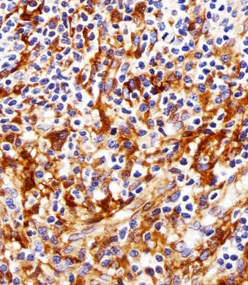
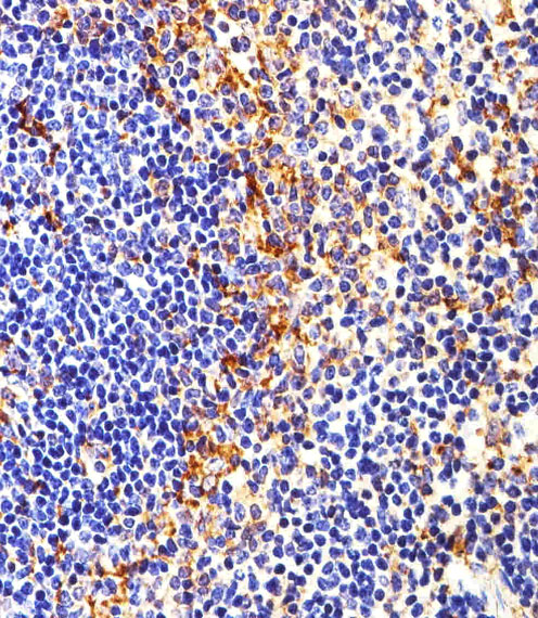
Anti-FYN options include paraffin-section IHC in human tonsil (M00684-2 image caption) and IF image data at 1:50 (M00684 image caption); M00684 lists human, mouse, and rat reactivity (catalog).

Real IHC data Immunohistochemical analysis of paraffin-embedded H. tonsil section using FYN. M00684-2 was diluted at 1:25 dilution. A peroxidase-conjugated goat anti-mouse IgG at 1:400 dilution was used as the secondary antibody, followed by DAB staining.
Anti-FYN Antibody
Cat # M00684-2
Real IF data Immunofluorescent analysis using the Antibody at 1:50 dilution.
Anti-Fyn Rabbit Monoclonal Antibody
Cat # M00684

M00684-2 is listed for human IHC-P (catalog) and has a paraffin-embedded human tonsil DAB image at 1:25 (M00684-2 image caption). M00684 is listed for IF/ICC in human, mouse, and rat (catalog), with an IF image at 1:50 that does not identify a specimen (M00684 image caption).

Which to pick: For tissue IHC, choose mouse monoclonal M00684-2 (catalog): its own image shows DAB staining of a paraffin-embedded human tonsil section at 1:25 (M00684-2 image caption). The caption does not report the fixative (M00684-2 image caption). For IF/ICC or listed reactivity across human, mouse, and rat, choose rabbit monoclonal M00684 (catalog); its IF image uses 1:50 but does not report a specimen or fixation method (M00684 image caption).

Each figure is that product's own IHC / IF validation image from its datasheet.

References

  1. UniProt Consortium. UniProt entry P06241 (FYN_HUMAN, Tyrosine-protein kinase Fyn).
  2. Human Protein Atlas. FYN tissue IHC expression (reliability: Approved).
  3. Human Protein Atlas. FYN subcellular location (ICC-IF): Mainly localized to the plasma membrane. In addition localized to the cytosol..
  4. Human Protein Atlas. FYN antibody validation summary (4 antibodies).
  5. Role of Fyn expression in predicting the sensitivity to platinum‑based chemotherapy in patients with ovarian serous carcinoma. Oncology letters 2024 — PMC11391251.
  6. FYN is overexpressed in human prostate cancer. BJU international 2009 — PMC2741693.
  7. Regulatory region genetic variation is associated with FYN expression in Alzheimer's disease. Neurobiology of aging 2017 — PMC5358011.
  8. Fyn expression is associated with the response of patients with locally advanced uterine cervical squamous cell carcinoma to neoadjuvant chemotherapy. Molecular and clinical oncology 2022 — PMC9468808.
  9. PubMed PMID:3099169 — UniProt-cited evidence.
  10. PubMed PMID:3526330 — UniProt-cited evidence.
  11. PubMed PMID:7822789 — UniProt-cited evidence.