Click image to see more details
-

-
-
-
-
+1
Product Info Summary
| SKU: | M00684-2 |
|---|---|
| Size: | 80 µl |
| Reactive Species: | Human |
| Host: | Mouse |
| Application: | Flow Cytometry, IHC-P, WB |
Customers Who Bought This Also Bought

Product info
Product Name
Anti-FYN Antibody
SKU/Catalog Number
M00684-2
Size
80 µl
Description
Boster Bio Anti-FYN Antibody (Catalog # M00684-2). Tested in IHC-P, Flow Cytometry, WB application(s). This antibody reacts with Human.
Storage & Handling
Maintain refrigerated at 2-8°C for up to 2 weeks. For long-term storage, store at -20°C in small aliquots to prevent freeze-thaw cycles.
Cite This Product
Anti-FYN Antibody (Boster Biological Technology, Pleasanton CA, USA, Catalog # M00684-2)
Host
Mouse
Contents
Purified monoclonal antibody supplied in PBS with 0.09% (W/V) sodium azide.
Clonality
Monoclonal
Clone Number
1302CT390.118.237
Isotype
IgG1,κ
Immunogen
This FYN antibody is generated from a mouse immunized with a recombinant preotein from human FYN.
Reactive Species
M00684-2 is reactive to FYN in Human
Calculated molecular weight
60.8 kDa
Background of FYN
Non-receptor tyrosine-protein kinase that plays a role in many biological processes including regulation of cell growth and survival, cell adhesion, integrin-mediated signaling, cytoskeletal remodeling, cell motility, immune response and axon guidance. Inactive FYN is phosphorylated on its C-terminal tail within the catalytic domain. Following activation by PKA, the protein subsequently associates with PTK2/FAK1, allowing PTK2/FAK1 phosphorylation, activation and targeting to focal adhesions. Involved in the regulation of cell adhesion and motility through phosphorylation of CTNNB1 (beta-catenin) and CTNND1 (delta- catenin). Regulates cytoskeletal remodeling by phosphorylating several proteins including the actin regulator WAS and the microtubule-associated proteins MAP2 and MAPT. Promotes cell survival by phosphorylating AGAP2/PIKE-A and preventing its apoptotic cleavage. Participates in signal transduction pathways that regulate the integrity of the glomerular slit diaphragm (an essential part of the glomerular filter of the kidney) by phosphorylating several slit diaphragm components including NPHS1, KIRREL and TRPC6. Plays a role in neural processes by phosphorylating DPYSL2, a multifunctional adapter protein within the central nervous system, ARHGAP32, a regulator for Rho family GTPases implicated in various neural functions, and SNCA, a small pre-synaptic protein. Participates in the downstream signaling pathways that lead to T-cell differentiation and proliferation following T-cell receptor (TCR) stimulation. Also participates in negative feedback regulation of TCR signaling through phosphorylation of PAG1, thereby promoting interaction between PAG1 and CSK and recruitment of CSK to lipid rafts. CSK maintains LCK and FYN in an inactive form. Promotes CD28-induced phosphorylation of VAV1.
Antibody Validation
Boster validates all antibodies on WB, IHC, ICC, Immunofluorescence, and ELISA with known positive control and negative samples to ensure specificity and high affinity, including thorough antibody incubations.
Application & Images
Applications
M00684-2 is guaranteed for Flow Cytometry, IHC-P, WB Boster Guarantee
Recommend Dilution
WB: 1:1000
IHC-P: 1:25
FC: 1:25
Validation Images & Assay Conditions
Click image to see more details
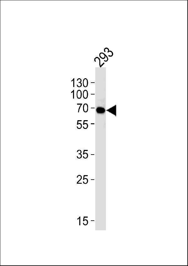
Western blot analysis of lysate from 293 cell line using FYN. M00684-2 was diluted at 1:1000 at each lane. A goat anti-mouse IgG H&L (HRP) at 1:3000 dilution was used as the secondary antibody. Lysate at 35μg per lane.

Click image to see more details
Immunohistochemical analysis of paraffin-embedded H. tonsil section using FYN. M00684-2 was diluted at 1:25 dilution. A peroxidase-conjugated goat anti-mouse IgG at 1:400 dilution was used as the secondary antibody, followed by DAB staining.

Click image to see more details
Immunohistochemical analysis of paraffin-embedded M. spleen section using FYN. M00684-2 was diluted at 1:25 dilution. A peroxidase-conjugated goat anti-mouse IgG at 1:400 dilution was used as the secondary antibody, followed by DAB staining.

Click image to see more details
Immunohistochemical analysis of paraffin-embedded R. spleen section using FYN. M00684-2 was diluted at 1:25 dilution. A peroxidase-conjugated goat anti-mouse IgG at 1:400 dilution was used as the secondary antibody, followed by DAB staining.

Click image to see more details
Flow cytometric analysis of Hela cells using FYN(green, Cat#M00684-2) compared to an isotype control of mouse IgG1(blue). M00684-2 was diluted at 1:25 dilution. An Alexa Fluor® 488 goat anti-mouse lgG at 1:400 dilution was used as the secondary antibody.
Specific Publications For Anti-FYN Antibody (M00684-2)
Loading publications
Recommended Resources
Here are featured tools and databases that you might find useful.
- Boster's Pathways Library
- Protein Databases
- Bioscience Research Protocol Resources
- Data Processing & Analysis Software
- Photo Editing Software
- Scientific Literature Resources
- Research Paper Management Tools
- Molecular Biology Software
- Primer Design Tools
- Bioinformatics Tools
- Phylogenetic Tree Analysis
Customer Reviews
Have you used Anti-FYN Antibody?
Share your experimental results or join a short interview to earn up to $1,000 in product credits or other rewards.
0 Reviews For Anti-FYN Antibody
Customer Q&As
Have a question?
Find answers in Q&As, reviews.
Can't find your answer?
Submit your question
