This website uses cookies to ensure you get the best experience on our website.


- Table of Contents
Plan chromogenic paraffin-section IHC with the catalog antibody (datasheet PA1578). Use high-staining cerebellar synaptic glomeruli and cortical neuropil as reference tissues (HPA tissue IHC), and interpret the pattern alongside GABRA1’s postsynaptic membrane location (UniProt).

Expected localisation, validated protocols, controls and antibodies — the at-a-glance facts below, then the full design guide.
| Expected localisation | Postsynaptic membrane (UniProt); neuropil (HPA tissue IHC) | |
| Staining pattern | Selective staining in neuropil and a subset of neurons (HPA tissue IHC) | |
| Antigen retrieval | EDTA pH 8.0 HIER, heat-mediated (datasheet PA1578) | |
| Positive control | Cerebellum+1 more · see all | |
| Negative control | Adipose tissue+4 more · see all |
| Fixation | Keep fixation consistent across sections. (standard IHC practice; not target-specific) | |
| Caveat | Regional variation: low neuronal staining in hippocampus and caudate (HPA tissue IHC) | |
| Regulation | No regulator in supplied record (UniProt) | |
| Isoform / epitope | 0 isoforms annotated; extracellular versus cytoplasmic epitope location matters (UniProt) |
The catalog antibody’s IHC-P protocol is accompanied by published GABRA1 IHC methods for mouse brain (PMC11421448) and rat hypothalamus (PMC8740267).
| Sample | Tissue sections; selected-image fixative not specified (standard IHC workflow) |
| Fixation | Image fixative and duration unreported (datasheet PA1578); verify before use. |
| Sectioning | 4–5 µm sections on charged slides (standard) |
| Deparaffinisation | Xylene, graded ethanol series to water (standard) |
| Antigen retrieval | Heat retrieval: EDTA pH 8.0 (datasheet PA1578); 20 min, 95–100 °C (standard) |
| Peroxidase block | 3% H2O2, 10 min, room temperature (standard) |
| Blocking | 10% goat serum (datasheet PA1578) |
| Primary antibody | Rabbit anti-GABRA1, 0.5-1μg/ml (datasheet PA1578) |
| Primary incubation | Overnight at 4 °C (datasheet PA1578) |
| Detection | HRP-conjugated secondary, DAB chromogen (datasheet PA1578) |
| Counterstain | Hematoxylin, blue, dehydrate and mount (standard) |
| Expected result | GABRA1-positive staining in synaptic glomeruli - capsule of cerebellum (HPA tissue IHC: High). HPA tissue profile: Selective expression in neuropil and subset of neurons. No signal in the no-primary control. |
GABRA1 is a membrane receptor subunit concentrated at postsynaptic sites, with four transmembrane segments (UniProt P14867 topology; UniProt P14867 subcellular). In IHC, expect staining in neuropil and a subset of neurons, especially cerebellar synaptic glomeruli and cerebral cortical neuropil (HPA tissue IHC: Enhanced; HPA tissue IHC: High in both sites). HPA calls the tissue pattern Enhanced because staining is consistent with RNA expression (HPA tissue IHC: reliability description).
| Strong staining outlines cerebellar synaptic glomeruli or fills cortical neuropil while nearby structures vary in intensity (HPA tissue IHC: High in both sites). | This matches the reported selective neural pattern (HPA tissue IHC: selective expression in neuropil and a subset of neurons). Assess signal within the named structures instead of requiring every neuronal cell body to stain; UniProt places GABRA1 mainly at GABAergic synapses and at lower concentration on extrasynaptic membrane (UniProt P14867 subcellular). |
| Predominantly nuclear staining appears in a paraffin section, without a convincing neural membrane or neuropil pattern. | Treat this as discordant with the expected IHC distribution, then check controls and morphology before calling it GABRA1 (UniProt P14867 subcellular; HPA tissue IHC: profile). HPA reports nucleoplasm only as an uncertain additional ICC-IF location and cautions that its subcellular summary uses antibodies targeting multiple genes (HPA subcellular: uncertain nucleoplasm; HPA subcellular: caution). |
| Strong, widespread staining appears in glandular or other non-neural cells where the selected HPA reference is negative. | For example, HPA reports adrenal glandular cells and bronchial respiratory epithelial cells as not detected (HPA tissue IHC: adrenal gland; HPA tissue IHC: bronchus). Such staining warrants checks for cross-reactivity or endogenous detection activity; it does not alone prove either cause (general IHC practice). Compare the same cell type and detection system in a negative control. |
| A uniform chromogen haze covers tissue compartments and obscures neuropil boundaries. | A field-wide haze cannot establish the selective pattern reported for GABRA1 (HPA tissue IHC: profile). Consider nonspecific antibody binding, incomplete blocking, or unquenched endogenous activity where the detection chemistry permits it (general IHC practice). Judge specificity by whether the background clears while the expected neural structures remain distinguishable. |
| Cerebellar synaptic glomeruli and cortical neuropil show no convincing staining (HPA tissue IHC: High in both sites). | An absent signal in these known-positive structures makes the run inconclusive (HPA tissue IHC: High in both sites; general IHC practice). Check section integrity, antibody and detection controls, then empirically review the IHC-P conditions used. The supplied sources do not report GABRA1-specific fixation sensitivity, so do not assign the failure to fixation. |
| Tissue and structure selected for scoring | Use cerebellar synaptic glomeruli and cortical neuropil as the clearest supplied positive references (HPA tissue IHC: High in both sites). Hippocampal and caudate neuronal cells are reported at low level, so faint staining there carries less weight than a high-level reference (HPA tissue IHC: Low in hippocampus and caudate). |
| Membrane topology and epitope position | GABRA1 has four transmembrane segments, an extracellular region at residues 28–253, and a cytoplasmic region at 334–421 (UniProt P14867 topology). Epitope location can guide antibody and permeabilisation choices in general IHC/IF practice; the supplied record gives no antibody epitope, so it cannot predict which retrieval condition or access route works. |
| Evidence tied to the antibody and application | Two listed antibodies, HPA055746 and CAB022502, have Enhanced IHC status, while HPA029363 has ICC approval and no listed IHC status (HPA antibodies: validation status). Match any interpretation to the antibody's stated application; the tissue-level Enhanced rating describes agreement between staining and RNA data, not universal performance for every reagent (HPA tissue IHC: reliability description). |
| IF/ICC Q: Is nucleoplasmic signal an expected GABRA1 result? | A: HPA supports plasma membrane as the main ICC-IF location but marks nucleoplasm uncertain, with a multiple-gene antibody caution (HPA subcellular: supported plasma membrane; HPA subcellular: uncertain nucleoplasm; HPA subcellular: caution). Interpret IF/ICC on its own evidence; this IHC section offers no IF protocol option. |
| Situation | Likely cause | Next action |
|---|---|---|
| The known-positive cerebellar or cortical structure is blank. | The run may have failed, or its IHC-P conditions may need empirical adjustment (HPA tissue IHC: High in both sites; general IHC practice). | Confirm the section contains the intended structure, inspect positive and detection controls, and review the antibody's IHC-P instructions and retrieval settings (general IHC practice). Do not infer a GABRA1-specific fixation effect from a blank slide. |
| All tissue compartments show similar brown staining. | Nonspecific binding or detection-system background can hide selective staining (general IHC practice). | Compare a no-primary control, check blocking and antibody concentration, and address endogenous activity appropriate to the detection chemistry (general IHC practice). Reassess whether cerebellar glomeruli or cortical neuropil remain enriched (HPA tissue IHC: High in both sites). |
| Nuclei dominate the stain. | Nuclear dominance conflicts with the principal membrane and synaptic locations (UniProt P14867 subcellular; HPA subcellular: supported plasma membrane). | Check counterstain, no-primary control, and antibody-specific validation before scoring nuclei as positive (general IHC practice). HPA's nucleoplasmic ICC-IF call is uncertain and cannot validate nuclear IHC on its own (HPA subcellular: uncertain nucleoplasm). |
| A reported negative cell type stains as strongly as cortical neuropil. | Cross-reactivity or endogenous detection activity is possible; the slide alone cannot distinguish them (general IHC practice). | Compare a matched no-primary control and verify cell identity and anatomy (general IHC practice). Use the exact HPA cell-type reference, such as adrenal glandular cells reported as not detected, rather than treating an entire organ as uniformly negative (HPA tissue IHC: adrenal gland). |
| Hippocampal or caudate neurons are faint while the positive reference is clear. | Low signal may fit the supplied tissue pattern: HPA rates neuronal cells in both sites low (HPA tissue IHC: Low in hippocampus and caudate). | Score those cells against matched background and the same-run high-level cerebellar or cortical reference (HPA tissue IHC: High in both sites; general IHC practice). Avoid escalating a low-level site into a failed run solely because it is weaker. |
| An ICC-IF image suggests nuclear staining, but IHC shows a neural membrane pattern. | The sources describe different applications; HPA's ICC-IF nucleoplasmic location is uncertain (HPA subcellular: uncertain nucleoplasm; HPA antibodies: validation status). | Keep the IHC call anchored to tissue morphology and IHC evidence (HPA tissue IHC: profile; HPA antibodies: IHC status). Review IF/ICC on its dedicated guide and with its own controls (general IF practice). |



Comprehensive Human Protein Atlas IHC scoring per tissue (reliability: Enhanced — High consistency between antibody staining and RNA expression data.). Rows are taken directly from the HPA tissue chart — click any row's HPA link to view the source.
| Tissue | Cell type | Level | Evidence | Source |
|---|---|---|---|---|
| Cerebellum | Synaptic glomeruli - capsule | High | Protein (IHC) | HPA → |
| Cerebral cortex | Neuropil | High | Protein (IHC) | HPA → |
| Tissue | Cell type | Level | Evidence | Source |
|---|---|---|---|---|
| Adipose tissue | Adipocytes | Not detected | Protein (IHC) | HPA → |
| Adrenal gland | Glandular cells | Not detected | Protein (IHC) | HPA → |
| Appendix | Glandular cells | Not detected | Protein (IHC) | HPA → |
| Bone marrow | Hematopoietic cells | Not detected | Protein (IHC) | HPA → |
| Breast | Adipocytes | Not detected | Protein (IHC) | HPA → |
Troubleshoot GABRA1 staining in paraffin sections by checking retrieval, controls, tissue distribution and membrane localisation before interpreting signal intensity.
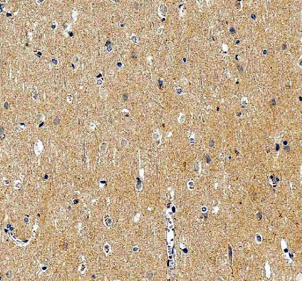
PA1578 has real paraffin-section IHC images from rat and human brain; the catalog lists human, mouse, and rat reactivity, with no IF data (PA1578 image captions; catalog reactivity and images).
PA1578 is listed for IHC and WB, with human, mouse, and rat reactivity (PA1578 catalog applications and reactivity). Its IHC images show rat brain tissue and a paraffin-embedded human brain section (PA1578 image captions).
Which to pick: Choose PA1578 for paraffin-section tissue IHC: its own captions show rat brain and paraffin-embedded human brain, but do not report the fixative (PA1578 image captions). For work across human, mouse, and rat, PA1578 lists all three as reactive; the supplied images document human and rat tissue only (PA1578 catalog reactivity; image captions). No IF/ICC application or image is supplied, so there is no supported IF/ICC pick here (PA1578 catalog applications and images).