GALNT3 / Polypeptide N-acetylgalactosaminyltransferase 3 · IHC design guide

Design Immunohistochemistry for GALNT3

Plan GALNT3 IHC in paraffin sections using the granular cytoplasmic tissue pattern (HPA tissue IHC). This guide covers fixation consistency, Golgi localization (UniProt), and the catalog antibody’s 1:50–1:200 IHC dilution (datasheet A04300).

Evidence assembled Oct 2026 · For research use; verify linked source records and product datasheet before use
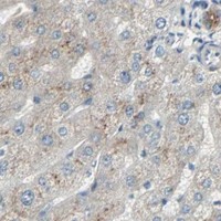
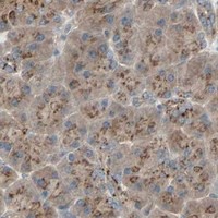
Immunohistochemistry protocol sheet for GALNT3 (IHC for GALNT3): expected localisation Golgi stack expected (UniProt); granular cytoplasm observed (HPA tissue IHC), antibody A04300, validated IHC image, and IHC protocol steps
Printable GALNT3 IHC protocol sheet — expected localisation Golgi stack expected (UniProt); granular cytoplasm observed (HPA tissue IHC), antibody A04300, controls and protocol steps. Open the full GALNT3 IHC guide →

GALNT3 Immunohistochemistry Experimental Design Guide

Expected localisation, validated protocols, controls and antibodies — the at-a-glance facts below, then the full design guide.

Must know before staining
Expected localisation Golgi stack expected (UniProt); granular cytoplasm observed (HPA tissue IHC)
Staining pattern Granular cytoplasmic staining in several tissues (HPA tissue IHC)
Antigen retrieval Citrate pH 6.0 HIER, heat-mediated (datasheet A04300)
Positive control ⓘ Adipose tissue+4 more · see all
Negative control ⓘ Lymph node+1 more · see all
Important caveats
Reasons your staining may differ from the expected pattern.
Fixation Keep fixation consistent across paraffin sections (standard IHC practice; not target-specific); Matched tissue-IHC evidence does not establish the fixation claim. Validate the specimen-specific method before use. (selected-SKU IHC image A04300)
Caveat Staining and RNA show medium consistency (HPA tissue IHC)
Regulation Stomach-enhanced RNA expression (HPA tissue RNA)
Isoform / epitope 2 isoforms; epitope coverage is unreported (UniProt)
Section 1

Recommended GALNT3 IHC & IF Protocols

The catalog antibody protocol uses citrate pH 6.0 heat retrieval (datasheet A04300). Three published GALNT3 IHC protocols provide tissue-specific starting conditions (PMC4984453; PMC6539655; PMC12822125).

Recommended immunohistochemistry (IHC-P) protocol parameters
SampleParaffin-embedded human colon carcinoma tissue; fixative not specified (datasheet A04300)
FixationImage fixative and duration unreported (datasheet A04300); verify before use.
Sectioning4–5 µm sections on charged slides (standard)
DeparaffinisationXylene, graded ethanol series to water (standard)
Antigen retrievalHeat retrieval: Citrate pH 6.0 (datasheet A04300); 20 min, 95–100 °C (standard)
Peroxidase block3% H2O2, 10 min, room temperature (standard)
Blocking10% normal serum of the secondary host, 30 min, room temperature (standard)
Primary antibodyRabbit anti-GALNT3, 1:50-1:200 (datasheet A04300)
Primary incubationOvernight at 4 °C (standard)
DetectionHRP-polymer secondary, DAB chromogen 5–10 min (standard)
CounterstainHematoxylin, blue, dehydrate and mount (standard)
Expected resultGALNT3-positive staining in adipocytes of adipose tissue (HPA tissue IHC: High). HPA tissue profile: Granular cytoplasmic expression in several tissues. No signal in the no-primary control.
💡Decision noteStart with citrate pH 6.0 heat retrieval for the catalog antibody (datasheet A04300); adjust retrieval to match a published protocol when reproducing its conditions.
Section 2

What Is the Expected GALNT3 Staining Pattern?

GALNT3 is a Golgi stack membrane protein enriched in the medial and trans Golgi, with a short cytoplasmic tail and a lumenal catalytic region (UniProt Q14435 topology). In paraffin sections, expect granular cytoplasmic staining in selected cells, especially glandular, respiratory epithelial and endocrine cells (HPA tissue IHC: Enhanced; medium consistency with RNA). Judge localisation and cell identity together; tissue-wide uniform staining is not the reported pattern (HPA tissue IHC).

What am I looking at on my slide?
Granular cytoplasmic staining in breast or cervical glandular cells, bronchial respiratory epithelial cells, or intestinal endocrine cells.This matches the reported positive cell populations and granular profile (HPA tissue IHC: High; Enhanced). A compact perinuclear distribution is compatible with Golgi localisation, but chromogenic IHC alone does not prove organelle identity (UniProt Q14435; general IHC practice).
Predominantly nuclear, cell-surface, or uniformly diffuse staining replaces the granular cytoplasmic pattern.These compartments do not match the reported Golgi localisation (UniProt Q14435; HPA subcellular ICC-IF: Golgi supported). Treat the result as suspect; review morphology, detection background and antibody specificity before scoring it as GALNT3 (general IHC practice).
Strong stain appears mainly in lymph-node germinal center cells or splenic red-pulp cells.Those populations were not detected in the supplied HPA tissue survey (HPA tissue IHC). Consider nonspecific antibody binding or endogenous detection activity; a single unexpected section cannot establish a new GALNT3 expression site (general IHC practice).
Weak, widespread haze obscures cell borders and the expected granules.That pattern is difficult to distinguish from nonspecific background and does not reproduce the reported granular cytoplasmic profile (HPA tissue IHC). Compare the negative control, then assess blocking, washing and detection conditions before interpreting weak cells (general IHC practice).
No signal is visible in a section containing a reported high-staining cell population.An absent stain in breast glandular cells or bronchial respiratory epithelial cells conflicts with the supplied reference pattern (HPA tissue IHC: High). First confirm those cells are present and preserved, then check retrieval, antibody and detection performance (general IHC practice).
💡Expected GALNT3 appearanceCall a result positive when the appropriate cells show distinct granular cytoplasmic staining, potentially concentrated near the nucleus, with a clearly readable chromogenic signal (HPA tissue IHC: High; UniProt Q14435 Golgi localisation); widespread nuclear stain or uniform haze is suspect (general IHC practice).
How each factor affects the staining
Golgi topology and apparent compartmentGALNT3 spans the Golgi stack membrane at residues 20–37; residues 38–633 are lumenal (UniProt Q14435 topology). Read punctate or granular cytoplasmic chromogen in tissue context; light microscopy cannot resolve membrane orientation (general IHC practice).
Cell population within a tissueHPA reports high staining in selected glandular, respiratory epithelial and endocrine populations, plus adipocytes (HPA tissue IHC). It reports low Sertoli-cell staining despite UniProt describing high testis expression overall (HPA tissue IHC; UniProt Q14435 tissue specificity). Score identified cells, not an entire organ by name.
Antibody validationThe listed rabbit antibody HPA007613 has Enhanced IHC status; HPA describes medium consistency between staining and RNA expression (HPA antibodies; HPA tissue IHC). This supports use of the reference pattern but leaves unexpected staining in a new specimen to be checked independently (general IHC practice).
Epitope and molecular formsUniProt lists two isoforms and glycosylation sites at residues 132, 297 and 484 (UniProt Q14435). No epitope map or isoform-specific IHC result is supplied, so these annotations cannot predict which form stains or justify a particular retrieval setting.
IF/ICC cross-checkHPA reports supported Golgi localisation in ICC-IF images from A-431 and RT-4 cells (HPA subcellular ICC-IF). That offers a compartment cross-check for ambiguous IHC staining; IF/ICC conditions and an IF protocol belong in the separate guide.
Why is my staining missing, weak or wrong?
SituationLikely causeNext action
A reported high-staining tissue gives no chromogenic signal.The relevant cell population may be absent from the section, or an IHC step may have failed (HPA tissue IHC: High; general IHC practice).Locate the expected cells on the counterstain; check a known-positive section and the catalog antibody’s IHC-P instructions before adjusting retrieval or dilution (general IHC practice).
Nuclei stain more strongly than cytoplasmic granules.The dominant compartment conflicts with Golgi localisation (UniProt Q14435; HPA subcellular ICC-IF).Review the negative control and section morphology; repeat with appropriate detection controls before calling nuclear staining specific (general IHC practice).
Germinal center or splenic red-pulp cells stain strongly.These cells were not detected in the supplied tissue survey; nonspecific binding or endogenous detection activity is plausible (HPA tissue IHC; general IHC practice).Compare reagent-omission controls, review blocking and detection chemistry, and seek an independent antibody or orthogonal check if the finding matters (general IHC practice).
A faint brown haze covers both positive and negative regions.Diffuse background may mask the reported granular cytoplasmic pattern (HPA tissue IHC; general IHC practice).Inspect the negative control; optimize blocking, washes, antibody concentration and chromogen development using the catalog IHC-P instructions (general IHC practice).
Testis appears weak although UniProt calls testis highly expressed.The statements refer to different scales: UniProt describes organ expression, while HPA reports low staining specifically in Sertoli cells (UniProt Q14435 tissue specificity; HPA tissue IHC).Identify the cells being scored and document their staining separately; do not use the organ-level statement as a Sertoli-cell positive-control threshold.
Granules are visible, but Golgi identity remains uncertain.Chromogenic IHC shows the reported granular cytoplasmic profile without resolving an organelle boundary (HPA tissue IHC; general IHC practice).Check whether the pattern is concentrated near nuclei and restricted to expected cells; consult the supported Golgi ICC-IF reference on the separate IF/ICC guide (HPA subcellular ICC-IF).

Sample controls for GALNT3 IHC & IF

🧪Run breast first and require staining in its glandular cells (HPA: High in breast glandular cells). Run lymph node as the negative tissue (HPA: Not detected in germinal center cells); on the breast slide, use adjacent stromal cells as an internal background comparison, without assuming they are GALNT3-negative.
Positive control tissue: Adipose tissue (Adipocytes, HPA High)
Negative control tissue: Lymph node (HPA Not detected)
ICC-IF cell lines (HPA subcellular resource): HPA ICC-IF images show GALNT3 in A-431, RT-4, with annotated localisation: Golgi apparatus (supported) (HPA subcellular).
Technical controls: Include a no-primary, secondary-only control, a concentration-matched nonimmune rabbit IgG control for the catalog rabbit pAb (caption: Rabbit pAb), and GALNT3-knockout tissue as a biological specificity control. For chromogenic staining, quench endogenous peroxidase and inspect the breast section for residual background before scoring glandular cells.
⚠️Feasibility: A target-specific fixation window or fixation effect is unreported, and the selected A04300 caption does not state a fixative (caption: fixative not stated). Retrieval dependence is unestablished; 10 mM citrate, pH 6.0, with high-pressure retrieval is a documented starting condition for paraffin-embedded colon carcinoma, not a validated requirement for breast (caption: A04300 colon carcinoma IHC). Frozen sections are not established as easier by the supplied evidence; IF/ICC images exist for A-431 and RT-4 (HPA: ICC-IF cell lines), while breast glandular lumens should be checked for deposited signal before calling cellular staining, which is expected to be Golgi-associated (HPA: Golgi apparatus supported; UniProt Q14435: Golgi stack membrane).

HPA tissue IHC evidence for GALNT3

Comprehensive Human Protein Atlas IHC scoring per tissue (reliability: Enhanced — Medium consistency between antibody staining and RNA expression data.). Rows are taken directly from the HPA tissue chart — click any row's HPA link to view the source.

Positive expression · recommended positive controls

TissueCell typeLevelEvidenceSource
Adipose tissue Adipocytes High Protein (IHC) HPA →
Appendix Endocrine cells High Protein (IHC) HPA →
Breast Glandular cells High Protein (IHC) HPA →
Bronchus Respiratory epithelial cells High Protein (IHC) HPA →
Cervix Glandular cells High Protein (IHC) HPA →

Undetected expression · recommended negative controls

TissueCell typeLevelEvidenceSource
Lymph node Germinal center cells Not detected Protein (IHC) HPA →
Spleen Cells in red pulp Not detected Protein (IHC) HPA →
Section 3

Advanced GALNT3 IHC Tips

Troubleshoot GALNT3 staining by checking retrieval, compartment, cell type and controls before comparing chromogenic IHC results across sections.

Which retrieval conditions should I try when GALNT3 staining is weak?
Start with heat-mediated retrieval in 10 mM citrate buffer, pH 6.0, before chromogenic IHC (datasheet A04300). The selected paraffin-section example used high-pressure retrieval with this buffer and stained human colon carcinoma at 1:100 (caption A04300); its fixative was not reported (caption A04300). If staining remains weak, compare a longer heating period or a second retrieval condition on adjacent sections while keeping antibody dilution and detection constant (standard IHC practice). Check that any increase in granular cytoplasmic signal preserves plausible Golgi-associated staining rather than increasing diffuse staining across the section (HPA tissue IHC; UniProt Q14435 localisation).
How should I troubleshoot variable GALNT3 staining between paraffin blocks?
GALNT3-specific sensitivity to fixation is unknown from the supplied evidence; the selected paraffin-section caption does not state a fixative (caption A04300). Record each block’s fixative, fixation duration and processing history, then compare similarly processed sections using 10 mM citrate, pH 6.0 retrieval (datasheet A04300; standard IHC practice). Include a reference section in each staining run so changes in retrieval or detection can be distinguished from differences between blocks (standard IHC practice). Judge preservation alongside staining: damaged morphology or uneven staining makes an apparent difference in GALNT3 expression difficult to interpret (standard IHC practice).
What staining pattern should count as GALNT3 localisation in tissue?
Expect a compact cytoplasmic pattern consistent with the Golgi: GALNT3 resides preferentially in medial and trans Golgi stacks (UniProt Q14435 localisation). Tissue IHC describes granular cytoplasmic expression, while subcellular imaging supports Golgi localisation (HPA tissue IHC; HPA subcellular). Examine signal at high magnification in intact cells and compare its position with nuclei and the broader cytoplasm; a perinuclear concentration is compatible with the expected compartment (UniProt Q14435 localisation; standard IHC practice). Diffuse nuclear or uniform cell-surface staining alone does not establish GALNT3 localisation, so check those patterns against controls before scoring them (UniProt Q14435 topology; standard IHC practice).
Could epitope position explain different GALNT3 staining patterns?
GALNT3 has 2 annotated isoforms, but the supplied record does not map their sequence differences or the catalog antibody’s epitope (UniProt Q14435 isoforms; supplied antibody evidence). Its membrane anchor spans residues 20–37, with residues 1–19 cytoplasmic and 38–633 lumenal (UniProt Q14435 topology). The lumenal region includes glycosylation sites at 132, 297 and 484 and a Ricin B-type lectin domain at 504–630 (UniProt Q14435 features). If antibodies disagree, obtain their epitope information and compare staining on adjacent sections before attributing the difference to an isoform or glycosylation (standard IHC practice).
How can I check GALNT3 localisation by IF alongside tissue IHC?
Use IF as a separate localisation check: GALNT3 is supported at the Golgi, and HPA lists ICC/IF images from A-431 and RT-4 cells (HPA subcellular). For tissue multiplexing, pair GALNT3 with a marker that identifies the expected cell population, such as endocrine cells in colon, and assess the signals within the same cells (HPA tissue IHC; standard IF practice). Choose fluorophores after inspecting unstained tissue for autofluorescence, and include single-label controls when channels overlap (standard IF practice). Permeabilise as needed for an epitope on the lumenal side of the Golgi membrane; the catalog antibody’s epitope side is unspecified, so verify accessibility experimentally (UniProt Q14435 topology; supplied antibody evidence; standard IF practice).
How do I distinguish GALNT3 signal from chromogenic background?
Include a no-primary control and inspect whether unwanted DAB appears in the same structures as the test section (standard IHC practice). A 3% hydrogen peroxide block before peroxidase detection can reduce signal from endogenous peroxidase; this is a general chromogenic IHC step, not GALNT3-specific evidence (standard IHC practice). Titrate the catalog antibody around the caption’s 1:100 dilution and keep DAB development comparable across sections (caption A04300; standard IHC practice). Prefer staining with granular cytoplasmic distribution in plausible cells over uniform surface deposits or broad staining unrelated to cell boundaries (HPA tissue IHC; UniProt Q14435 localisation; standard IHC practice).
How should GALNT3 IHC be scored across tissue samples? ⚠ ANSWER MARKED FOR VERIFICATION
Define the cell population and compartment before scoring; HPA reports high staining in colon endocrine cells and granular cytoplasmic expression across several tissues (HPA tissue IHC). Report the percentage of positive cells plus an intensity-based H-score, or positive-cell density per mm² when cell abundance itself is the question (standard IHC practice). Use the same threshold, exposure-free visual assessment conditions and sampled area for all chromogenic sections, with blinded review where feasible (standard IHC practice). Normalise counts to the relevant intact cell population or viable tissue area, and exclude necrotic or poorly preserved regions from the denominator (standard IHC practice).
When should apparent GALNT3 positivity be treated as artefact?
A credible result combines granular cytoplasmic staining with a plausible Golgi distribution and cell identity (HPA tissue IHC; HPA subcellular; UniProt Q14435 localisation). High staining in colon endocrine cells is supported, whereas germinal-center cells in lymph node and red-pulp cells in spleen were reported as not detected (HPA tissue IHC). Recheck isolated nuclear staining, unexpected cell populations, strong section-edge deposits and necrotic areas before calling them positive (UniProt Q14435 localisation; HPA tissue IHC; standard IHC practice). Compare suspicious deposits with a no-primary control and the peroxidase-blocked section to assess detection background or endogenous enzyme activity (standard IHC practice).
Boster reagents

Best GALNT3 / Polypeptide N-acetylgalactosaminyltransferase 3 IHC Antibodies

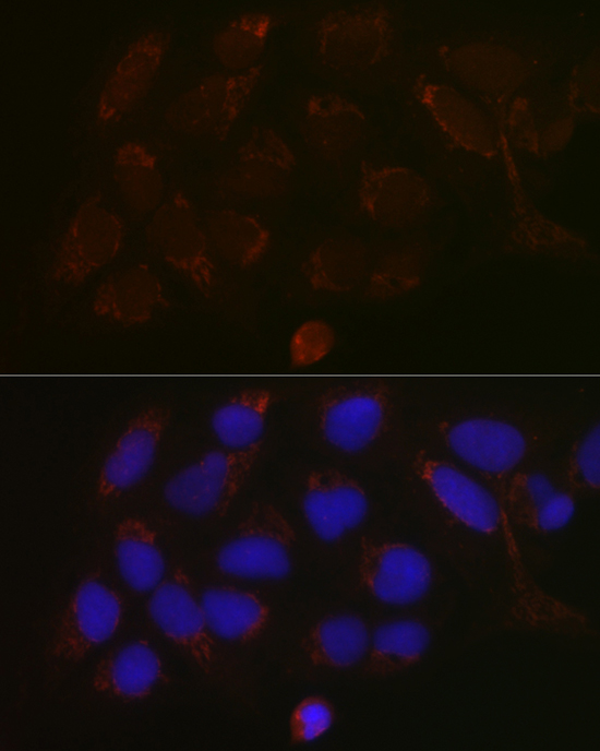
Anti-GALNT3 antibody A04300 has IHC data from paraffin-embedded human colon carcinoma and IF data from U-2 OS cells (catalog image captions); the catalog lists Human, Mouse and Rat reactivity (catalog: reactivity).

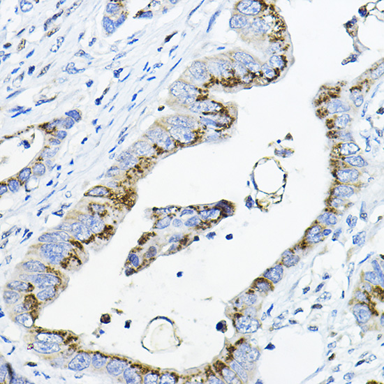
Real IHC data Immunohistochemistry of paraffin-embedded human colon carcinoma using GALNT3 Rabbit pAb at dilution of 1:100 .Perform high pressure antigen retrieval with 10 mM citrate buffer pH 6.0 before commencing with IHC staining protocol.
Anti-GALNT3 Antibody
Cat # A04300

A04300 will render with an IHC image of paraffin-embedded human colon carcinoma stained at 1:100 after high-pressure retrieval in 10 mM citrate buffer, pH 6.0 (A04300 IHC image caption). Its IF image shows U-2 OS cells stained at 1:100 with DAPI nuclear staining (A04300 IF image caption).

Which to pick: Choose A04300 for paraffin-section IHC: its own image documents that preparation and retrieval conditions, but does not report the fixative (A04300 IHC image caption). Choose A04300 for IF/ICC because ICC/IF is listed as an application and its IF image shows staining in U-2 OS cells (catalog: applications; A04300 IF image caption). For Human, Mouse or Rat samples, A04300 is the listed rabbit polyclonal option; the supplied IHC and IF images document human samples only (catalog: reactivity and host; A04300 IHC and IF image captions).

Each figure is that product's own IHC / IF validation image from its datasheet.

References

  1. UniProt Consortium. UniProt entry Q14435 (GALT3_HUMAN, Polypeptide N-acetylgalactosaminyltransferase 3).
  2. Human Protein Atlas. GALNT3 tissue IHC expression (reliability: Enhanced).
  3. Human Protein Atlas. GALNT3 subcellular location (ICC-IF): Localized to the Golgi apparatus..
  4. Human Protein Atlas. GALNT3 antibody validation summary (1 antibodies).
  5. Loss of N-acetylgalactosaminyltransferase 3 in poorly differentiated pancreatic cancer: augmented aggressiveness and aberrant ErbB family glycosylation. British journal of cancer 2016 — PMC4984453.
  6. The polypeptide GALNT6 Displays Redundant Functions upon Suppression of its Closest Homolog GALNT3 in Mediating Aberrant O-Glycosylation, Associated with Ovarian Cancer Progression. International journal of molecular sciences 2019 — PMC6539655.
  7. GALNT3 is a novel target driving lymphomagenesis via O-glycosylation of FGFR2. Cell communication and signaling : CCS 2025 — PMC12822125.
  8. A machine learning approach to identify predictive molecular markers for cisplatin chemosensitivity following surgical resection in ovarian cancer. Scientific reports 2021 — PMC8377048.
  9. PubMed PMID:8663203 — UniProt-cited evidence.
  10. PubMed PMID:15815621 — UniProt-cited evidence.
  11. PubMed PMID:15489334 — UniProt-cited evidence.