This website uses cookies to ensure you get the best experience on our website.


- Table of Contents
Plan chromogenic GAR1 IHC in paraffin sections using the observed nuclear tissue pattern (HPA tissue IHC). Start with the catalog antibody’s 2–5 μg/ml IHC range (datasheet A07049-1), and compare high-staining bone marrow hematopoietic cells with adipose tissue adipocytes reported as undetected (HPA tissue IHC).

Expected localisation, validated protocols, controls and antibodies — the at-a-glance facts below, then the full design guide.
| Expected localisation | Nuclear staining observed in tissue (HPA tissue IHC) | |
| Staining pattern | Widespread nuclear staining across tissues (HPA tissue IHC) | |
| Antigen retrieval | EDTA pH 8.0 HIER, heat-mediated (datasheet A07049-1) | |
| Positive control | Adrenal gland+4 more · see all | |
| Negative control | Adipose tissue |
| Fixation | Keep fixation conditions consistent across sections. (standard IHC practice; not target-specific) | |
| Caveat | Adipocytes stain high in breast but are undetected in adipose tissue (HPA tissue IHC) | |
| Regulation | Low tissue specificity (HPA tissue RNA) | |
| Isoform / epitope | 2 isoforms; assess epitope coverage for each (UniProt) |
The catalog antibody’s IHC-P protocol (datasheet A07049-1) is accompanied by a published protocol for paraffin-embedded HCC tissue (PMC8541795).
| Sample | Paraffin-embedded human breast cancer tissue; fixative not specified (datasheet A07049-1) |
| Fixation | Image fixative and duration unreported (datasheet A07049-1); verify before use. |
| Sectioning | 4–5 µm sections on charged slides (standard) |
| Deparaffinisation | Xylene, graded ethanol series to water (standard) |
| Antigen retrieval | Heat retrieval: EDTA pH 8.0 (datasheet A07049-1); 20 min, 95–100 °C (standard) |
| Peroxidase block | 3% H2O2, 10 min, room temperature (standard) |
| Blocking | 10% goat serum (datasheet A07049-1) |
| Primary antibody | Rabbit anti-GAR1, 2-5 μg/ml (datasheet A07049-1) |
| Primary incubation | Overnight at 4 °C (datasheet A07049-1) |
| Detection | Streptavidin-biotin complex (SABC), DAB chromogen (datasheet A07049-1) |
| Counterstain | Hematoxylin, blue, dehydrate and mount (standard) |
| Expected result | GAR1-positive staining in glandular cells of adrenal gland (HPA tissue IHC: High). HPA tissue profile: Ubiquitous nuclear expression. No signal in the no-primary control. |
GAR1 is a nuclear protein concentrated in nucleoli and also found in Cajal bodies (UniProt Q9NY12). In paraffin-section IHC, expect nuclear staining across many cell types; HPA describes ubiquitous nuclear expression, with high staining in several listed tissues (HPA tissue IHC). HPA rates the tissue pattern Supported, with medium consistency between antibody staining and RNA expression (HPA tissue IHC). GAR1 has no transmembrane segment (UniProt Q9NY12 topology).
| Nuclear chromogen in glandular cells of appendix, adrenal gland, or cervix. | This fits the reported high staining in those cell populations and the ubiquitous nuclear profile (HPA tissue IHC). Assess the nuclei within each named population; neighboring cells need not have identical intensity (general IHC interpretation). |
| Nuclear staining with more intense punctate areas. | A nucleolar emphasis is biologically plausible because UniProt places GAR1 in nucleoli (UniProt Q9NY12). Routine chromogenic sections may not resolve nucleolar substructures; HPA's fibrillar-center assignment comes from ICC-IF (HPA subcellular). |
| Predominantly cytoplasmic, membranous, or extracellular staining, with little nuclear signal. | That distribution conflicts with GAR1's nuclear localization and lack of a transmembrane segment (UniProt Q9NY12). Treat it as suspect staining; inspect controls and detection conditions before assigning a biological explanation (general IHC practice). |
| Strong staining in an unexpected cell population, including adipocytes in adipose tissue. | HPA reports adipocytes as not detected in adipose tissue, yet high in breast; location therefore matters (HPA tissue IHC). Check morphology and controls for cross-reactivity or endogenous detection activity before calling the result GAR1-positive (general IHC practice). |
| No nuclear signal in a section expected to provide a positive comparison. | Bone-marrow hematopoietic cells and bronchial respiratory epithelium are listed as high (HPA tissue IHC). An absent result there weakens interpretation of a test section; review tissue integrity, controls, retrieval, and detection as general IHC checks. |
| Tissue and cell context | HPA reports low tissue RNA specificity and ubiquitous nuclear protein expression, but individual listed cell populations differ: several are High, while adipocytes in adipose tissue are Not detected (HPA tissue IHC). Compare the same cell population and tissue context. |
| Antibody evidence | The listed rabbit polyclonal antibody HPA059098 has Supported IHC status (HPA antibodies). HPA rates the overall tissue pattern Supported, with medium antibody-staining/RNA consistency (HPA tissue IHC); these labels support comparison, not certainty for every cell. |
| Compartment and topology | Nucleolus and Cajal-body localization, together with no transmembrane segment, favor an intranuclear reading (UniProt Q9NY12). The record gives no basis for interpreting a membrane outline as expected GAR1 staining. |
| Isoforms and processing | UniProt lists two isoforms and a 1–217 chain, with no signal peptide or propeptide (UniProt Q9NY12). No antibody epitope is supplied, so isoform-specific staining cannot be predicted from this record. |
| IF/ICC: where should signal appear? | HPA reports a supported nucleoli fibrillar-center location and an uncertain nucleoplasm location in ICC-IF images from A-431, PC-3, and U2OS (HPA subcellular). These IF details help interpret localization; they do not define an IHC protocol. |
| Situation | Likely cause | Next action |
|---|---|---|
| A listed high-staining tissue has no nuclear chromogen. | The result conflicts with that cell population's HPA IHC pattern (HPA tissue IHC); the immediate cause is undetermined. | Confirm the expected cells are present, then review the IHC-validated antibody, retrieval, primary incubation, and detection controls (general IHC practice). |
| Color appears mainly outside nuclei. | That compartment conflicts with the reported nuclear localization (UniProt Q9NY12); nonspecific staining or a detection artifact is possible (general IHC interpretation). | Compare a no-primary control and an HPA-listed positive tissue; adjust general blocking or detection conditions if the controls implicate background (general IHC practice). |
| Chromogen is diffuse across tissue or obscures nuclear boundaries. | Broad background limits compartment assessment; excess detection signal or inadequate blocking may contribute (general IHC practice). | Inspect the no-primary control, reduce overly strong detection or primary concentration, and preserve a counterstain that leaves nuclei readable (general IHC practice). |
| Adipocytes in adipose tissue stain strongly. | HPA lists them as Not detected there, although breast adipocytes are High (HPA tissue IHC); tissue context prevents a universal adipocyte rule. | Verify tissue identity and cell morphology; compare a no-primary control and nuclear staining in an HPA-listed positive population (general IHC practice; HPA tissue IHC). |
| Only weak nuclear color is visible in a listed High population. | HPA's High category describes its reported staining, not a guaranteed intensity in every preparation (HPA tissue IHC); the cause of weak color remains unresolved. | Check section quality and positive controls, then review retrieval and detection settings using standard IHC optimization (general IHC practice). |
| Puncta cannot be assigned confidently to nucleoli. | UniProt places GAR1 in nucleoli, but HPA's fibrillar-center detail was observed by ICC-IF (UniProt Q9NY12; HPA subcellular). Chromogenic resolution may be insufficient (general microscopy practice). | Score convincing nuclear staining at the resolution available; reserve the finer subnuclear call for the separate IF/ICC assessment (general microscopy practice). |


Comprehensive Human Protein Atlas IHC scoring per tissue (reliability: Supported — Medium consistency between antibody staining and RNA expression data.). Rows are taken directly from the HPA tissue chart — click any row's HPA link to view the source.
| Tissue | Cell type | Level | Evidence | Source |
|---|---|---|---|---|
| Adrenal gland | Glandular cells | High | Protein (IHC) | HPA → |
| Appendix | Glandular cells | High | Protein (IHC) | HPA → |
| Bone marrow | Hematopoietic cells | High | Protein (IHC) | HPA → |
| Breast | Adipocytes | High | Protein (IHC) | HPA → |
| Bronchus | Respiratory epithelial cells | High | Protein (IHC) | HPA → |
| Tissue | Cell type | Level | Evidence | Source |
|---|---|---|---|---|
| Adipose tissue | Adipocytes | Not detected | Protein (IHC) | HPA → |
Troubleshoot GAR1 staining in paraffin sections by assessing retrieval, nuclear localisation, background and scoring; the IF guidance addresses the separate secondary application (datasheet A07049-1; UniProt Q9NY12).
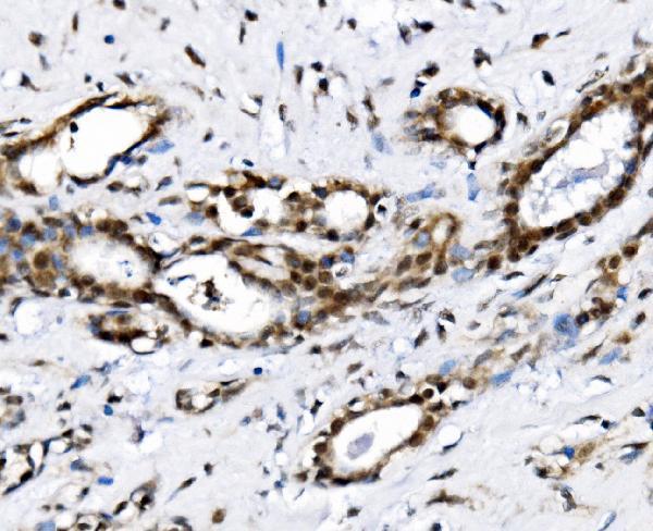
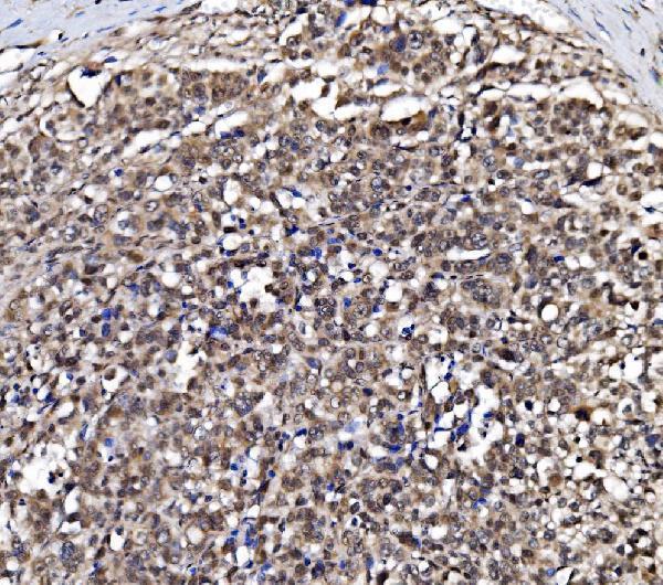
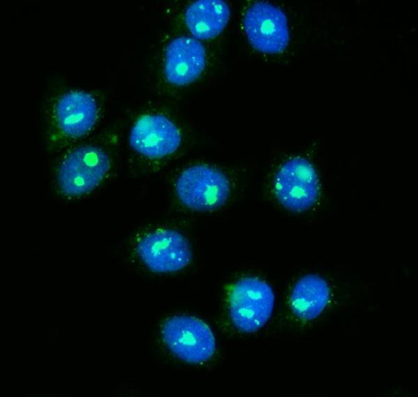
A07049-1 has IHC images from human paraffin sections and an IF/ICC image from MCF-7 cells (catalog image captions); listed reactivity is human (catalog: reactivity).
A07049-1 has IHC images from human breast cancer, gastric carcinoma, liver cancer, and ovarian adenoma paraffin sections (catalog: IHC image captions). A07049-1 also has an IF/ICC image from MCF-7 cells (catalog: IF image caption).
Which to pick: For tissue IHC, choose A07049-1 for human paraffin sections; its captions do not report the fixative (catalog: IHC image captions). For IF/ICC, A07049-1 has an MCF-7 image and a listed concentration of 5 μg/ml (catalog: IF image caption; datasheet: IF dilution). No cross-species option is supported: A07049-1 lists human reactivity only, and its clonality is unspecified (catalog: reactivity; clone field).