This website uses cookies to ensure you get the best experience on our website.


- Table of Contents
Plan chromogenic IHC for GBP4 in paraffin sections using the catalog antibody at 1:100–1:300 (datasheet: A11011). Assess cytoplasmic staining in glandular or kidney tubular cells, with adipocytes as a low-signal comparison (HPA tissue IHC).

Expected localisation, validated protocols, controls and antibodies — the at-a-glance facts below, then the full design guide.
| Expected localisation | Cytoplasmic staining in several tissues (HPA tissue IHC) | |
| Staining pattern | Cytoplasmic staining in glandular and kidney tubular cells (HPA tissue IHC) | |
| Antigen retrieval | Tris-EDTA pH 9.0 HIER, 95–98 °C, 20 min (rule: nuclear antigen) | |
| Positive control | Adrenal gland+4 more · see all | |
| Negative control | Adipose tissue+2 more · see all |
| Fixation | Keep fixation consistent across sections (standard IHC practice; not target-specific) | |
| Caveat | GBP1/2/5 heterodimers can shift GBP4 location (UniProt) | |
| Regulation | IFN-inducible (UniProt) | |
| Isoform / epitope | No annotated isoforms; one 1–640 chain (UniProt) |
The catalog antibody’s IHC-P protocol is accompanied by 2 published GBP4 IHC methods for ovarian and pancreatic cancer tissue (PMC8155613; PMC11306721).
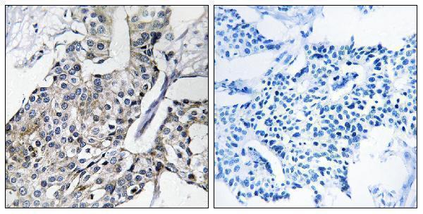
| Sample | Paraffin-embedded human breast carcinoma tissue; fixative not specified (datasheet A11011) |
| Fixation | Image fixative and duration unreported (datasheet A11011); verify before use. |
| Sectioning | 4–5 µm sections on charged slides (standard) |
| Deparaffinisation | Xylene, graded ethanol series to water (standard) |
| Antigen retrieval | Heat-induced epitope retrieval in Tris-EDTA buffer, pH 9.0, 20 min at 95–98 °C (standard rule: nuclear antigen) |
| Peroxidase block | 3% H2O2, 10 min, room temperature (standard) |
| Blocking | 10% normal serum of the secondary host, 30 min, room temperature (standard) |
| Primary antibody | Rabbit monoclonal (clone 3A8) anti-GBP4, 1:100-1:300 (datasheet A11011) |
| Primary incubation | Overnight at 4 °C (standard) |
| Detection | HRP-polymer secondary, DAB chromogen 5–10 min (standard) |
| Counterstain | Hematoxylin, blue, dehydrate and mount (standard) |
| Expected result | GBP4-positive staining in glandular cells of adrenal gland (HPA tissue IHC: High). HPA tissue profile: Cytoplasmic expression in several tissues. No signal in the no-primary control. |
GBP4 is reported in the cytoplasm, perinuclear region, Golgi membrane and nucleus, with no transmembrane segment (UniProt Q96PP9 localization and topology). In paraffin sections, expect cytoplasmic staining in HPA-positive glandular cells, kidney tubules and lymph node non-germinal center cells (HPA tissue IHC). HPA rates its tissue IHC evidence Enhanced, with medium consistency between antibody staining and RNA expression (HPA tissue IHC reliability).
| Cytoplasmic staining in colon or duodenal glandular cells, kidney tubules, or lymph node non-germinal center cells (HPA tissue IHC). | This matches the reported high-staining cell populations (HPA tissue IHC). Assess the named cells against nearby cells and the counterstain; a perinuclear accent is compatible with GBP4 localization but is not required to call a paraffin section positive (UniProt Q96PP9 localization). |
| Dominant staining in an unexpected compartment, with little staining in the expected cytoplasm. | Check section morphology and detection artefact before assigning that pattern to GBP4. HPA reports cytoplasmic tissue staining, while UniProt also lists nuclear and Golgi-associated localization; nuclear signal alone cannot be ruled out categorically from these sources (HPA tissue IHC; UniProt Q96PP9 localization). |
| Strong signal in adipocytes, hippocampal glial cells, or ovarian stromal cells. | These named populations were not detected in HPA tissue IHC (HPA tissue IHC). Review antibody specificity and endogenous chromogenic activity, especially if the positive-control cell pattern is absent. A signal in another, unlisted cell population is not automatically cross-reactivity: HPA reports low tissue RNA specificity (HPA tissue IHC). |
| Diffuse chromogen across cells and extracellular space, without a readable cellular pattern. | Treat the distribution as background until controls establish otherwise. Review blocking, washes, primary-antibody concentration and detection controls as general IHC practice; HPA's cytoplasmic tissue profile does not validate uniform extracellular staining (HPA tissue IHC). |
| No staining in the expected cells of a known-positive section. | Colon glandular cells and kidney tubule cells are reported as high-staining examples (HPA tissue IHC). First check that those cells are present and interpretable; then review the antibody's IHC-P instructions and detection controls. A blank section alone cannot establish GBP4 absence. |
| Cell population and comparison tissue | HPA reports high staining in several glandular populations, kidney tubules and lymph node non-germinal center cells, but no detection in three specified cell populations (HPA tissue IHC). Score the named cells; a whole-tissue positive or negative label can hide that distinction. |
| Intracellular distribution | Golgi, cytoplasmic, perinuclear and nuclear locations are listed by UniProt; GBP1, GBP2 and GBP5 heterodimers can alter GBP4 location (UniProt Q96PP9 localization and subunit). HPA tissue IHC describes cytoplasmic expression, so do not require a sharply defined Golgi pattern in chromogenic sections (HPA tissue IHC). |
| Strength of the evidence | The tissue IHC profile is Enhanced, with medium staining-to-RNA consistency (HPA tissue IHC reliability). Antibody HPA030101 has Enhanced IHC validation, whereas HPA062039 has Supported ICC validation (HPA antibody validation); these are application-specific assessments. |
| Chromogenic detection controls | Endogenous enzyme activity can create colour unrelated to primary-antibody binding (general IHC practice). Check a suitable detection control when staining is widespread or appears in HPA not-detected cells; that observation does not, by itself, identify the cause (HPA tissue IHC). |
| Situation | Likely cause | Next action |
|---|---|---|
| The expected cells stain weakly in a positive-control paraffin section. | The reported HPA level is an observed tissue pattern, not a promised intensity under every staining setup (HPA tissue IHC). The run may lack adequate detectable contrast. | Verify cell identity and counterstain, then review the catalog antibody's IHC-P instructions for retrieval, dilution and detection settings. Adjust one general IHC variable at a time against the same positive control; no GBP4-specific retrieval setting is supplied here. |
| Colour appears chiefly outside cells or uniformly across the section. | A widespread pattern does not match HPA's cytoplasmic tissue profile (HPA tissue IHC); nonspecific detection or residual chromogen can obscure cellular boundaries (general IHC practice). | Inspect the no-primary detection control, blocking and washes. Reassess staining only where intact cells and their counterstained boundaries can be identified (general IHC practice). |
| Adipocytes, hippocampal glial cells or ovarian stromal cells appear strongly positive. | HPA records these specific populations as not detected (HPA tissue IHC). Cross-reactivity or endogenous detection activity is possible, but the staining alone cannot distinguish them. | Compare a positive tissue and a no-primary control in the same run. If unexpected staining persists only with primary antibody, examine IHC validation and repeat at an appropriately optimized antibody dilution (general IHC practice). |
| The section looks predominantly nuclear, without convincing cytoplasmic staining. | UniProt includes nuclear localization, but HPA describes cytoplasmic staining in several tissues (UniProt Q96PP9 localization; HPA tissue IHC). A nuclear-only slide therefore needs context, not an automatic positive call. | Check morphology, counterstain and detection controls, then compare the same cell type in an HPA high-staining tissue. Record nuclear staining separately from the expected cytoplasmic pattern (HPA tissue IHC). |
| A tissue seems negative despite visible staining elsewhere on the slide. | HPA levels refer to particular cell populations; low-staining cells include hepatocytes and splenic red-pulp cells (HPA tissue IHC). Section-wide scoring can misclassify a sparse cell-specific pattern. | Identify and score the relevant cells individually, then compare with a named high-staining control population. Do not infer failure solely from weak signal in an HPA low-staining cell type (HPA tissue IHC). |
| Can the paraffin IHC pattern be used as the IF/ICC protocol or its expected image? | HPA ICC-IF places GBP4 mainly at the Golgi (supported) and additionally at the plasma membrane (uncertain); its image cell lines are HEL, RT-4 and U2OS (HPA subcellular ICC-IF). Those observations use a different application from tissue IHC. | Use the separate IF/ICC guide for that application. Interpret a Golgi-enriched IF signal with the ICC evidence, and treat plasma-membrane signal cautiously; do not turn the tissue IHC pattern into an IF/ICC protocol (HPA subcellular ICC-IF). |
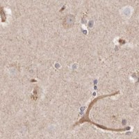
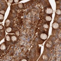


Comprehensive Human Protein Atlas IHC scoring per tissue (reliability: Enhanced — Medium consistency between antibody staining and RNA expression data.). Rows are taken directly from the HPA tissue chart — click any row's HPA link to view the source.
| Tissue | Cell type | Level | Evidence | Source |
|---|---|---|---|---|
| Adrenal gland | Glandular cells | High | Protein (IHC) | HPA → |
| Colon | Glandular cells | High | Protein (IHC) | HPA → |
| Duodenum | Glandular cells | High | Protein (IHC) | HPA → |
| Endometrium | Glandular cells | Medium | Protein (IHC) | HPA → |
| Gallbladder | Glandular cells | High | Protein (IHC) | HPA → |
Use compartment, cell type, and matched controls to troubleshoot GBP4 staining in paraffin sections; assess IF separately.
Anti-GBP4 A11011 has an IHC image from paraffin-embedded human breast carcinoma (catalog image caption); IF is listed, but no IF image is provided (catalog applications; catalog IF image alts).
A11011 will render with an IHC image of paraffin-embedded human breast carcinoma, including a peptide-blocked comparison (catalog image caption). IHC and IF are listed applications, and Human and Mouse are listed reactivities; no IF image is provided (catalog applications; catalog reactivity; catalog IF image alts).
Which to pick: For tissue IHC, choose A11011: its own image shows paraffin-embedded human breast carcinoma (catalog image caption), and its IHC dilution is 1:100–1:300 (datasheet: IHC dilution). The fixative is unreported (catalog image caption). For IF/ICC or human–mouse work, A11011 is the only listed option: it is rabbit clone 3A8, lists IF as an application and Human and Mouse as reactivities, but provides no IF image or ICC validation (catalog host and clone; catalog applications; catalog reactivity; catalog IF image alts).