This website uses cookies to ensure you get the best experience on our website.


- Table of Contents
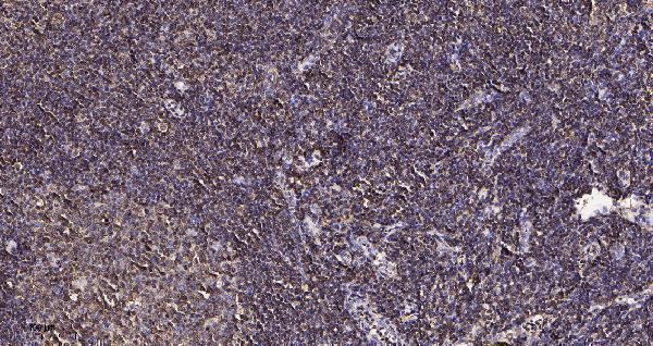
Plan GCSAM paraffin IHC around cytoplasmic immune-cell staining, using tonsil germinal-center cells as a positive control (HPA tissue IHC). Start catalog antibody M12995-1 within its 1:100–1:300 IHC dilution range (datasheet M12995-1).

Expected localisation, validated protocols, controls and antibodies — the at-a-glance facts below, then the full design guide.
| Expected localisation | Cytoplasm in tissue immune cells (HPA tissue IHC) | |
| Staining pattern | Cytoplasmic immune-cell staining; tonsil GC cells medium (HPA tissue IHC) | |
| Antigen retrieval | EDTA pH 9.0 HIER, heat-mediated (datasheet M12995-1) | |
| Positive control | Cerebellum+4 more · see all | |
| Negative control | Adipose tissue+4 more · see all |
| Fixation | Keep fixation consistent across paraffin sections. (standard IHC practice; not target-specific) | |
| Caveat | Antibody staining and RNA expression show low consistency (HPA tissue IHC) | |
| Regulation | GC enriched; IL6 alters localisation (UniProt) | |
| Isoform / epitope | 3 isoforms; no transmembrane segment; epitope impact unknown (UniProt) |
The catalog antibody’s IHC-P protocol (datasheet M12995-1) is paired with one published FFPE tissue protocol (PMC10901781 methods).
| Sample | Paraffin-embedded human tonsil tissue; fixative not specified (datasheet M12995-1) |
| Fixation | Image fixative and duration unreported (datasheet M12995-1); verify before use. |
| Sectioning | 4–5 µm sections on charged slides (standard) |
| Deparaffinisation | Xylene, graded ethanol series to water (standard) |
| Antigen retrieval | Heat retrieval: EDTA pH 9.0, 20min (datasheet M12995-1) |
| Peroxidase block | 3% H2O2, 10 min, room temperature (standard) |
| Blocking | 10% normal serum of the secondary host, 30 min, room temperature (standard) |
| Primary antibody | Rabbit anti-GCSAM, 1:100-1:300 (datasheet M12995-1) |
| Primary incubation | Overnight at 4 °C (standard) |
| Detection | HRP-polymer secondary, DAB chromogen 5–10 min (standard) |
| Counterstain | Hematoxylin, blue, dehydrate and mount (standard) |
| Expected result | GCSAM-positive staining in cells in molecular layer of cerebellum (HPA tissue IHC: Medium). HPA tissue profile: Cytoplasmic expression in immune cells. No signal in the no-primary control. |
Expect GCSAM chiefly in the cytoplasm of germinal center lymphocytes, with cell membrane localization also possible (UniProt Q8N6F7 localization; HPA: cytoplasmic expression in immune cells). Tonsil germinal center cells show medium staining (HPA: tonsil). GCSAM has no transmembrane segment (UniProt Q8N6F7 topology). HPA rates its tissue IHC antibody Approved but reports low consistency between staining and RNA expression (HPA: tissue IHC reliability).
| Distinct cytoplasmic staining in tonsil germinal center cells. | This matches the reported tissue pattern; medium intensity is the HPA reference observation, not a required intensity for every section (HPA: tonsil, Medium; HPA: cytoplasmic expression in immune cells). Assess the stained cells against the germinal center on the same slide (general IHC practice). |
| Predominantly nuclear staining, with little cytoplasmic signal. | An isolated nuclear pattern conflicts with the reported cytoplasmic and cell membrane locations (UniProt Q8N6F7 localization; HPA: cytoplasmic expression in immune cells). Treat it as a possible staining artefact and review morphology, counterstain, controls and antibody specificity before assigning it to GCSAM (general IHC practice). |
| Strong staining in adipocytes or bone marrow hematopoietic cells. | Those cells were not detected in HPA tissue IHC, so prominent staining warrants a check for cross-reactivity or endogenous detection activity (HPA: adipocytes and bone marrow hematopoietic cells, Not detected; general IHC practice). Other nonimmune staining cannot automatically be dismissed: HPA reports medium staining in several nonimmune cell populations (HPA: tissue IHC positives). |
| Diffuse color covers cells and surrounding tissue without clear cell boundaries. | That appearance does not resolve the reported cytoplasmic immune cell pattern (HPA: tissue IHC profile). Evaluate it as background using a no-primary control and the section morphology; incomplete blocking, residual detection reagent or an overly strong detection reaction are general IHC possibilities, not documented GCSAM-specific effects (general IHC practice). |
| No staining in tonsil germinal center cells. | This misses an HPA-observed positive population, but one blank section does not establish absence of GCSAM (HPA: tonsil germinal center cells, Medium). Confirm that the germinal center is present, then check the positive control and detection workflow (general IHC practice). HPA's Approved rating includes a low staining–RNA consistency caveat (HPA: tissue IHC reliability). |
| Location and topology | GCSAM is annotated in cytoplasm and at the cell membrane, without a transmembrane segment (UniProt Q8N6F7 localization and topology). Read membrane-associated color in the context of a cell's cytoplasmic staining and morphology; topology alone cannot identify an antibody epitope (general IHC interpretation). |
| Tissue and antibody evidence | The tissue IHC profile is Approved, yet HPA notes low consistency between antibody staining and RNA expression; the listed antibody, HPA002473, is IHC Approved, not IHC Enhanced (HPA: tissue IHC reliability; HPA: antibody validation). Treat the listed positive and negative cells as observed references, not absolute specificity guarantees. |
| Isoforms and modifications | UniProt lists 3 isoforms and phosphorylation sites at residues 99 and 148 (UniProt Q8N6F7 isoforms and modified residues). The supplied sources do not map the IHC antibody's epitope or show isoform- or phosphorylation-dependent staining; do not interpret intensity differences as either effect. |
| Retrieval and processing | No target-specific antigen retrieval response or fixation sensitivity is supplied. UniProt annotates one 1–178 chain, no signal peptide and no propeptide (UniProt Q8N6F7 processing). Those annotations do not predict which retrieval condition will work in paraffin sections; optimize retrieval against controls (general IHC practice). |
| IF/ICC: What pattern is supported? | UniProt lists cytoplasm and cell membrane, with movement to podosome-like structures after IL6 treatment (UniProt Q8N6F7 localization). HPA provides no main ICC-IF location or cell-line images, and HPA002473 has no ICC validation listed (HPA: subcellular record; HPA: antibody validation). Therefore, no observed IF/ICC pattern can be promised here. |
| Situation | Likely cause | Next action |
|---|---|---|
| Tonsil germinal centers appear negative. | The positive cells may be absent from the section, or the IHC detection workflow may have failed; HPA observed medium staining in tonsil germinal center cells (HPA: tonsil; general IHC practice). | Verify germinal center morphology and an on-run positive control. Review the catalog antibody's IHC-P instructions for the applied dilution and retrieval conditions, then change one workflow variable at a time (general IHC practice). |
| The slide shows nuclear-only color. | The result conflicts with reported cytoplasmic and cell membrane localization; nonspecific staining or misread counterstain is possible (UniProt Q8N6F7 localization; HPA: tissue IHC profile; general IHC practice). | Compare with the counterstain and a no-primary control. Reassess antibody concentration and detection exposure before scoring nuclear color as GCSAM (general IHC practice). |
| Adipocytes or bone marrow hematopoietic cells stain strongly. | HPA reports these populations as not detected; cross-reactivity or endogenous detection activity is plausible, although HPA staining is not an absolute absence test (HPA: adipose tissue and bone marrow; general IHC practice). | Run no-primary and detection-only controls, inspect the cell identity and check the antibody's IHC validation. Record the discrepancy rather than treating it as a confirmed GCSAM-positive population (general IHC practice). |
| Color is diffuse across the section. | A tissue-wide haze obscures the reported cytoplasmic immune cell pattern (HPA: tissue IHC profile). Excess detection reaction or insufficient blocking are general IHC possibilities, without target-specific evidence here (general IHC practice). | Check a no-primary control, wash steps, blocking and chromogen development. Adjust the detection workflow using the same positive tissue as a reference (general IHC practice; HPA: tonsil). |
| Only a faint signal appears in tonsil. | HPA's reference is medium staining in germinal center cells, but section quality and workflow can affect visibility; no GCSAM-specific fixation effect is documented in the supplied sources (HPA: tonsil; general IHC practice). | Confirm the correct cell population and working positive control. Check retrieval, antibody dilution and detection against the catalog IHC-P instructions without assuming that fixation caused the weak result (general IHC practice). |
| An IF/ICC image seems inconsistent with the IHC slide. | HPA lists no ICC-IF images or main subcellular location, and the listed antibody has no ICC validation; UniProt's localization is a protein annotation rather than an image-based ICC result (HPA: subcellular record; HPA: antibody validation; UniProt Q8N6F7 localization). | Interpret the paraffin IHC slide against its own tissue controls and HPA tissue observations. Use the separate IF/ICC guide for fluorescence assay design (HPA: tissue IHC profile; general assay practice). |


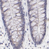
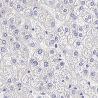

Comprehensive Human Protein Atlas IHC scoring per tissue (reliability: Approved — Low consistency between antibody staining and RNA expression data.). Rows are taken directly from the HPA tissue chart — click any row's HPA link to view the source.
| Tissue | Cell type | Level | Evidence | Source |
|---|---|---|---|---|
| Cerebellum | Cells in molecular layer | Medium | Protein (IHC) | HPA → |
| Cerebral cortex | Neuronal cells | Medium | Protein (IHC) | HPA → |
| Hippocampus | Neuronal cells | Medium | Protein (IHC) | HPA → |
| Pancreas | Pancreatic endocrine cells | Medium | Protein (IHC) | HPA → |
| Placenta | Trophoblastic cells | Medium | Protein (IHC) | HPA → |
| Tissue | Cell type | Level | Evidence | Source |
|---|---|---|---|---|
| Adipose tissue | Adipocytes | Not detected | Protein (IHC) | HPA → |
| Adrenal gland | Glandular cells | Not detected | Protein (IHC) | HPA → |
| Bone marrow | Hematopoietic cells | Not detected | Protein (IHC) | HPA → |
| Esophagus | Squamous epithelial cells | Not detected | Protein (IHC) | HPA → |
| Heart muscle | Cardiomyocytes | Not detected | Protein (IHC) | HPA → |
Troubleshoot GCSAM staining in paraffin sections by checking retrieval, cell identity, compartment, controls, and scoring before interpreting signal (datasheet M12995-1; UniProt Q8N6F7).
Two human-reactive anti-GCSAM antibodies have IHC images from paraffin-embedded tissue and are listed for IF/ICC; no IF images are supplied (catalog: reactivity, applications, image captions, IF image alts).
M12995-1 is listed for human IHC and IF/ICC, with an IHC image from paraffin-embedded human tonsil (catalog: applications, reactivity, IHC caption). M12995 has the same listed applications and human reactivity, with an IHC image from paraffin-embedded human bladder cancer (catalog: applications, reactivity, IHC caption).
Which to pick: For tissue IHC, M12995-1 offers the more detailed starting point: its paraffin-embedded human tonsil caption reports primary antibody at 1:200 and EDTA pH 9.0 retrieval; the fixative is unreported (M12995-1 IHC caption). For IF/ICC, either rabbit monoclonal is listed for human samples, though neither has a supplied IF image; M12995 also has a paraffin-embedded human bladder cancer IHC image with fixative unreported (catalog: host, applications, reactivity, IF image alts; M12995 IHC caption). Neither SKU has stated cross-species reactivity (catalog: both list human reactivity only).