This website uses cookies to ensure you get the best experience on our website.


- Table of Contents
Plan chromogenic paraffin IHC for GFI1 and assess nuclear staining (HPA tissue IHC). Start with 0.5–1 μg/mL of the catalog antibody (datasheet A00888-1), and interpret results in light of HPA's uncertain staining reliability (HPA tissue IHC).

Expected localisation, validated protocols, controls and antibodies — the at-a-glance facts below, then the full design guide.
| Expected localisation | General nuclear expression (HPA tissue IHC) | |
| Staining pattern | General nuclear staining across tissues (HPA tissue IHC) | |
| Antigen retrieval | Citrate pH 6 HIER, heat-mediated (datasheet A00888-1) | |
| Positive control | Bone marrow+4 more · see all | |
| Negative control | None in HPA (detected in all 45 tissues); use no-primary + isotype controls |
| Fixation | Keep fixation consistent across sections (standard IHC practice; not target-specific) | |
| Caveat | Staining has low consistency with RNA expression (HPA tissue IHC) | |
| Regulation | Marrow/lymphoid RNA group enrichment (HPA tissue IHC: RNA) | |
| Isoform / epitope | No isoforms or processing annotated; epitope impact unknown (UniProt) |
The catalog antibody protocol uses citrate retrieval (datasheet A00888-1); the published cartilage and knee joint IHC protocol uses enzymatic retrieval (PMC12332208).
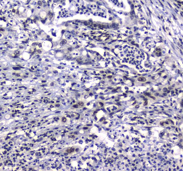
| Sample | Paraffin-embedded human mammary cancer tissue; fixative not specified (datasheet A00888-1) |
| Fixation | Image fixative and duration unreported (datasheet A00888-1); verify before use. |
| Sectioning | 4–5 µm sections on charged slides (standard) |
| Deparaffinisation | Xylene, graded ethanol series to water (standard) |
| Antigen retrieval | Heat retrieval: Citrate pH 6, 20 min (datasheet A00888-1) |
| Peroxidase block | 3% H2O2, 10 min, room temperature (standard) |
| Blocking | 10% goat serum (datasheet A00888-1) |
| Primary antibody | Rabbit anti-GFI1, 1μg/ml (datasheet A00888-1) |
| Primary incubation | Overnight at 4 °C (datasheet A00888-1) |
| Detection | Streptavidin-biotin complex (SABC), DAB chromogen (datasheet A00888-1) |
| Counterstain | Hematoxylin, blue, dehydrate and mount (standard) |
| Expected result | GFI1-positive staining in hematopoietic cells of bone marrow (HPA tissue IHC: High). HPA tissue profile: General nuclear expression. No signal in the no-primary control. |
GFI1 is a nuclear protein with no transmembrane segment; it can colocalize with PIAS3 and RUNX1T1 in nuclear dots (UniProt Q99684). In paraffin-section IHC, look for nuclear staining in hematopoietic cells of bone marrow and in other cell populations scored High by HPA (HPA: tissue IHC). Treat the pattern as provisional: HPA rates its tissue staining Uncertain because antibody staining and RNA expression have low consistency (HPA: reliability Uncertain).
| Clear nuclear staining in bone marrow hematopoietic cells, with limited staining outside nuclei. | This matches GFI1's annotated nuclear location and HPA's High staining in those cells (UniProt Q99684; HPA: High in bone marrow hematopoietic cells). Nuclear dots are plausible, but a dotted pattern is not required for a positive IHC result (UniProt Q99684: colocalization in nuclear dots). |
| Predominantly cytoplasmic, membranous, or extracellular staining with little nuclear signal. | That distribution conflicts with the annotated nuclear location and absence of a transmembrane segment (UniProt Q99684). Consider nonspecific staining or detection artefact before calling it GFI1; HPA's Uncertain tissue-IHC reliability adds reason to verify the pattern (HPA: reliability Uncertain). |
| Strong staining in a cell population expected to show Low staining, such as pancreatic exocrine glandular cells. | This differs from the reported Low pattern for that population (HPA: Low in pancreatic exocrine glandular cells). Check whether the signal is nuclear and survives controls; cross-reactivity or endogenous detection activity is possible, but the HPA rating does not establish the cause (HPA: reliability Uncertain; standard IHC practice). |
| Broad, diffuse color across nuclei and surrounding tissue, without a discernible cell-specific pattern. | Diffuse background cannot establish nuclear GFI1 staining (UniProt Q99684: nucleus). Examine the no-primary control and optimize detection and blocking conditions using standard IHC practice; HPA reports a general nuclear profile but rates its tissue-IHC evidence Uncertain (HPA: tissue IHC profile and reliability). |
| No nuclear signal in bone marrow hematopoietic cells. | This misses a reported High-staining population, so first assess whether the IHC run worked (HPA: High in bone marrow hematopoietic cells; standard IHC practice). Review a concurrently processed positive section, antibody dilution, detection reagents, and the counterstain before interpreting absence of GFI1 (standard IHC practice). |
| Expected compartment | GFI1 is annotated in the nucleus, sometimes colocalizing in nuclear dots, and has no transmembrane segment (UniProt Q99684). Score nuclear signal first; a membrane-dominant pattern needs independent verification (UniProt Q99684; standard IHC practice). |
| Cell population and tissue | HPA scores hematopoietic cells in bone marrow High, while pancreatic exocrine glandular cells and adipocytes are Low (HPA: tissue IHC). Compare like cell populations rather than treating every cell in a section as an equivalent positive control (standard IHC practice). |
| Strength of IHC evidence | HPA calls the tissue pattern Uncertain because staining and RNA expression show low consistency; its listed antibody CAB011197 has IHC status Uncertain (HPA: tissue reliability; HPA: CAB011197 IHC Uncertain). Interpret intensity and unexpected positives cautiously. |
| RNA versus stained cells | HPA reports group-enriched RNA in bone marrow and lymphoid tissue, alongside High protein staining in several listed cell populations outside those groups (HPA: RNA specificity; HPA: tissue IHC). RNA enrichment alone does not resolve whether an individual stained nucleus is specific (standard IHC interpretation). |
| Situation | Likely cause | Next action |
|---|---|---|
| Bone marrow section lacks nuclear staining. | The result conflicts with HPA's High call for hematopoietic cells, but a failed IHC run and true sample variation require different checks (HPA: High in bone marrow hematopoietic cells; standard IHC practice). | Inspect a concurrently stained positive section, then check section quality, primary dilution, retrieval conditions, and detection reagents; use the catalog antibody's IHC-P protocol for its specified settings (standard IHC practice). |
| Color is strongest in cytoplasm or along cell borders. | This conflicts with GFI1's nuclear annotation and lack of a transmembrane segment (UniProt Q99684). Nonspecific binding or detection background is possible (standard IHC practice). | Compare the no-primary control and nuclear counterstain; reassess primary and detection conditions before scoring the signal as GFI1 (standard IHC practice). |
| Many unrelated structures stain diffusely. | A diffuse deposit does not reproduce HPA's general nuclear profile and may reflect assay background (HPA: tissue IHC profile; standard IHC practice). | Check the no-primary control, blocking and washes, then reduce detection background according to the reagents' instructions; score only discernible cellular staining (standard IHC practice). |
| A Low-scored cell population shows intense nuclear staining. | The observation differs from HPA's Low call, while HPA's Uncertain reliability prevents a definitive specificity judgment from that comparison alone (HPA: tissue IHC; HPA: reliability Uncertain). | Confirm cell identity and compare a reported High population in the same run; consider independent antibody or orthogonal evidence before accepting the unexpected positive (standard IHC practice). |
| Two sections give different staining intensity. | HPA reports low consistency between antibody staining and RNA expression, so its tissue scores are an uncertain benchmark; routine IHC processing differences may also affect comparisons (HPA: reliability Uncertain; standard IHC practice). | Compare matched cell populations and controls across runs, and review the recorded retrieval, dilution, detection, and counterstain conditions before assigning a biological difference (standard IHC practice). |
| Can IF/ICC establish the same GFI1 pattern? | HPA provides no main ICC-IF location or cell-line ICC-IF images for GFI1; its tissue-IHC pattern is rated Uncertain (HPA: subcellular record; HPA: reliability Uncertain). | Use the separate IF/ICC guide for that assay. Here, treat nuclear localization as the UniProt expectation and assess the paraffin-section IHC result against its own controls (UniProt Q99684: nucleus; standard IHC practice). |



Comprehensive Human Protein Atlas IHC scoring per tissue (reliability: Uncertain — Low consistency between antibody staining and RNA expression data.). Rows are taken directly from the HPA tissue chart — click any row's HPA link to view the source.
| Tissue | Cell type | Level | Evidence | Source |
|---|---|---|---|---|
| Bone marrow | Hematopoietic cells | High | Protein (IHC) | HPA → |
| Cerebellum | Cells in granular layer | High | Protein (IHC) | HPA → |
| Cerebral cortex | Neuronal cells | High | Protein (IHC) | HPA → |
| Colon | Glandular cells | High | Protein (IHC) | HPA → |
| Duodenum | Glandular cells | High | Protein (IHC) | HPA → |
| Tissue | Cell type | Level | Evidence | Source |
|---|---|---|---|---|
| None in HPA: GFI1 is detected in all 45 scored tissues. Use a no-primary (secondary-only) and an isotype control instead. | ||||
GFI1 is a nuclear transcription repressor (UniProt Q99684). Interpret chromogenic staining cautiously because tissue IHC shows low consistency with RNA expression (HPA: reliability uncertain).
Two anti-GFI1 antibodies have real IHC images from human paraffin sections: mammary cancer and rectal cancer tissue (catalog IHC captions). Both list human, mouse and rat reactivity (catalog).
A00888-1 lists IHC for human, mouse and rat and shows GFI1 staining in human mammary cancer paraffin sections (catalog applications, reactivity and IHC caption). A00888-3 lists IHC for human tissue and shows GFI1 staining in human rectal cancer paraffin sections (catalog dilution entry and IHC caption).
Which to pick: For tissue IHC, choose A00888-1 for the documented citrate pH 6 retrieval or A00888-3 for the documented EDTA pH 8 retrieval; both captions describe paraffin sections and leave the fixative unreported (catalog IHC captions). Neither SKU lists IF/ICC or provides an IF figure, so neither has documented IF/ICC support here (catalog applications and image records). For cross-species work, A00888-1 is the clearer IHC starting point because it is polyclonal and lists human, mouse and rat reactivity, while A00888-3 specifies human for its IHC dilution; the supplied IHC images for both show human tissue only (catalog dilution entries and IHC captions).