GLI2 / Transcription activator GLI2 · IHC design guide

Design Immunohistochemistry for GLI2

Plan GLI2 paraffin-section IHC around the broad nuclear tissue pattern (HPA tissue IHC). The guide covers chromogenic staining with the IHC-validated antibody (datasheet A00701-5) and interpretation when staining and RNA levels disagree (HPA tissue IHC).

Evidence assembled Oct 2026 · For research use; verify linked source records and product datasheet before use
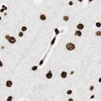
Immunohistochemistry protocol sheet for GLI2 (IHC for GLI2): expected localisation Broad nuclear tissue staining (HPA tissue IHC), antibody A00701-5, validated IHC image, and IHC protocol steps
Printable GLI2 IHC protocol sheet — expected localisation Broad nuclear tissue staining (HPA tissue IHC), antibody A00701-5, controls and protocol steps. Open the full GLI2 IHC guide →

GLI2 Immunohistochemistry Experimental Design Guide

Expected localisation, validated protocols, controls and antibodies — the at-a-glance facts below, then the full design guide.

Must know before staining
Expected localisation Broad nuclear tissue staining (HPA tissue IHC)
Staining pattern Nuclear staining across many tissue cell types (HPA tissue IHC)
Antigen retrieval Citrate pH 6 HIER, heat-mediated (datasheet A00701-5)
Positive control ⓘ Adipose tissue+4 more · see all
Negative control ⓘ None in HPA (detected in all 43 tissues); use no-primary + isotype controls
Important caveats
Reasons your staining may differ from the expected pattern.
Fixation Matched tissue-IHC evidence does not establish the fixation claim. Validate the specimen-specific method before use. (selected-SKU IHC image A00701-5)
Caveat Antibody staining and RNA show low consistency (HPA tissue IHC)
Regulation SMO signaling can shift GLI2 localisation (UniProt)
Isoform / epitope 5 isoforms plus GLI2R processing; map the epitope (UniProt)
Section 1

Recommended GLI2 IHC & IF Protocols

The catalog antibody’s IHC-P protocol (datasheet: A00701-5) is followed by published GLI2 protocols for paraffin-embedded liver specimens (PMC3565946; PMC3632642).

Recommended immunohistochemistry (IHC-P) protocol parameters
SampleParaffin-embedded human mammary cancer tissues; fixative not specified (datasheet A00701-5)
FixationImage fixative and duration unreported (datasheet A00701-5); verify before use.
Sectioning4–5 µm sections on charged slides (standard)
DeparaffinisationXylene, graded ethanol series to water (standard)
Antigen retrievalHeat retrieval: Citrate pH 6, 20 min (datasheet A00701-5)
Peroxidase block3% H2O2, 10 min, room temperature (standard)
Blocking10% goat serum (datasheet A00701-5)
Primary antibodyRabbit anti-GLI2, 0.5-1μg/ml (datasheet A00701-5)
Primary incubationOvernight at 4 °C (datasheet A00701-5)
DetectionStreptavidin-biotin complex (SABC), DAB chromogen (datasheet A00701-5)
CounterstainHematoxylin, blue, dehydrate and mount (standard)
Expected resultGLI2-positive staining in adipocytes of adipose tissue (HPA tissue IHC: High). HPA tissue profile: Ubiquitous nuclear expression. No signal in the no-primary control.
💡Decision noteStart with heat-mediated citrate retrieval at pH 6 (datasheet: A00701-5); both included studies report citrate retrieval (PMC3565946; PMC3632642).
Section 2

What Is the Expected GLI2 Staining Pattern?

GLI2 is a nuclear transcription factor that can also occur in the cytoplasm and cilium (UniProt P10070). In paraffin-section IHC, expect predominantly nuclear staining across many cell types; HPA describes a ubiquitous nuclear pattern and high staining in several tissues (HPA tissue IHC). Interpret intensity cautiously: its IHC reliability is Approved, but antibody staining has low consistency with RNA expression (HPA tissue IHC). GLI2 has no transmembrane segment (UniProt P10070 topology).

What am I looking at on my slide?
Predominantly nuclear staining in the expected cells.This matches the broad tissue pattern reported for GLI2 (HPA tissue IHC: ubiquitous nuclear expression). High staining is reported in adipocytes in adipose tissue and breast, adrenal and appendix glandular cells, bone marrow hematopoietic cells, bronchial respiratory epithelial cells, caudate glial cells, and cerebellar granular-layer cells (HPA tissue IHC). Compare the relevant cell population with its surrounding tissue.
Cytoplasmic signal predominates, or a sharp membrane rim replaces nuclear staining.Cytoplasmic GLI2 can be biological: full-length protein may be sequestered there by SUFU without smoothened signaling (UniProt P10070). A membrane-rim pattern has no support from GLI2 topology, which lacks a transmembrane segment (UniProt P10070). Check whether nuclei retain specific signal and whether the pattern persists with an appropriate staining control (general IHC practice).
An unexpected cell population stains more strongly than the intended population.Treat this as a specificity question, not proof of cross-reactivity: HPA describes broadly distributed nuclear staining and supplies no negative tissue reference here (HPA tissue IHC). Review morphology and compare the same compartment in a documented high-staining population. Signal in an unexpected population can also reflect nonspecific antibody binding or endogenous detection activity (general IHC practice).
Diffuse chromogen covers cells and surrounding tissue without clear nuclear boundaries.That appearance cannot establish GLI2 localization. Examine a control lacking primary antibody for detection-system background and inspect staining outside cells (general IHC practice). HPA's reported tissue pattern is nuclear, while UniProt also permits cytoplasmic localization; neither supports uniform staining across tissue structures as a specific positive result (HPA tissue IHC; UniProt P10070).
A documented high-staining tissue has no discernible nuclear signal.First confirm that the section contains the relevant cell type: HPA levels refer to named populations, such as adipocytes or glandular cells, rather than every cell in an organ (HPA tissue IHC). Check section quality, antigen retrieval, antibody incubation, detection, and counterstain using routine IHC controls (general IHC practice). One blank section does not establish biological absence.
💡Expected GLI2 appearanceCall a convincing IHC positive when nuclei are clearly stained in the relevant cells, potentially strongly in HPA high-staining populations; diffuse haze or an isolated membrane rim is suspect (HPA tissue IHC; UniProt P10070 topology).
How each factor affects the staining
Cell population and intensityHPA reports high staining in eight named tissue–cell combinations, while its overall profile is ubiquitous nuclear expression; use those combinations as reference patterns, not a universal intensity threshold (HPA tissue IHC).
Signaling state and compartmentFull-length GLI2 may remain cytoplasmic through SUFU interaction without smoothened signaling; nuclear enrichment is therefore informative, but cytoplasmic signal alone is not automatically an artefact (UniProt P10070).
Antibody evidenceIHC is Approved for CAB008558; HPA074275 is Supported for ICC, with no IHC status supplied. HPA also reports low consistency between tissue staining and RNA expression, so keep the validation caveat visible (HPA antibodies; HPA tissue IHC).
Protein formsUniProt lists five isoforms and a full-length activator plus a repressor form. The supplied records give no epitope position or form-specific IHC result, so they cannot predict which forms an individual antibody detects (UniProt P10070).
IF/ICC Q: What localization should an IF image show?A: HPA reports mainly nucleoli, with supported nucleoplasm, centrosome, and basal-body localization (HPA subcellular ICC-IF). This is an IF observation; do not require those fine structures to be resolved in chromogenic paraffin-section IHC.
Why is my staining missing, weak or wrong?
SituationLikely causeNext action
No nuclear staining in a selected positive tissue.The section may miss the cell population scored High by HPA, or a routine IHC step may have failed (HPA tissue IHC; general IHC practice).Locate the named population first, then review section quality, retrieval, incubation, detection, and a working staining control (general IHC practice). Do not call the tissue GLI2-negative from this result alone.
Strong cytoplasmic staining with little nuclear staining.SUFU can sequester full-length GLI2 in cytoplasm without smoothened signaling; background remains another possibility (UniProt P10070; general IHC practice).Assess cellular boundaries and nuclear counterstain, compare a documented high-staining population, and inspect a control lacking primary antibody (HPA tissue IHC; general IHC practice).
Crisp cell-surface rims dominate the slide.A cell-surface membrane pattern is unsupported by GLI2's lack of a transmembrane segment (UniProt P10070 topology).Check for tissue-edge or detection artefacts and repeat interpretation against nuclear staining in relevant cells and a control lacking primary antibody (HPA tissue IHC; general IHC practice).
Brown signal appears in unexpected cells or persists without primary antibody.Nonspecific binding or endogenous detection activity can create apparent positives (general IHC practice); HPA supplies no negative tissue comparator in this payload (HPA tissue IHC).Compare the control and test section within the same cell types; investigate blocking and endogenous-enzyme control steps appropriate to the detection system (general IHC practice).
Diffuse staining obscures nuclei.Excess background or inadequate visual separation of chromogen and counterstain can prevent compartment scoring (general IHC practice).Review controls, wash and detection conditions, and counterstain intensity before assigning nuclear positivity (general IHC practice). Record the result as uninterpretable if boundaries remain unclear.
IF shows nucleolar puncta while IHC looks broadly nuclear.HPA reports supported nucleolar and additional subcellular IF locations, while its tissue IHC profile is broadly nuclear (HPA subcellular ICC-IF; HPA tissue IHC).Score each application at its demonstrated resolution. For paraffin-section IHC, document nuclear staining and cell identity; reserve finer subcellular claims for the separate IF/ICC assessment (HPA tissue IHC; HPA subcellular ICC-IF).

Sample controls for GLI2 IHC & IF

🧪Run breast tissue first and score its adipocytes, which HPA rates High for GLI2 (HPA: High in breast adipocytes). HPA detects GLI2 in all 43 scored tissues, so there is no supported negative tissue; no-primary and isotype controls carry the negative comparison, while erythrocytes within the positive section should remain unstained after peroxidase blocking (HPA: no negative tissue rows; standard IHC practice).
Positive control tissue: Adipose tissue (Adipocytes, HPA High)
Negative control tissue: None in HPA: GLI2 is detected in all 43 scored tissues. Use a no-primary (secondary-only) and an isotype control instead.
ICC-IF cell lines (HPA subcellular resource): HPA ICC-IF images show GLI2 in A-549, BJ [Human fibroblast], U2OS, ASC52telo, RPTEC/TERT1, Sperm, with annotated localisation: Nucleoli (supported) (HPA subcellular).
Technical controls: Include a no-primary, secondary-only section and a rabbit IgG control matched to the catalog antibody’s format and concentration (caption: rabbit anti-GLI2; standard IHC practice). Confirm specificity with GLI2-knockout material or a validated peptide-block control, and quench endogenous peroxidase before DAB detection, particularly when assessing erythrocytes (standard IHC practice).
⚠️Feasibility: The selected paraffin-section caption reports heat retrieval in citrate buffer at pH 6 for 20 minutes, but gives no fixative or target-specific fixation window or effect (caption: A00701-5). The supplied evidence does not establish whether frozen sections or IF are easier; its matched tissue protocol is paraffin IHC, while HPA provides ICC-IF localization images (caption: A00701-5; HPA: subcellular). In breast adipose areas, paraffin processing can leave empty lipid vacuoles, so score cellular staining rather than the vacuoles (standard IHC practice).

HPA tissue IHC evidence for GLI2

Comprehensive Human Protein Atlas IHC scoring per tissue (reliability: Approved — Low consistency between antibody staining and RNA expression data.). Rows are taken directly from the HPA tissue chart — click any row's HPA link to view the source.

Positive expression · recommended positive controls

TissueCell typeLevelEvidenceSource
Adipose tissue Adipocytes High Protein (IHC) HPA →
Adrenal gland Glandular cells High Protein (IHC) HPA →
Appendix Glandular cells High Protein (IHC) HPA →
Bone marrow Hematopoietic cells High Protein (IHC) HPA →
Breast Adipocytes High Protein (IHC) HPA →

Undetected expression · recommended negative controls

TissueCell typeLevelEvidenceSource
None in HPA: GLI2 is detected in all 43 scored tissues. Use a no-primary (secondary-only) and an isotype control instead.
Section 3

Advanced GLI2 IHC Tips

GLI2 can appear in the nucleus and cytoplasm (UniProt P10070); interpret chromogenic staining by compartment, cell type and controls (standard IHC practice).

How should I retrieve GLI2 antigen when staining paraffin sections?
Use heat-mediated citrate buffer at pH 6 for antigen retrieval in paraffin sections (datasheet A00701-5). The selected tissue-IHC example heated sections for 20 minutes before blocking with 10% goat serum (caption A00701-5). Keep section thickness, heating conditions and cooling consistent across cases so differences in nuclear staining remain comparable (standard IHC practice). If staining is weak, vary heating time on adjacent sections while retaining the documented citrate buffer as the reference condition (standard IHC practice). Compare each trial with a no-primary control, since excessive retrieval can damage morphology or increase background (standard IHC practice).
Could fixation explain weak GLI2 staining in my paraffin sections?
The selected GLI2 tissue-IHC caption describes paraffin sections but does not state their fixative, so target-specific fixation sensitivity is unknown (caption A00701-5). Record each specimen’s fixative and fixation duration before comparing staining intensity across cases (standard IHC practice). On adjacent sections, apply the documented citrate pH 6, 20-minute retrieval condition consistently to assess whether technical variation explains weak signal (caption A00701-5; standard IHC practice). Include a previously staining section and a no-primary control in the same run (standard IHC practice). Do not treat an HPA tissue pattern or GLI2’s reported modifications as evidence that a particular fixative preserves its epitope (HPA tissue IHC; UniProt P10070).
How should I interpret nuclear, cytoplasmic and punctate GLI2 staining?
Score nuclear and cytoplasmic signal separately: GLI2 is reported in both compartments, and full-length GLI2 can be held in the cytoplasm by SUFU without smoothened signaling (UniProt P10070). The HPA tissue-IHC profile describes ubiquitous nuclear expression, while its subcellular imaging supports nucleolar, nucleoplasmic, centrosomal and basal-body locations (HPA tissue IHC; HPA subcellular). Confirm that puncta lie within intact cells before assigning them to centrosomes or basal bodies (standard IHC practice). GLI2 is also associated with cilia, but chromogenic puncta alone do not establish ciliary identity (UniProt P10070; standard IHC practice). Compare compartment scores with matched controls and preserved morphology (standard IHC practice).
Can this stain distinguish GLI2 isoforms or its activator and repressor forms?
Do not assign an isoform from this stain without an epitope map and isoform-specific validation: the record lists 5 GLI2 isoforms (UniProt P10070; standard IHC practice). GLI2 also has a full-length activator and a processed repressor form, so antibody recognition depends on whether the epitope remains in each form (UniProt P10070; standard IHC practice). No epitope position is supplied for the selected antibody (caption A00701-5). Preserve the documented citrate pH 6, 20-minute retrieval condition while comparing staining patterns, to limit retrieval as a variable (caption A00701-5; standard IHC practice). Report compartment and cell-level staining without labeling either form unless independently verified (standard IHC practice).
How can IF help resolve ambiguous GLI2 staining in these sections?
For a separate IF assessment, multiplex GLI2 with an independently validated marker for the cell type being assessed and a nuclear counterstain (standard IF practice). Choose spectrally separated fluorophores after checking the section’s autofluorescence, and use a far-red channel for a weak signal when background permits (standard IF practice). Permeabilise for intracellular access because GLI2 is reported in nuclei and cytoplasm and has no transmembrane segment; the antibody’s precise epitope is unspecified (UniProt P10070; caption A00701-5; standard IF practice). Include single-label and no-primary controls to assess bleed-through and background (standard IF practice). Treat the paraffin-section citrate pH 6, 20-minute example as IHC evidence, not IF validation (caption A00701-5).
What should I check when GLI2 DAB staining looks diffuse or excessive?
First compare the stained section with a no-primary control to identify signal arising from detection reagents rather than GLI2 binding (standard IHC practice). The selected example used 1 μg/mL primary antibody overnight at 4°C, followed by a biotinylated secondary and DAB development (caption A00701-5). If both sections stain, check endogenous peroxidase and endogenous biotin contributions before changing the primary concentration (caption A00701-5; standard IHC practice). If background appears only with primary antibody, adjust blocking, washes or antibody concentration while keeping the citrate pH 6 retrieval reference (caption A00701-5; standard IHC practice). Assess nuclear and cytoplasmic signal separately because both locations are reported for GLI2 (UniProt P10070).
How should I quantify GLI2 staining across paraffin-section samples? ⚠ ANSWER MARKED FOR VERIFICATION
Define the tissue region and cell population before scoring, then apply identical inclusion rules to every section (standard IHC practice). Report the percentage of positive cells and, where intensity is reproducible, an H-score based on the percentages in each intensity category (standard IHC practice). Score nuclear and cytoplasmic staining separately because GLI2 is reported in both compartments (UniProt P10070). For spatial comparisons, count positive cells per mm² and normalise to viable tissue area or the relevant cell count, as appropriate (standard IHC practice). Keep retrieval at citrate pH 6 for 20 minutes across compared sections and document thresholds before analysis (caption A00701-5; standard IHC practice).
How can I distinguish genuine GLI2 staining from tissue artefact?
Look for reproducible signal in intact cells with plausible nuclear or cytoplasmic localisation, rather than judging a dark DAB deposit alone (UniProt P10070; standard IHC practice). Compare cell identity with the sampled compartment: HPA reports high nuclear staining in breast adipocytes, while the selected example shows staining in human mammary cancer tissue (HPA tissue IHC; caption A00701-5). Exclude section edges, folds and necrotic regions from interpretation when they show disproportionate staining (standard IHC practice). A no-primary control helps reveal endogenous enzyme or detection-system signal (standard IHC practice). Interpret diffuse positivity cautiously because HPA calls its tissue-IHC reliability Approved but reports low consistency with RNA expression (HPA tissue IHC).
Boster reagents

Best GLI2 / Transcription activator GLI2 IHC Antibodies

Both anti-GLI2 antibodies have paraffin-section IHC images from human tissue; A00701-5 also has mouse and rat brain IHC images, and A00701-6 has human U2OS IF data (catalog image captions).

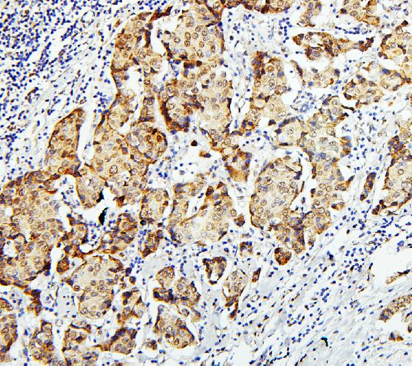
Real IHC data IHC analysis of GLI2 using anti-GLI2 antibody (A00701-5). GLI2 was detected in paraffin-embedded section of human mammary cancer tissues. Heat mediated antigen retrieval was performed in citrate buffer (pH6, epitope retrieval solution) for 20 mins. The tissue section was blocked with 10% goat serum. The tissue section was then incubated with 1μg/ml rabbit anti-GLI2 Antibody (A00701-5) overnight at 4°C. Biotinylated goat anti-rabbit IgG was used as secondary antibody and incubated for 30 minutes at 37°C. The tissue section was developed using Strepavidin-Biotin-Complex (SABC)(Catalog # SA1022) with DAB as the chromogen.
Anti-Zinc finger protein GLI2 GLI2 Antibody ®
Cat # A00701-5
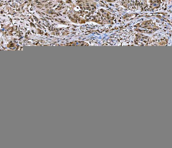
Real IHC data IHC analysis of GLI2 using anti-GLI2 antibody (A00701-6). GLI2 was detected in a paraffin-embedded section of human esophageal squamous carcinoma tissue. Heat mediated antigen retrieval was performed in EDTA buffer (pH 8.0, epitope retrieval solution). The tissue section was blocked with 10% goat serum. The tissue section was then incubated with 2 μg/ml rabbit anti-GLI2 Antibody (A00701-6) overnight at 4°C. Peroxidase Conjugated Goat Anti-rabbit IgG was used as secondary antibody and incubated for 30 minutes at 37°C. The tissue section was developed using HRP Conjugated Rabbit IgG Super Vision Assay Kit (Catalog # SV0002) with DAB as the chromogen.
Anti-GLI2 Antibody ®
Cat # A00701-6

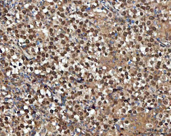
A00701-5 will render with human mammary cancer IHC; its additional captions show human intestinal cancer and mouse and rat brain IHC (A00701-5 IHC captions). A00701-6 will render with human esophageal squamous carcinoma IHC; its additional captions show human testicular germ cell tumor and tonsil IHC, plus U2OS IF (A00701-6 IHC and IF captions).

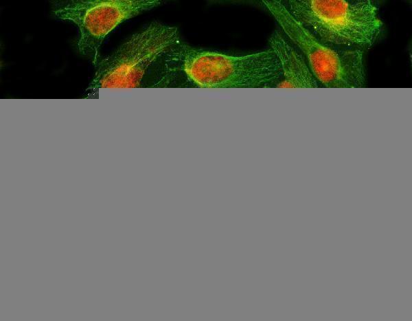
Which to pick: For paraffin-section IHC across human, mouse and rat samples, choose A00701-5: its captions show all three species with citrate retrieval at pH 6 for 20 minutes; the fixative is unreported (A00701-5 IHC captions). For human tissue IHC, A00701-6 is another option, with illustrated EDTA retrieval at pH 8.0 and an unreported fixative (A00701-6 IHC captions). For human IF/ICC, choose A00701-6, which lists both applications and has a U2OS IF image; clonality is unspecified for both SKUs (catalog applications, IF caption and clone fields).

Each figure is that product's own IHC / IF validation image from its datasheet.

References

  1. UniProt Consortium. UniProt entry P10070 (GLI2_HUMAN, Transcription activator GLI2).
  2. Human Protein Atlas. GLI2 tissue IHC expression (reliability: Approved).
  3. Human Protein Atlas. GLI2 subcellular location (ICC-IF): Mainly localized to the nucleoli. In addition localized to the nucleoplasm, centrosome and basal body..
  4. Human Protein Atlas. GLI2 antibody validation summary (2 antibodies).
  5. Expression of glioma-associated oncogene 2 (Gli 2) is correlated with poor prognosis in patients with hepatocellular carcinoma undergoing hepatectomy. World journal of surgical oncology 2013 — PMC3565946.
  6. Hedgehog pathway and pediatric nonalcoholic fatty liver disease. Hepatology (Baltimore, Md.) 2013 — PMC3637920.
  7. Signals from dying hepatocytes trigger growth of liver progenitors. Gut 2010 — PMC3632642.
  8. Arsenic inhibits hedgehog signaling during P19 cell differentiation. Toxicology and applied pharmacology 2014 — PMC4255151.
  9. PubMed PMID:9557682 — UniProt-cited evidence.
  10. PubMed PMID:15994174 — UniProt-cited evidence.
  11. PubMed PMID:15815621 — UniProt-cited evidence.