Click image to see more details
-

-
-
-
-
+6
Product Info Summary
| SKU: | A00701-5 |
|---|---|
| Size: | 100 μg/vial |
| Reactive Species: | Human, Mouse, Rat |
| Host: | Rabbit |
| Application: | ELISA, IHC, WB |
Customers Who Bought This Also Bought

Product info
Product Name
Anti-Zinc finger protein GLI2 GLI2 Antibody Picoband®
SKU/Catalog Number
A00701-5
Size
100 μg/vial
Form
Lyophilized
Description
Boster Bio Anti-Zinc finger protein GLI2 GLI2 Antibody catalog # A00701-5. Tested in ELISA, IHC, WB applications. This antibody reacts with Human, Mouse, Rat. The brand Picoband indicates this is a premium antibody that guarantees superior quality, high affinity, and strong signals with minimal background in Western blot applications. Only our best-performing antibodies are designated as Picoband, ensuring unmatched performance.
Storage & Handling
Store at -20˚C for one year from date of receipt. After reconstitution, at 4˚C for one month. It can also be aliquotted and stored frozen at -20˚C for six months. Avoid repeated freeze-thaw cycles.
Cite This Product
Anti-Zinc finger protein GLI2 GLI2 Antibody Picoband® (Boster Biological Technology, Pleasanton CA, USA, Catalog # A00701-5)
Host
Rabbit
Contents
Each vial contains 4mg Trehalose, 0.9mg NaCl, 0.2mg Na2HPO4, 0.05mg NaN3.
Clonality
Polyclonal
Isotype
Rabbit IgG
Immunogen
E.coli-derived human GLI2 recombinant protein (Position: K721-D1457).
Cross-reactivity
No cross-reactivity with other proteins.
Reactive Species
A00701-5 is reactive to GLI2 in Human, Mouse, Rat
Observed Molecular Weight
180-200 kDa
Calculated molecular weight
167.8 kDa
Background of GLI2
GLI2 (Gli-Kruppel Family Member 2), also called ONCOGENE GLI2, is a protein that in humans is encoded by the GLI2 gene. Sequencing of GLI cDNA clones showed the presence of 5 tandem zinc fingers connected by histidine-cysteine links, which indicated that the gene belongs to the family of zinc finger genes related to Kruppel (Kr). The Drosophila gene Kr is a member of the gap class of segmentation genes; thoracic and anterior abdominal segments fail to form in Kr mutant embryos. By fluorescence in situ hybridization, Matsumoto et al. refined the assignment of the GLI2 gene to chromosome 2q14. Roessler et al. showed that GLI2-delta-N exhibited potent transcriptional activity in vivo: overexpression in mouse skin led to the formation of hedgehog-independent epithelial downgrowths resembling basal cell carcinomas, which in humans are associated with constitutive hedgehog signaling.
Antibody Validation
Boster validates all antibodies on WB, IHC, ICC, Immunofluorescence, and ELISA with known positive control and negative samples to ensure specificity and high affinity, including thorough antibody incubations.
Application & Images
Applications
A00701-5 is guaranteed for ELISA, IHC, WB Boster Guarantee
Recommend Dilution
| Application | Dilution | Species |
|---|---|---|
| Western blot | 0.1-0.5μg/ml | |
| Immunohistochemistry (Paraffin-embedded Section) | 0.5-1μg/ml | |
| ELISA | 0.1-0.5μg/ml |
Tested application
Suggested blocking solution with 5% non-fat milk or BSA; (*)Recommended protein loading: 20-40 µg per lane
Use TE buffer pH 9.0 for antigen retrieval; (*) citrate buffer pH 6.0 is an alternative.
Validation Images & Assay Conditions
Click image to see more details
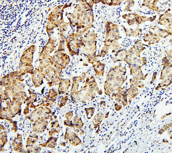
IHC analysis of GLI2 using anti-GLI2 antibody (A00701-5).
GLI2 was detected in paraffin-embedded section of human mammary cancer tissues. Heat mediated antigen retrieval was performed in citrate buffer (pH6, epitope retrieval solution) for 20 mins. The tissue section was blocked with 10% goat serum. The tissue section was then incubated with 1μg/ml rabbit anti-GLI2 Antibody (A00701-5) overnight at 4°C. Biotinylated goat anti-rabbit IgG was used as secondary antibody and incubated for 30 minutes at 37°C. The tissue section was developed using Strepavidin-Biotin-Complex (SABC)(Catalog # SA1022) with DAB as the chromogen.

Click image to see more details
ROC1 regulation of tumor cell growth through hedgehog signaling. a , b Western blot. Stable ROC1-overexpressed and transient ROC1 siRNA-transfected 5637 ( a ) and T24 ( b ) cells were grown and subjected to western blot analysis of cyclin D1 and Cdc25c expression. c , d qRT-PCR. Stable ROC1-overexpressed and transient ROC1 siRNA-transfected 5637 and T24 cells were grown and subjected to qRT-PCR analysis of Gli1 and PTCH1. e , f Western blot. Transient ROC1 siRNA-transfected 5637 cells were treated with SAG ( e ), stable ROC1-overexpressed T24 cells were treated with the hedgehog signaling pathway inhibitor GDC0449 ( f ), and then the cells were subjected to western blot analysis of Gli1 and Gli2. Bars, SEM; * P < 0.05, ** P < 0.01, *** P < 0.001
Index in PubMed under a CC BY license. PMID: 33499884

Click image to see more details
ROC1 modulation of SUFU protein levels. a , b Western blot. Expression of SUFU protein in 5637 ( a ) and T24 ( b ) cells after transfection with siROC1 or plasmid-ROC1 for 48, 72, 96, and 120 h. c qPCR. Bladder cancer 5637 cells were cotransfected with siRNA targeting ROC1 and SUFU and then subjected to qRT-PCR analysis of Gli1 and PTCH1 mRNA. d Western blot. Bladder cancer 5637 cells were cotransfected with siRNA targeting ROC1 and SUFU and then subjected to western blot analysis of Gli2 and SUFU protein. e , f Immunoprecipitation. Immunoprecipitation of SUFU from 5637 cells ( e ) and T24 cells ( f ) that were transfected with either siROC1 or pROC1. Nonspecific rabbit immunoglobulin G (IgG) was used as a negative control. Cell lysates were subjected to western blot analysis. Bars, SEM; * P < 0.05, *** P < 0.001
Index in PubMed under a CC BY license. PMID: 33499884

Click image to see more details
ROC1 regulation of SUFU ubiquitination for degradation. a , b Western blot. ROC1-knocked down and ROC1-overexpressed 5637 ( a ) and T24 ( b ) cells were treated with cycloheximide (CHX) for the indicated time points and then subjected to western blot analysis of SUFU protein. The graph shows the quantified data of the western blots depicted in the bottom panel. c , d Coimmunoprecipitation. Detection of ubiquitylated SUFU in 5637 ( c ) and T24 ( d ) cells cotransfected with HA-tagged ubiquitin (Ub) along with either siROC1 or pROC1 by western blot analysis of the cell lysates. Immunoprecipitation of HA antibody of nonspecific rabbit immunoglobulin G (IgG) was used as a negative control. e , f Western blot. 5637 ( e ) and T24 ( f ) cells were treated with the CRL inhibitor MLN4924 at different concentrations and then subjected to western blot analysis of Gli2, cyclin D1, Cdc25c, and SUFU proteins
Index in PubMed under a CC BY license. PMID: 33499884

Click image to see more details
ROC1 expression in human bladder cancer tissues. a The expression of ROC1, SUFU, and Gli2 proteins was immunohistochemically analyzed in human bladder cancer tissues. Representative immunohistochemical images of low-grade or high-grade cancer are shown. b Association of ROC1, SUFU, and Gli2 expression with the clinicopathological grade. The expression levels were divided into two categories (low vs. high) according to their immunoreactivity scores and were associated with the cancer pathological grade (low-grade vs. high-grade) by using the χ 2 test. Scale bar, 50 µm
Index in PubMed under a CC BY license. PMID: 33499884

Click image to see more details
IHC analysis of GLI2 using anti-GLI2 antibody (A00701-5).
GLI2 was detected in paraffin-embedded section of mouse brain tissues. Heat mediated antigen retrieval was performed in citrate buffer (pH6, epitope retrieval solution) for 20 mins. The tissue section was blocked with 10% goat serum. The tissue section was then incubated with 1μg/ml rabbit anti-GLI2 Antibody (A00701-5) overnight at 4°C. Biotinylated goat anti-rabbit IgG was used as secondary antibody and incubated for 30 minutes at 37°C. The tissue section was developed using Strepavidin-Biotin-Complex (SABC)(Catalog # SA1022) with DAB as the chromogen.

Click image to see more details
IHC analysis of GLI2 using anti-GLI2 antibody (A00701-5).
GLI2 was detected in paraffin-embedded section of rat brain tissues. Heat mediated antigen retrieval was performed in citrate buffer (pH6, epitope retrieval solution) for 20 mins. The tissue section was blocked with 10% goat serum. The tissue section was then incubated with 1μg/ml rabbit anti-GLI2 Antibody (A00701-5) overnight at 4°C. Biotinylated goat anti-rabbit IgG was used as secondary antibody and incubated for 30 minutes at 37°C. The tissue section was developed using Strepavidin-Biotin-Complex (SABC)(Catalog # SA1022) with DAB as the chromogen.

Click image to see more details
IHC analysis of GLI2 using anti-GLI2 antibody (A00701-5).
GLI2 was detected in paraffin-embedded section of human intestinal cancer tissues. Heat mediated antigen retrieval was performed in citrate buffer (pH6, epitope retrieval solution) for 20 mins. The tissue section was blocked with 10% goat serum. The tissue section was then incubated with 1μg/ml rabbit anti-GLI2 Antibody (A00701-5) overnight at 4°C. Biotinylated goat anti-rabbit IgG was used as secondary antibody and incubated for 30 minutes at 37°C. The tissue section was developed using Strepavidin-Biotin-Complex (SABC)(Catalog # SA1022) with DAB as the chromogen.

Click image to see more details
Western blot analysis of GLI2 using anti-GLI2 antibody (A00701-5).
Electrophoresis was performed on a 5-20% SDS-PAGE gel at 70V (Stacking gel) / 90V (Resolving gel) for 2-3 hours. The sample well of each lane was loaded with 50ug of sample under reducing conditions.
Lane 1: human U2OS whole cell lysates,
Lane 2: human A549 whole cell lysates,
Lane 3: human PC-3 whole cell lysates,
Lane 4: human HEK293 whole cell lysates,
Lane 5: human Hela whole cell lysates.
After Electrophoresis, proteins were transferred to a Nitrocellulose membrane at 150mA for 50-90 minutes. Blocked the membrane with 5% Non-fat Milk/ TBS for 1.5 hour at RT. The membrane was incubated with rabbit anti-GLI2 antigen affinity purified polyclonal antibody (Catalog # A00701-5) at 0.5 μg/mL overnight at 4°C, then washed with TBS-0.1%Tween 3 times with 5 minutes each and probed with a goat anti-rabbit IgG-HRP secondary antibody at a dilution of 1:10000 for 1.5 hour at RT. The signal is developed using an Enhanced Chemiluminescent detection (ECL) kit (Catalog # EK1002) with Tanon 5200 system. A specific band was detected for GLI2 at approximately 210KD. The expected band size for GLI2 is at 168KD.

Click image to see more details
Western blot analysis of GLI2 using anti-GLI2 antibody (A00701-5).
Electrophoresis was performed on a 5-20% SDS-PAGE gel at 70V (Stacking gel) / 90V (Resolving gel) for 2-3 hours. The sample well of each lane was loaded with 50ug of sample under reducing conditions.
Lane 1: rat thymus tissue lysates,
Lane 2: mouse thymus tissue lysates,
Lane 3: mouse Neuro-2a whole cell lysates.
After Electrophoresis, proteins were transferred to a Nitrocellulose membrane at 150mA for 50-90 minutes. Blocked the membrane with 5% Non-fat Milk/ TBS for 1.5 hour at RT. The membrane was incubated with rabbit anti-GLI2 antigen affinity purified polyclonal antibody (Catalog # A00701-5) at 0.5 μg/mL overnight at 4°C, then washed with TBS-0.1%Tween 3 times with 5 minutes each and probed with a goat anti-rabbit IgG-HRP secondary antibody at a dilution of 1:10000 for 1.5 hour at RT. The signal is developed using an Enhanced Chemiluminescent detection (ECL) kit (Catalog # EK1002) with Tanon 5200 system. A specific band was detected for GLI2 at approximately 210KD. The expected band size for GLI2 is at 168KD.
Specific Publications For Anti-Zinc finger protein GLI2 GLI2 Antibody Picoband® (A00701-5)
Loading publications
Recommended Resources
Here are featured tools and databases that you might find useful.
- Boster's Pathways Library
- Protein Databases
- Bioscience Research Protocol Resources
- Data Processing & Analysis Software
- Photo Editing Software
- Scientific Literature Resources
- Research Paper Management Tools
- Molecular Biology Software
- Primer Design Tools
- Bioinformatics Tools
- Phylogenetic Tree Analysis
Customer Reviews
Have you used Anti-Zinc finger protein GLI2 GLI2 Antibody Picoband®?
Share your experimental results or join a short interview to earn up to $1,000 in product credits or other rewards.
0 Reviews For Anti-Zinc finger protein GLI2 GLI2 Antibody Picoband®
Customer Q&As
Have a question?
Find answers in Q&As, reviews.
Can't find your answer?
Submit your question
