This website uses cookies to ensure you get the best experience on our website.
- Table of Contents
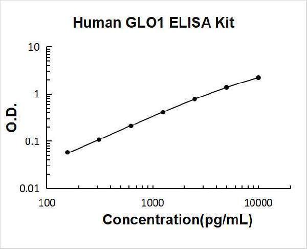
1 Q&As
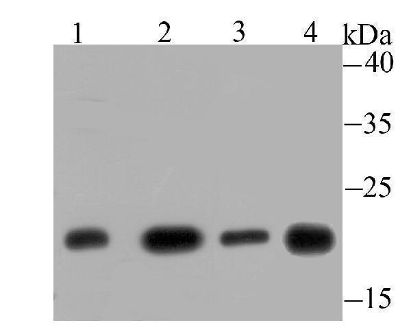
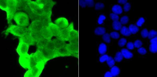
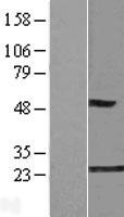
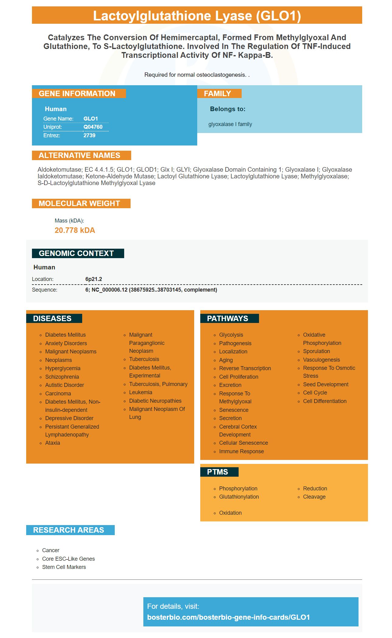
Facts about Lactoylglutathione lyase.

Required for normal osteoclastogenesis. .
| Human | |
|---|---|
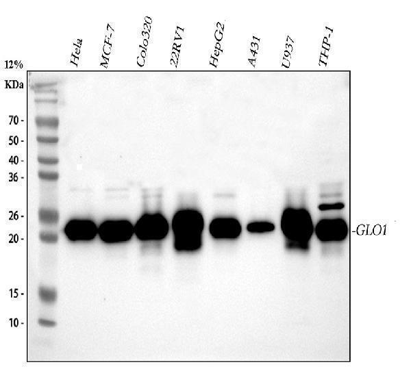
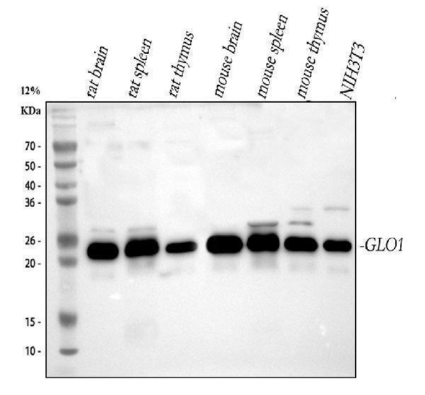
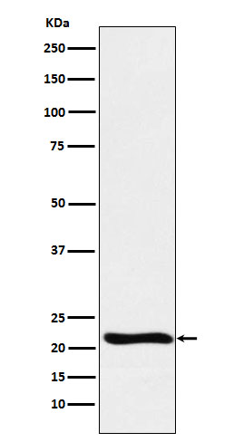
| Gene Name: | GLO1 |
| Uniprot: | Q04760 |
| Entrez: | 2739 |

| Belongs to: |
|---|
| glyoxalase I family |

Aldoketomutase; EC 4.4.1.5; GLO1; GLOD1; Glx I; GLYI; glyoxalase domain containing 1; Glyoxalase I; glyoxalase Ialdoketomutase; Ketone-aldehyde mutase; lactoyl glutathione lyase; lactoylglutathione lyase; Methylglyoxalase; S-D-lactoylglutathione methylglyoxal lyase
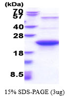
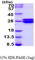
Mass (kDA):
20.778 kDA

| Human | |
|---|---|
| Location: | 6p21.2 |
| Sequence: | 6; NC_000006.12 (38675925..38703145, complement) |


PMID: 7684374 by Kim N.-S., et al. Human glyoxalase I. cDNA cloning, expression, and sequence similarity to glyoxalase I from Pseudomonas putida.
PMID: 8449929 by Ranganathan S., et al. Cloning and characterization of human colon glyoxalase-I.