GNA11 / Guanine nucleotide-binding protein subunit alpha-11 · IHC design guide

Design Immunohistochemistry for GNA11

Plan GNA11 paraffin-section IHC around membranous and cytoplasmic staining, with colon or appendix glandular cells as positive comparators (HPA tissue IHC). The IHC-validated antibody A03817 has a 1:50–1:200 dilution range (datasheet A03817); compare staining with a no-primary control (standard IHC practice).

Evidence assembled Oct 2026 · For research use; verify linked source records and product datasheet before use
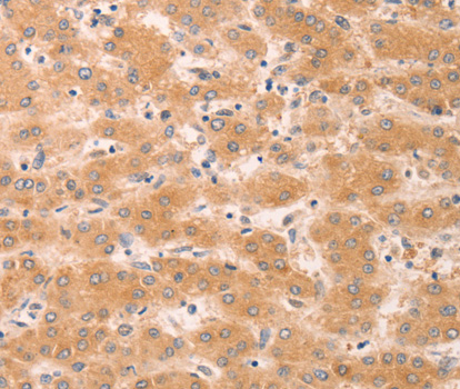
Immunohistochemistry protocol sheet for GNA11 (IHC for GNA11): expected localisation Membranous and cytoplasmic tissue staining (HPA tissue IHC), antibody A03817, validated IHC image, and IHC protocol steps
Printable GNA11 IHC protocol sheet — expected localisation Membranous and cytoplasmic tissue staining (HPA tissue IHC), antibody A03817, controls and protocol steps. Open the full GNA11 IHC guide →

GNA11 Immunohistochemistry Experimental Design Guide

Expected localisation, validated protocols, controls and antibodies — the at-a-glance facts below, then the full design guide.

Must know before staining
Expected localisation Membranous and cytoplasmic tissue staining (HPA tissue IHC)
Staining pattern Glandular cells show membranous and cytoplasmic staining (HPA tissue IHC)
Antigen retrieval Citrate pH 6.0 HIER, heat-mediated (datasheet A03817)
Positive control ⓘ Appendix+4 more · see all
Negative control ⓘ Adipose tissue+4 more · see all
Important caveats
Reasons your staining may differ from the expected pattern.
Fixation Keep fixation consistent across sections (standard IHC practice; not target-specific)
Caveat HPA staining may include protein from another gene (HPA tissue IHC)
Regulation No expression inducer reported (UniProt)
Isoform / epitope No isoforms reported; one 1–359 chain (UniProt)
Section 1

Recommended GNA11 IHC & IF Protocols

The catalog antibody’s IHC-P protocol (datasheet A03817) is paired with one published GNA11 tissue microarray protocol (PMC8462091).

Recommended immunohistochemistry (IHC-P) protocol parameters
SampleParaffin-embedded human esophageal cancer tissue; fixative not specified (datasheet A03817)
FixationImage fixative and duration unreported (datasheet A03817); verify before use.
Sectioning4–5 µm sections on charged slides (standard)
DeparaffinisationXylene, graded ethanol series to water (standard)
Antigen retrievalHeat retrieval: Citrate pH 6.0 (datasheet A03817); 20 min, 95–100 °C (standard)
Peroxidase block3% H2O2, 10 min, room temperature (standard)
Blocking10% normal serum of the secondary host, 30 min, room temperature (standard)
Primary antibodyRabbit anti-GNA11, 1:50-1:200 (datasheet A03817)
Primary incubationOvernight at 4 °C (standard)
DetectionHRP-polymer secondary, DAB chromogen 5–10 min (standard)
CounterstainHematoxylin, blue, dehydrate and mount (standard)
Expected resultGNA11-positive staining in glandular cells of appendix (HPA tissue IHC: Medium). HPA tissue profile: Membranous and cytoplasmic expression in several tissues. No signal in the no-primary control.
💡Decision noteStart with heat-mediated citrate retrieval at pH 6.0 (datasheet A03817); the published protocol does not specify retrieval conditions (PMC8462091).
Section 2

What Is the Expected GNA11 Staining Pattern?

GNA11 is reported at the cell membrane and in the cytoplasm, with no transmembrane segment; testicular cells are a reported cytoplasmic exception (UniProt P29992 localisation and topology). In paraffin sections, expect membranous and cytoplasmic staining in the cell populations reported by HPA, including glandular cells in appendix and colon (HPA: Medium). Interpret these patterns cautiously: HPA rates the tissue staining Approved but warns that the antibody targets proteins from more than one gene (HPA: reliability description).

What am I looking at on my slide?
Membranous and cytoplasmic signal in appendix or colon glandular cells.This matches the reported compartments and a Medium staining level in those cells (HPA: tissue IHC). Compare the signal with adjacent structures and controls; an HPA match supports the expected pattern but does not establish GNA11 specificity because the tissue antibody may recognize another gene product (HPA: reliability description).
Predominantly nuclear staining, without a discernible membrane or cytoplasmic component.The compartment differs from the reported membrane and cytoplasmic localisation (UniProt P29992; HPA: tissue IHC). Treat it as a suspect pattern and check the counterstain, detection controls and antibody conditions before scoring it as GNA11. Testicular cells are a cytoplasmic exception, not evidence for nuclear staining (UniProt P29992).
Strong signal in adipocytes or bone marrow hematopoietic cells, especially with little staining in an HPA positive comparison.HPA reports these populations as Not detected and reports Medium staining in appendix glandular cells (HPA: tissue IHC). Cross-reactivity or endogenous detection activity is possible (general IHC practice). Review morphology and controls before calling this GNA11; an HPA negative population is a comparator, not proof that every positive cell is false.
Uniform colour across cells, stroma and the section edge, with no clear cell boundaries.That distribution is difficult to reconcile with the reported membranous and cytoplasmic cellular pattern (HPA: tissue IHC). In chromogenic IHC, incomplete blocking, concentrated primary antibody or detection background can produce diffuse staining (general IHC practice). Examine a no-primary control and titrate the assay before interpreting intensity.
No discernible signal in appendix or colon glandular cells on an otherwise intact section.Both are reported at Medium intensity (HPA: tissue IHC), so absence of signal prompts a technical check. Confirm tissue identity and cell preservation, then review retrieval, primary antibody dilution and detection controls (general IHC practice). A failed positive comparator cannot establish that a test sample lacks GNA11.
💡Expected GNA11 appearanceA plausible positive shows membranous and cytoplasmic colour in appendix or colon glandular cells at approximately the HPA Medium level; isolated nuclear colour or diffuse cell independent colour is suspect (HPA: tissue IHC; UniProt P29992 localisation).
How each factor affects the staining
Compartment and topologyUniProt places GNA11 at the cell membrane and in the cytoplasm and reports no transmembrane segment (UniProt P29992). Score the observed cellular compartments rather than requiring a sharply outlined, membrane only rim (HPA: membranous and cytoplasmic tissue profile).
Cell and tissue contextHPA reports Medium staining in appendix, colon, gallbladder and prostate glandular cells, but Not detected in adipocytes (HPA: tissue IHC). Select a positive comparator by its reported cell population, then assess the same population on the slide.
Testicular localisationUniProt reports expression in testis and exclusively cytoplasmic localisation in testicular cells (UniProt P29992). A membrane rim is therefore not required when interpreting those cells; the supplied HPA tissue examples do not assign a testicular IHC intensity.
Specificity and validationThe tissue staining is rated Approved, with a caution that the antibody targets protein from more than one gene (HPA: reliability description). HPA048886 is also listed as IHC Approved, not Enhanced (HPA: antibody validation). Concordant location alone cannot resolve cross-reactivity.
IF/ICC: what pattern can be expected?Membrane and cytoplasmic localisation is the UniProt expectation (UniProt P29992). HPA supplies no main ICC-IF location or ICC-IF images, and the listed antibody has no ICC status (HPA: subcellular and antibody records). An IF/ICC staining pattern is therefore unverified here.
Why is my staining missing, weak or wrong?
SituationLikely causeNext action
Expected glandular cells appear blank.Retrieval, primary antibody exposure or detection may be inadequate (general IHC practice); appendix and colon glandular cells are reported Medium (HPA: tissue IHC).Run a reported positive tissue alongside the sample. Check retrieval and detection controls, then optimise primary antibody dilution within its documented IHC conditions (general IHC practice).
The whole section has diffuse chromogenic colour.Detection background, incomplete blocking or excessive primary antibody can obscure cellular staining (general IHC practice).Inspect a no-primary control, review blocking and washes, and titrate the primary antibody (general IHC practice). Reassess whether distinct membrane and cytoplasmic signal remains (HPA: tissue IHC profile).
Signal is nuclear only.The location conflicts with the reported membrane and cytoplasmic pattern (UniProt P29992; HPA: tissue IHC).Check cell boundaries against the counterstain and compare with a positive control; review antibody dilution and detection background before assigning a GNA11 score (general IHC practice).
Adipocytes or bone marrow hematopoietic cells stain strongly.Those populations are Not detected in HPA tissue IHC; cross-reactivity or endogenous detection activity is possible (HPA: tissue IHC; general IHC practice).Compare a no-primary control and the expected positive cells. Investigate endogenous chromogen activity and antibody specificity before attributing this signal to GNA11 (general IHC practice; HPA: specificity caution).
Staining is cytoplasmic with little membrane outline in testicular cells.UniProt reports exclusively cytoplasmic localisation in testicular cells (UniProt P29992).Assess cytoplasmic localisation and appropriate controls rather than rejecting the section for lacking a membrane rim (UniProt P29992; general IHC practice). Do not infer an expected testicular intensity from the supplied HPA examples.
A plausible positive pattern needs a specificity call.HPA cautions that its Approved tissue antibody targets protein from more than one gene; the listed IHC antibody is Approved without Enhanced status (HPA: reliability and antibody records).Report the compartment and cell population observed, and retain the specificity caveat. Use an independent specificity check before making a GNA11 specific attribution (general IHC practice; HPA: reliability description).

Sample controls for GNA11 IHC & IF

🧪Run colon first and score glandular cells for GNA11 staining (HPA: Medium in colon glandular cells). Use adipose tissue adipocytes as the negative tissue (HPA: Not detected in adipocytes); on the colon slide, neighboring cells without staining can show local background, but their GNA11-negative status is unverified (HPA: colon row specifies glandular cells only).
Positive control tissue: Appendix (Glandular cells, HPA Medium)
Negative control tissue: Adipose tissue (HPA Not detected)
ICC-IF cell lines (HPA subcellular resource): HPA carries no ICC-IF cell line for GNA11; derive a cell-line control from the positive tissue's cell type (Glandular cells) and confirm it by RNA or western blot first.
Technical controls: Include a no-primary, secondary-only control and concentration-matched rabbit IgG control (selected IHC caption: rabbit pAb), plus GNA11 knockout material or an antigen-peptide block if available. Quench endogenous peroxidase and inspect unstained colon sections for pigment or other background before scoring chromogenic signal (standard IHC practice).
⚠️Feasibility: A GNA11-specific fixation window or fixation effect is unreported, and the selected paraffin-section caption does not state its fixative (selected IHC caption: fixative not stated). High-pressure retrieval with 10 mM citrate at pH 6.0 and antibody dilution 1:150 were reported for esophageal cancer tissue; their performance in colon requires validation (selected IHC caption). Whether frozen sections or IF/ICC are easier is unreported; HPA supplies no ICC-IF images for this gene (HPA subcellular payload: no cell lines with ICC-IF images).

HPA tissue IHC evidence for GNA11

Comprehensive Human Protein Atlas IHC scoring per tissue (reliability: Approved — Caution, targets protein from more than one gene. Pending external verification.). Rows are taken directly from the HPA tissue chart — click any row's HPA link to view the source.

Positive expression · recommended positive controls

TissueCell typeLevelEvidenceSource
Appendix Glandular cells Medium Protein (IHC) HPA →
Caudate Glial cells Medium Protein (IHC) HPA →
Cerebellum Purkinje cells Medium Protein (IHC) HPA →
Colon Glandular cells Medium Protein (IHC) HPA →
Endometrium Cells in endometrial stroma Low Protein (IHC) HPA →

Undetected expression · recommended negative controls

TissueCell typeLevelEvidenceSource
Adipose tissue Adipocytes Not detected Protein (IHC) HPA →
Bone marrow Hematopoietic cells Not detected Protein (IHC) HPA →
Bronchus Respiratory epithelial cells Not detected Protein (IHC) HPA →
Esophagus Squamous epithelial cells Not detected Protein (IHC) HPA →
Heart muscle Cardiomyocytes Not detected Protein (IHC) HPA →
Section 3

Advanced GNA11 IHC Tips

Troubleshoot GNA11 staining in paraffin sections by checking retrieval, compartment pattern, controls and cell-specific scoring.

What retrieval should I try first if GNA11 staining is weak?
Start with heat-mediated citrate retrieval at 10 mM and pH 6.0 for paraffin sections (datasheet A03817). The selected antibody image used high-pressure retrieval in that buffer before staining human esophageal cancer at 1:150 (datasheet A03817). If staining remains weak, compare longer and shorter heating within the same citrate method while holding detection and antibody concentration constant (standard IHC practice). Include a positive control section in each run, because absent staining in both test and control sections points toward a processing or reagent problem (standard IHC practice). Any alternative buffer or pH should be evaluated only as a fallback against that control (standard IHC practice).
Could fixation explain variable GNA11 staining between paraffin blocks?
GNA11-specific fixation sensitivity is unknown from the supplied evidence; the selected tissue image identifies paraffin embedding but does not state its fixative (datasheet A03817). Record fixative, fixation duration and processing history for each block before comparing staining, because those variables can change antigen accessibility in IHC (standard IHC practice). When possible, compare sections processed together with the same citrate pH 6.0 retrieval and antibody conditions (datasheet A03817; standard IHC practice). A control section that stains consistently helps separate block-dependent processing variation from a failed staining run (standard IHC practice). Do not assign a GNA11-specific fixation effect without a controlled comparison (standard IHC practice).
Should GNA11 appear at the membrane or in the cytoplasm?
Assess both membranous and cytoplasmic chromogen patterns: GNA11 is annotated in both compartments, and tissue IHC reports both patterns (UniProt P29992 localisation; HPA tissue IHC profile). GNA11 has no transmembrane segment, so a membrane-associated pattern does not imply that the protein spans the membrane (UniProt P29992 topology). In testicular cells, the annotated localisation is exclusively cytoplasmic (UniProt P29992 localisation). Inspect staining at cell boundaries and within cytoplasm on the same section, using morphology and a nuclear counterstain to avoid calling dark nuclei positive (standard IHC practice). Score each compartment separately when patterns differ between cell populations (standard IHC practice).
How should I assess an unexpected GNA11 epitope pattern?
The supplied record lists 0 isoforms and one chain spanning residues 1–359, so it does not support an isoform-specific explanation for staining differences (UniProt P29992 processing and isoforms). Its G-alpha domain spans residues 38–359, but the antibody epitope is not supplied; do not assign the stain to a particular domain surface (UniProt P29992 domains; datasheet A03817). GNA11 has no annotated glycosylation sites or transmembrane segment in this record (UniProt P29992 glycosylation and topology). If an unusual pattern persists, compare staining with an independently validated GNA11 antibody or another specificity control under matched section conditions (standard IHC practice). Record the antibody identity and retrieval settings alongside the result (standard IHC practice).
How can I check a GNA11 IHC pattern by multiplex IF?
Treat multiplex IF as a separate assay requiring its own validation; the supplied antibody image documents paraffin-section chromogenic IHC, with no IF result provided (datasheet A03817). Pair GNA11 with an epithelial marker such as cytokeratin when examining glandular cells, which are reported positive in colon and appendix IHC (HPA tissue IHC; standard IF practice). Choose fluorophores after inspecting unstained tissue autofluorescence, and place the weaker signal in a spectrally cleaner channel (standard IF practice). Because GNA11 is annotated at the cell membrane and in cytoplasm, optimise permeabilisation for antibody access to the relevant epitope side; that epitope side is unspecified here (UniProt P29992 localisation and topology; standard IF practice). Include single-stain and secondary-only controls to assess bleed-through and background (standard IF practice).
What should I change when GNA11 chromogenic staining is diffuse?
First examine a no-primary control and a positive control section alongside the affected tissue to distinguish detection background from reproducible cellular staining (standard IHC practice). Check deparaffinisation, peroxidase blocking, protein blocking, wash stringency and DAB development before increasing antibody concentration (standard IHC practice). The selected paraffin image used the catalog antibody at 1:150 after citrate pH 6.0 retrieval; that setting is a reference point, not proof of optimal conditions for every block (datasheet A03817; standard IHC practice). Diffuse precipitate, staining confined to section folds, or signal in the no-primary control should be excluded from GNA11 scoring (standard IHC practice). Reassess cell boundaries and cytoplasm after background is controlled (UniProt P29992 localisation; standard IHC practice).
How should I quantify GNA11 staining across tissue sections? ⚠ ANSWER MARKED FOR VERIFICATION
Define the cell population and scoring compartment before measuring GNA11, because membranous and cytoplasmic expression are both reported (HPA tissue IHC profile). For a chromogenic series, record the percentage of positive target cells and intensity, then calculate an H-score from intensity-weighted percentages using one prespecified scoring scale (standard IHC practice). Keep membrane and cytoplasm scores separate, and report the number of evaluable cells or tissue area behind each estimate (standard IHC practice). Normalise positive-cell counts to all evaluable cells of the same type, or staining density to evaluable tissue area in mm² (standard IHC practice). Apply identical thresholds, counterstaining and imaging settings across compared sections (standard IHC practice).
How do I distinguish true GNA11 positivity from artefact?
Give greatest weight to reproducible membrane or cytoplasmic staining in intact cells, consistent with the annotated and tissue IHC patterns (UniProt P29992 localisation; HPA tissue IHC profile). Treat isolated nuclear signal, section-edge intensification, necrotic deposits and staining that survives a no-primary control as potential artefacts requiring further controls (standard IHC practice). Check endogenous peroxidase blocking when DAB signal appears in unexpected cells, and compare each suspect area with adjacent intact tissue (standard IHC practice). A normal esophageal squamous epithelium result cannot directly negate the selected esophageal cancer image: they describe different specimens (HPA tissue IHC; datasheet A03817). Interpret specificity cautiously because the HPA antibody is flagged as targeting proteins from more than one gene (HPA reliability description).
Boster reagents

Best GNA11 / Guanine nucleotide-binding protein subunit alpha-11 IHC Antibodies

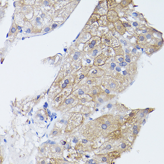
The catalog includes GNA11 antibodies with real paraffin-section IHC data from human esophageal cancer (A03817 image caption) and cell IF data from mouse NIH/3T3 cells (M03817 image caption).

Real IHC data Immunohistochemistry of paraffin-embedded human esophageal cancer using GNA11 Rabbit pAb at dilution of 1:150 .Perform high pressure antigen retrieval with 10 mM citrate buffer pH 6.0 before commencing with IHC staining protocol.
Anti-GNA11 Antibody
Cat # A03817
Real IF data Immunofluorescent analysis of 4% paraformaldehyde-fixed, 0.1% Triton X-100 permeabilized NIH/3T3 (mouse embryonic fibroblast cell line) cells labeling GNA11 with M03817 at 1/25 dilution, followed by Dylight® 488-conjugated goat anti-rabbit IgG secondary antibody at 1/200 dilution (green). Immunofluorescence image showing cytoplasm and membrane staining on NIH/3T3 cell line. The nuclear counter stain is DAPI (blue).
Anti-GNA11 Antibody (Center)
Cat # M03817

A03817 will render with human esophageal cancer paraffin-section IHC at 1:150 after citrate pH 6.0 retrieval (A03817 image caption). M03817 will render with mouse NIH/3T3 cell IF at 1:25 after paraformaldehyde fixation and Triton X-100 permeabilisation (M03817 image caption).

Which to pick: Choose A03817 for tissue IHC: it is a rabbit polyclonal antibody with IHC listed and human, mouse, and rat reactivity; its paraffin-section figure shows human tissue, but the fixative is unreported (A03817 catalog; A03817 image caption). Choose M03817 for cell IF/ICC: it is rabbit polyclonal with IF listed and human and mouse reactivity, and its figure shows mouse NIH/3T3 cells (M03817 catalog; M03817 image caption). For cross-species tissue work, A03817 has the broader listed reactivity, including rat, while its displayed IHC evidence is human only (A03817 catalog; A03817 image caption).

Each figure is that product's own IHC / IF validation image from its datasheet.

References

  1. UniProt Consortium. UniProt entry P29992 (GNA11_HUMAN, Guanine nucleotide-binding protein subunit alpha-11).
  2. Human Protein Atlas. GNA11 tissue IHC expression (reliability: Approved).
  3. Human Protein Atlas. GNA11 subcellular location (ICC-IF): Highest expression in PODO/TERT256: 63.7 nTPM.
  4. Human Protein Atlas. GNA11 antibody validation summary (1 antibodies).
  5. Identification and Evaluation of Autoantibody to a Novel Tumor-Associated Antigen GNA11 as a Biomarker in Esophageal Squamous Cell Carcinoma. Frontiers in oncology 2021 — PMC8462091.
  6. Mass Spectrometry-Based Profiling of Histone Post-Translational Modifications in Uveal Melanoma Tissues, Human Melanocytes, and Uveal Melanoma Cell Lines - A Pilot Study. Investigative ophthalmology & visual science 2024 — PMC10868634.
  7. SF3B1 and EIF1AX mutations occur in primary leptomeningeal melanocytic neoplasms; yet another similarity to uveal melanomas. Acta neuropathologica communications 2016 — PMC4714515.
  8. Oncogenic GNAQ and GNA11 mutations in uveal melanoma in Chinese. PloS one 2014 — PMC4184906.
  9. PubMed PMID:1902575 — UniProt-cited evidence.
  10. PubMed PMID:15057824 — UniProt-cited evidence.
  11. PubMed PMID:15489334 — UniProt-cited evidence.