This website uses cookies to ensure you get the best experience on our website.


- Table of Contents
Plan GNAQ staining in paraffin sections using colon or prostate glandular cells as reference tissues (HPA tissue IHC). This guide covers fixation consistency, a primary antibody range of 0.5–1 μg/mL (datasheet PB9707), chromogenic detection, and interpretation of uncertain tissue staining (HPA tissue IHC).

Expected localisation, validated protocols, controls and antibodies — the at-a-glance facts below, then the full design guide.
| Expected localisation | Cytoplasmic and membranous staining (HPA tissue IHC) | |
| Staining pattern | Variable cytoplasmic and membranous staining in glandular cells (HPA tissue IHC) | |
| Antigen retrieval | EDTA pH 8.0 HIER, heat-mediated (datasheet PB9707) | |
| Positive control | Appendix+4 more · see all | |
| Negative control | Adipose tissue+4 more · see all |
| Fixation | Matched tissue-IHC evidence does not establish the fixation claim. Validate the specimen-specific method before use. (selected-SKU IHC image PB9707) | |
| Caveat | Staining is uncertain; the antibody may target more than one gene (HPA tissue IHC) | |
| Regulation | Reduced in rheumatoid arthritis PBLs (UniProt) | |
| Isoform / epitope | No annotated isoforms; one chain spans residues 1–359 (UniProt) |
The catalog antibody uses EDTA retrieval (datasheet PB9707); the three published GNAQ IHC protocols below cover sheep testis and epididymis, skin, and hen uterovaginal junction (PMC5088417; PMC10392076; PMC8082553).
| Sample | Paraffin-embedded rat ovary tissue; fixative not specified (datasheet PB9707) |
| Fixation | Image fixative and duration unreported (datasheet PB9707); verify before use. |
| Sectioning | 4–5 µm sections on charged slides (standard) |
| Deparaffinisation | Xylene, graded ethanol series to water (standard) |
| Antigen retrieval | Heat retrieval: EDTA pH 8.0 (datasheet PB9707); 20 min, 95–100 °C (standard) |
| Peroxidase block | 3% H2O2, 10 min, room temperature (standard) |
| Blocking | 10% goat serum (datasheet PB9707) |
| Primary antibody | Rabbit anti-GNAQ, 0.5-1μg/ml (datasheet PB9707) |
| Primary incubation | Overnight at 4 °C (datasheet PB9707) |
| Detection | Streptavidin-biotin complex (SABC), DAB chromogen (datasheet PB9707) |
| Counterstain | Hematoxylin, blue, dehydrate and mount (standard) |
| Expected result | GNAQ-positive staining in glandular cells of appendix (HPA tissue IHC: Medium). HPA tissue profile: Cytoplasmic and membranous expression at variable levels in several tissues. No signal in the no-primary control. |
GNAQ is associated with the plasma membrane through a lipid anchor and has no transmembrane segment; UniProt also lists Golgi, nuclear and nuclear-membrane locations (UniProt P50148 topology and subcellular location). In tissue IHC, expect variable cytoplasmic and membranous staining, including medium staining in colon and prostate glandular cells (HPA tissue IHC). Interpret cautiously: HPA rates tissue IHC Uncertain because antibody staining and RNA data have low consistency.
| Membranous staining with some cytoplasmic signal in colon or prostate glandular cells. | This fits the reported tissue pattern; both cell populations show medium staining (HPA tissue IHC). Compare the two compartments separately when scoring. A plausible pattern supports interpretation, but does not independently establish antibody specificity: HPA rates tissue IHC Uncertain and cautions that antibodies may target protein from more than one gene (HPA tissue IHC). |
| Strong nuclear-only staining, with no convincing membranous or cytoplasmic signal. | Recheck the interpretation before calling this the expected tissue pattern: HPA describes tissue IHC as cytoplasmic and membranous (HPA tissue IHC). Nuclear staining is not automatically an artifact, since UniProt lists nucleus and nuclear membrane (UniProt P50148 subcellular location). Nuclear speckles come from ICC-IF evidence and have uncertain support (HPA subcellular). |
| Prominent staining in cells reported as undetected, such as adipocytes or lung alveolar cells. | Those cell types are reported as Not detected in tissue IHC (HPA tissue IHC). Check the cell identity and compare a control section. Persistent signal could reflect cross-reactivity or, with enzyme-based chromogenic detection, endogenous activity (general IHC practice). The HPA tissue rating is Uncertain, so a mismatch warrants investigation rather than an automatic biological conclusion. |
| Diffuse color covers tissue and blank areas without a clear cellular boundary. | This is difficult to score as GNAQ because the reported pattern is cellular, cytoplasmic and membranous (HPA tissue IHC). Background from detection reagents or nonspecific binding can obscure compartment assignment (general IHC practice). Compare the no-primary control and review blocking, washing and detection before assigning an intensity score. |
| No staining in colon or prostate glandular cells while the section otherwise appears interpretable. | These are useful comparison cells because medium staining was observed in both (HPA tissue IHC). An absent result may reflect assay performance or a different sample result; the HPA tissue assessment is Uncertain (HPA tissue IHC). Review the catalog antibody's IHC-P conditions and controls before calling the tissue GNAQ-negative (general IHC practice). |
| Tissue IHC evidence | Colon and prostate glandular cells are medium, while adipocytes and lung alveolar cells are Not detected (HPA tissue IHC). These are comparison patterns, not definitive controls: HPA rates the tissue result Uncertain and flags possible recognition of protein from more than one gene (HPA tissue IHC). |
| Membrane association and compartment | A lipid anchor associates GNAQ with the plasma membrane despite the absence of a transmembrane segment (UniProt P50148 topology and subcellular location). UniProt also lists Golgi, nucleus and nuclear membrane; tissue IHC primarily describes cytoplasmic and membranous expression (UniProt P50148 subcellular location; HPA tissue IHC). |
| Expression across tissues | UniProt describes predominant expression in ovary, prostate, testis and colon (UniProt P50148 tissue specificity). HPA reports low tissue RNA specificity and variable tissue IHC staining (HPA tissue IHC). Use the observed cell-level IHC pattern when reading a section, while keeping HPA's Uncertain reliability rating in view. |
| IF/ICC Q&A: should its pattern set the tissue IHC call? | No. Plasma-membrane localization is supported in ICC-IF, whereas nuclear speckles and cytosol are uncertain (HPA subcellular). Tissue IHC reports cytoplasmic and membranous staining and has Uncertain reliability (HPA tissue IHC). Read each application against its own evidence; this section supplies no IF/ICC protocol. |
| Situation | Likely cause | Next action |
|---|---|---|
| Colon and prostate glandular cells have no convincing signal. | The observed medium tissue pattern has not been reproduced (HPA tissue IHC); the cause cannot be assigned from this slide alone. | Check that the catalog antibody's IHC-P conditions were followed, and review the positive control, retrieval, dilution and detection records (general IHC practice). HPA's Uncertain rating limits how strongly one negative section can be interpreted (HPA tissue IHC). |
| The entire section has diffuse chromogen, including areas without identifiable cells. | Detection background or nonspecific binding may be obscuring the reported cellular pattern (general IHC practice; HPA tissue IHC). | Compare a no-primary control; inspect reagent blocking, wash quality and detection exposure (general IHC practice). Score GNAQ only where a cell and its compartment can be identified, using the cytoplasmic and membranous pattern as the comparison (HPA tissue IHC). |
| Lung alveolar cells or adipocytes stain as strongly as the proposed positive cells. | Both cell types are reported as Not detected (HPA tissue IHC). A discrepancy may arise from cell misidentification, cross-reactivity or chromogenic endogenous activity (general IHC practice). | Confirm morphology, inspect the no-primary control and compare a reported medium-staining cell population (general IHC practice; HPA tissue IHC). Treat persistent staining as unresolved because HPA flags possible recognition of protein from more than one gene (HPA tissue IHC). |
| A nuclear-only pattern dominates the section. | It differs from the reported tissue IHC pattern (HPA tissue IHC), although nuclear and nuclear-membrane localization appear in UniProt (UniProt P50148 subcellular location). | Check the counterstain and control section, then record nuclear staining separately from membrane and cytoplasm (general IHC practice). Do not use uncertain ICC-IF nuclear-speckle localization as confirmation of tissue IHC specificity (HPA subcellular). |
| Staining varies between fields or between the selected comparison tissues. | Variable levels across tissues are part of the HPA tissue description (HPA tissue IHC); uneven processing can also affect IHC appearance (general IHC practice). | Assess cell type and compartment in each field, then compare section quality and controls before interpreting the variation (general IHC practice). Document intensity by cell population rather than assigning one score to the whole section. |




Comprehensive Human Protein Atlas IHC scoring per tissue (reliability: Uncertain — Low consistency between antibody staining and RNA expression data. Caution, targets protein from more than one gene. Pending external verification.). Rows are taken directly from the HPA tissue chart — click any row's HPA link to view the source.
| Tissue | Cell type | Level | Evidence | Source |
|---|---|---|---|---|
| Appendix | Glandular cells | Medium | Protein (IHC) | HPA → |
| Caudate | Glial cells | Medium | Protein (IHC) | HPA → |
| Cerebellum | Purkinje cells | Medium | Protein (IHC) | HPA → |
| Colon | Glandular cells | Medium | Protein (IHC) | HPA → |
| Endometrium | Cells in endometrial stroma | Low | Protein (IHC) | HPA → |
| Tissue | Cell type | Level | Evidence | Source |
|---|---|---|---|---|
| Adipose tissue | Adipocytes | Not detected | Protein (IHC) | HPA → |
| Bone marrow | Hematopoietic cells | Not detected | Protein (IHC) | HPA → |
| Bronchus | Respiratory epithelial cells | Not detected | Protein (IHC) | HPA → |
| Esophagus | Squamous epithelial cells | Not detected | Protein (IHC) | HPA → |
| Heart muscle | Cardiomyocytes | Not detected | Protein (IHC) | HPA → |
Troubleshoot GNAQ staining in paraffin sections by checking retrieval, compartment pattern, controls and cell-specific scoring.
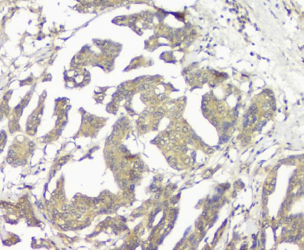
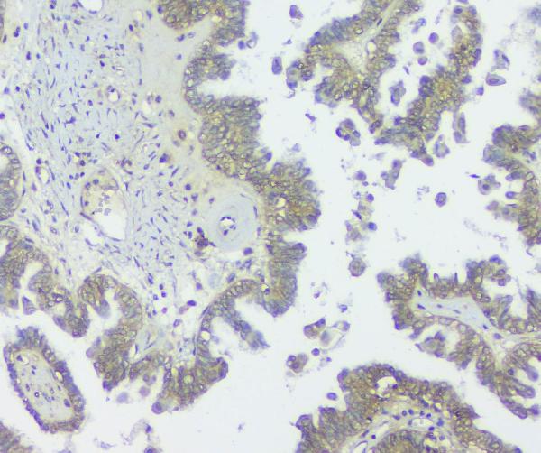
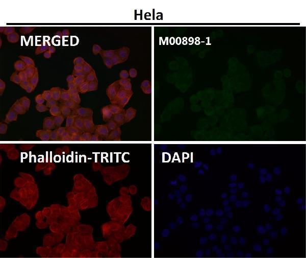
The catalog shows GNAQ IHC in paraffin sections of rat ovary, mouse testis, human ovarian cancer and human prostatic cancer (PB9707 and M00898 IHC captions), plus IF in A431 cells (PB9707 IF caption).
PB9707 has IHC images from rat ovary, mouse testis and human prostatic cancer paraffin sections, plus IF in A431 cells (PB9707 image captions); M00898 has IHC images from human ovarian cancer and mouse testis paraffin sections (M00898 IHC captions). M00898-1 lists IHC and IF/ICC applications, but its supplied figure shows IF without identifying a sample (M00898-1 applications and IF caption).
Which to pick: For tissue IHC, PB9707 has a rabbit antibody figure in rat ovary paraffin sections, while M00898 has a mouse monoclonal figure in human ovarian cancer paraffin sections; neither caption reports the fixative (PB9707 and M00898 IHC captions; catalog hosts and clone). For IF/ICC, PB9707 has an A431 cell IF image and a listed 2 μg/ml concentration; M00898-1 has an IF image at 1:50 with no sample identified (PB9707 IF caption and datasheet; M00898-1 IF caption). For cross-species work, both PB9707 and M00898 list human, mouse and rat reactivity, while only M00898 also lists monkey reactivity (catalog reactivity).