This website uses cookies to ensure you get the best experience on our website.


- Table of Contents
Plan paraffin-section GNL3L IHC with the catalog antibody’s 1:100–1:300 dilution range (datasheet A09415). Assess nuclear and cytoplasmic staining across cell types, with high staining in adrenal gland glandular cells and no detected staining in lymph-node germinal-center cells as tissue references (HPA tissue IHC).

Expected localisation, validated protocols, controls and antibodies — the at-a-glance facts below, then the full design guide.
| Expected localisation | Nuclear and cytoplasmic tissue staining (HPA tissue IHC) | |
| Staining pattern | Many cell types show cytoplasmic and nuclear staining (HPA tissue IHC) | |
| Antigen retrieval | Tris-EDTA pH 9.0 HIER, heat-mediated (datasheet A09415) | |
| Positive control | Adrenal gland+4 more · see all | |
| Negative control | Lymph node |
| Fixation | Keep fixation consistent across sections (standard IHC practice; not target-specific) | |
| Caveat | Germinal-center cells are undetected despite broad staining (HPA tissue IHC) | |
| Regulation | No expression regulation annotated (UniProt) | |
| Isoform / epitope | No annotated isoforms or cleavage to shift epitopes (UniProt) |
The catalog antibody protocol (datasheet A09415) is followed by three published GNL3L IHC protocols using human esophageal specimens (PMC12378702; PMC11087641; PMC8154413).
| Sample | Paraffin-embedded human brain tissue; fixative not specified (datasheet A09415) |
| Fixation | Image fixative and duration unreported (datasheet A09415); verify before use. |
| Sectioning | 4–5 µm sections on charged slides (standard) |
| Deparaffinisation | Xylene, graded ethanol series to water (standard) |
| Antigen retrieval | Heat retrieval: Tris-EDTA pH 9.0 (datasheet A09415); 20 min, 95–100 °C (standard) |
| Peroxidase block | 3% H2O2, 10 min, room temperature (standard) |
| Blocking | 10% normal serum of the secondary host, 30 min, room temperature (standard) |
| Primary antibody | Rabbit anti-GNL3L, 1:100-1:300 (datasheet A09415) |
| Primary incubation | Overnight at 4 °C (standard) |
| Detection | HRP-polymer secondary, DAB chromogen 5–10 min (standard) |
| Counterstain | Hematoxylin, blue, dehydrate and mount (standard) |
| Expected result | GNL3L-positive staining in glandular cells of adrenal gland (HPA tissue IHC: High). HPA tissue profile: Ubiquitous cytoplasmic and nuclear expression. No signal in the no-primary control. |
GNL3L is a nuclear and nucleolar protein with no transmembrane segment (UniProt Q9NVN8: localization and topology). Tissue IHC shows widespread nuclear and cytoplasmic staining, with high staining in several glandular, squamous epithelial, and neural cell populations (HPA tissue IHC). HPA rates the tissue pattern Supported, with medium agreement between staining and RNA data and external verification pending (HPA tissue IHC).
| Nuclear staining, sometimes with cytoplasmic staining, in adrenal glandular or cervical squamous epithelial cells. | This fits the reported tissue pattern; both cell populations have High staining (HPA tissue IHC). A nucleolar accent would also agree with the subcellular reference, which comes from ICC-IF rather than tissue IHC (HPA subcellular ICC-IF). |
| Predominantly cell-surface staining, with little nuclear signal. | Treat a membrane-dominant pattern as suspect: GNL3L has no transmembrane segment and is assigned to the nucleus and nucleolus (UniProt Q9NVN8: topology and localization). Recheck the signal against a known-positive tissue before assigning it to GNL3L (general IHC practice). |
| Strong staining in lymph-node germinal center cells. | That conflicts with the reported Not detected result for these cells (HPA tissue IHC). Consider cross-reactivity or endogenous chromogenic activity, and assess control sections; the HPA result is a reference pattern, not proof that every germinal center must be negative (HPA tissue IHC; general IHC practice). |
| Uniform chromogen across cells and surrounding tissue, with no clear cellular boundaries. | Diffuse background does not support a compartment assignment (general IHC practice). Check blocking, washes, and the detection-only control; interpret any remaining signal against the nuclear and cytoplasmic tissue pattern (HPA tissue IHC). |
| No signal in adrenal glandular cells or cervical squamous epithelial cells. | Both are reported High examples, so an absent signal calls for a technical check (HPA tissue IHC). Review tissue integrity, antibody application, retrieval conditions, and detection controls using the chosen assay workflow (general IHC practice). |
| Tissue and cell population | Intensity depends on the cells being assessed: adrenal glandular and cervical squamous epithelial cells are High; endometrial glandular cells are Medium; lung alveolar type I cells are Low; lymph-node germinal center cells are Not detected (HPA tissue IHC). Score the named population rather than the whole section (general IHC practice). |
| Compartment reference | Tissue IHC is described as ubiquitously cytoplasmic and nuclear (HPA tissue IHC). ICC-IF places GNL3L mainly in nucleoli, with additional nucleoplasm and cytosol (HPA subcellular ICC-IF). Use that finer location as supporting context, while allowing the tissue IHC pattern to guide slide interpretation. |
| Antibody evidence | The two listed antibodies, HPA036314 and HPA036315, are each Supported for IHC; HPA036315 is also Supported for ICC (HPA antibodies). The tissue-level Supported rating has medium staining–RNA consistency and awaits external verification (HPA tissue IHC). Neither designation guarantees specificity in an individual section. |
| Topology and processing | The record gives a 582-aa chain, no signal peptide or propeptide, no transmembrane segment, and no annotated glycosylation sites or isoforms (UniProt Q9NVN8). These features provide no basis to predict a membrane or secreted staining pattern; they do not establish epitope accessibility after fixation. |
| IF/ICC Q&A | Q: What pattern should IF/ICC show? A: Mainly nucleoli, with additional nucleoplasmic and cytosolic signal (HPA subcellular ICC-IF). HPA lists images from A-431, U-251MG, and U2OS and rates HPA036315 Supported for ICC (HPA subcellular ICC-IF; HPA antibodies). |
| Situation | Likely cause | Next action |
|---|---|---|
| A High reference population has no chromogenic signal. | The result disagrees with the reported High staining in adrenal glandular or cervical squamous epithelial cells (HPA tissue IHC); the cause cannot be identified from the pattern alone. | Confirm tissue identity and integrity, then inspect retrieval, primary-antibody application, and detection controls within the chosen IHC workflow (general IHC practice). |
| The slide shows cytoplasmic staining but little obvious nucleolar detail. | Tissue IHC reports nuclear and cytoplasmic expression, while the mainly nucleolar assignment comes from ICC-IF (HPA tissue IHC; HPA subcellular ICC-IF). The two assays need not resolve compartments equally (general microscopy practice). | Judge whether nuclear staining and the expected cell distribution are present; use the ICC-IF reference for finer localization without requiring visible nucleoli in every paraffin section (HPA tissue IHC; HPA subcellular ICC-IF). |
| Chromogen appears concentrated at cell borders. | A membrane-dominant pattern conflicts with the nuclear/nucleolar assignment and lack of a transmembrane segment (UniProt Q9NVN8: localization and topology). | Compare a known-positive population and control sections, and check whether the apparent border signal follows tissue edges or extends beyond cells (HPA tissue IHC; general IHC practice). |
| Diffuse brown staining obscures cell boundaries. | Nonspecific binding or endogenous detection activity can produce background in chromogenic IHC (general IHC practice); appearance alone cannot identify which applies. | Inspect a detection-only control, review blocking and washes, and score only signal that remains clearly cellular and compartmentalized (general IHC practice). |
| Germinal center cells stain strongly while nearby cells also look positive. | Germinal center cells are reported Not detected, despite the broader ubiquitous tissue profile (HPA tissue IHC). Strong staining there may reflect cross-reactivity or endogenous activity (general IHC practice). | Identify the stained cell population on the counterstained section and compare control sections before treating the germinal center signal as target-specific (general IHC practice). |
| Signal intensity differs between sections of the same reference tissue. | The HPA tissue profile describes cell populations, and its Supported rating has medium agreement with RNA data; it does not establish a target-specific fixation effect (HPA tissue IHC). Section or workflow differences require separate assessment (general IHC practice). | Compare the same cell population across sections and review section quality, retrieval, and detection records before assigning a biological intensity difference (general IHC practice). |


Comprehensive Human Protein Atlas IHC scoring per tissue (reliability: Supported — Medium consistency between antibody staining and RNA expression data. Pending external verification.). Rows are taken directly from the HPA tissue chart — click any row's HPA link to view the source.
| Tissue | Cell type | Level | Evidence | Source |
|---|---|---|---|---|
| Adrenal gland | Glandular cells | High | Protein (IHC) | HPA → |
| Cerebellum | Cells in molecular layer | High | Protein (IHC) | HPA → |
| Cerebral cortex | Glial cells | High | Protein (IHC) | HPA → |
| Cervix | Squamous epithelial cells | High | Protein (IHC) | HPA → |
| Endometrium | Glandular cells | Medium | Protein (IHC) | HPA → |
| Tissue | Cell type | Level | Evidence | Source |
|---|---|---|---|---|
| Lymph node | Germinal center cells | Not detected | Protein (IHC) | HPA → |
Troubleshoot GNL3L staining by checking retrieval, controls, and cellular compartment before comparing signal across paraffin sections (datasheet A09415; HPA subcellular).
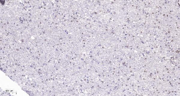
A09415 has an IHC image from paraffin-embedded human brain (IHC caption). IF is listed without an image, and Human, Mouse, Rat and Cat reactivity is listed (catalog applications/reactivity/IF images).
A09415 will render with its IHC figure from paraffin-embedded human brain, showing Tris-EDTA pH 9.0 retrieval and primary antibody at 1:200 overnight at 4°C (A09415 IHC caption). Its catalog lists IHC and IF applications and Human, Mouse, Rat and Cat reactivity; no IF image is supplied (A09415 catalog applications/reactivity/IF images).
Which to pick: For paraffin-section chromogenic IHC, pick A09415 based on its own human brain IHC figure; the fixative is unreported (A09415 IHC caption). For IF, A09415 is listed at 1:50, while ICC and IF imaging are undocumented (A09415 catalog applications/dilution/IF images). For cross-species work, A09415 lists Human, Mouse, Rat and Cat reactivity, although its IHC figure documents only human brain; clonality is unreported (A09415 catalog reactivity/clone; IHC caption).