This website uses cookies to ensure you get the best experience on our website.


- Table of Contents
Plan GPR158 paraffin-section IHC using a catalog antibody at 1:100–1:300 and a peptide-blocked control (datasheet: A11699, A30819 IHC). Interpret nuclear and cytoplasmic CNS staining cautiously because tissue-IHC reliability is uncertain (HPA tissue IHC).

Expected localisation, validated protocols, controls and antibodies — the at-a-glance facts below, then the full design guide.
| Expected localisation | Nuclear and cytoplasmic staining in CNS (HPA tissue IHC) | |
| Staining pattern | CNS cells show nuclear and cytoplasmic staining (HPA tissue IHC) | |
| Antigen retrieval | Tris-EDTA pH 9.0 HIER, 95–98 °C, 20 min (rule: nuclear antigen) | |
| Positive control | Cerebral cortex+2 more · see all | |
| Negative control | Adipose tissue+4 more · see all |
| Fixation | Keep paraffin-section fixation consistent. (standard IHC practice; not target-specific) | |
| Caveat | Presumed off-target staining; reliability uncertain (HPA tissue IHC) | |
| Regulation | Expression regulation unreported (UniProt) | |
| Isoform / epitope | No isoforms annotated; extracellular vs cytoplasmic epitope matters (UniProt) |
Compare the catalog antibody’s IHC-P protocol with one published chromogenic IHC protocol for mouse and human prostate sections (PMC4333349).
| Sample | Paraffin-embedded human brain tissue; fixative not specified (datasheet A11699) |
| Fixation | Image fixative and duration unreported (datasheet A11699); verify before use. |
| Sectioning | 4–5 µm sections on charged slides (standard) |
| Deparaffinisation | Xylene, graded ethanol series to water (standard) |
| Antigen retrieval | Heat-induced epitope retrieval in Tris-EDTA buffer, pH 9.0, 20 min at 95–98 °C (standard rule: nuclear antigen) |
| Peroxidase block | 3% H2O2, 10 min, room temperature (standard) |
| Blocking | 10% normal serum of the secondary host, 30 min, room temperature (standard) |
| Primary antibody | Rabbit anti-GPR158, 1:100-1:300 (datasheet A11699) |
| Primary incubation | Overnight at 4 °C (standard) |
| Detection | HRP-polymer secondary, DAB chromogen 5–10 min (standard) |
| Counterstain | Hematoxylin, blue, dehydrate and mount (standard) |
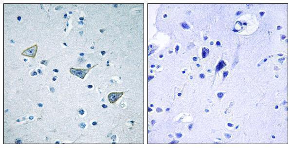
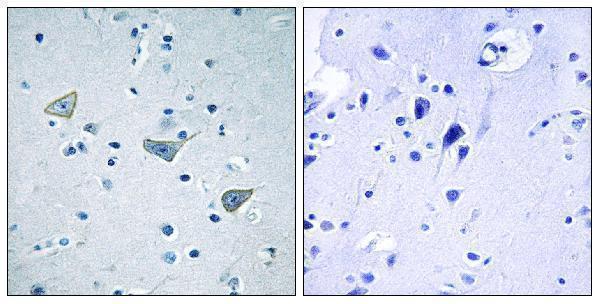
| Expected result | GPR158-positive staining in neuronal cells of cerebral cortex (HPA tissue IHC: High). HPA tissue profile: Nuclear and cytoplasmic expression in CNS. No signal in the no-primary control. |
GPR158 is a seven-pass receptor mainly assigned to postsynaptic membranes, with a smaller presynaptic pool and reported nuclear localisation (UniProt Q5T848). In tissue IHC, expect staining in CNS neuronal cells: high in cerebral cortex and medium in hippocampus and the cerebellar granular layer (HPA tissue IHC). HPA describes nuclear and cytoplasmic CNS staining, but rates its tissue IHC evidence Uncertain because staining and RNA data have low consistency and presumed off-target binding was disregarded (HPA tissue IHC).
| Neuronal staining in cerebral cortex, with weaker staining in hippocampus or the cerebellar granular layer. | This follows HPA's reported high cortical and medium hippocampal and granular-layer staining (HPA tissue IHC). Interpret membrane-associated signal in light of GPR158's predominantly postsynaptic localisation (UniProt Q5T848); assess nuclear or cytoplasmic signal cautiously because HPA reports both while rating the IHC evidence Uncertain (HPA tissue IHC). |
| Predominantly nuclear signal with little neuronal membrane-associated staining. | A nuclear pool is reported, and HPA describes nuclear CNS staining (UniProt Q5T848; HPA tissue IHC). Nuclear-only staining does not establish specificity: the receptor is mainly assigned to the postsynaptic membrane, and HPA's tissue IHC reliability is Uncertain (UniProt Q5T848; HPA tissue IHC). Compare controls before calling it artefact. |
| Strong signal in adipocytes, glandular cells or hematopoietic cells while CNS neurons are weak. | HPA reports no detection in adipocytes of adipose tissue, glandular cells of adrenal gland or appendix, and bone marrow hematopoietic cells (HPA tissue IHC). Such a reversal raises concern for nonspecific antibody binding or endogenous chromogenic activity; check the staining distribution and detection controls (general IHC practice). |
| Uniform colour across cells, extracellular spaces and slide background. | This distribution cannot be read as receptor localisation without cellular boundaries (general IHC practice). Uneven reagent coverage, excess detection background or retained chromogen can produce diffuse colour (general IHC practice). Compare a no-primary control and inspect tissue morphology before assigning any positive cells (general IHC practice). |
| No detectable signal in cerebral cortex neurons. | Cortex is HPA's strongest listed tissue observation, at high staining in neuronal cells (HPA tissue IHC). A blank result calls for checking section integrity, retrieval, antibody conditions and detection performance (general IHC practice). It does not by itself establish absent GPR158, particularly given HPA's Uncertain IHC reliability (HPA tissue IHC). |
| Compartment and topology | GPR158 has 7 transmembrane segments, an extracellular region at residues 24–417 and a cytoplasmic tail at 665–1215 (UniProt Q5T848 topology). Interpret a membrane-associated pattern against this architecture; the observed stain also depends on which region the antibody recognises, which is unspecified here. |
| Protein processing and glycosylation | UniProt lists a signal peptide at residues 1–23, a chain beginning at residue 24, and five glycosylation sites in the extracellular region (UniProt Q5T848). These annotations identify regions to consider when an antibody epitope is known; they do not establish shedding, retrieval needs or fixation sensitivity. |
| Tissue choice | Cerebral cortex neurons provide the strongest listed IHC-positive comparison; hippocampal neurons and cerebellar granular-layer cells are listed at medium staining (HPA tissue IHC). HPA lists several cell types as not detected, including adipose adipocytes and bone marrow hematopoietic cells (HPA tissue IHC). Compare like cell types when scoring. |
| Strength of validation | HPA rates tissue IHC Uncertain, citing low agreement between antibody staining and RNA expression and presumed off-target binding that was disregarded (HPA tissue IHC). Its listed antibody HPA013185 is also IHC Uncertain (HPA antibodies). Treat apparent positives as provisional until their distribution and controls agree. |
| Detection background | Endogenous enzyme activity and nonspecific detection can create chromogenic colour unrelated to antigen (general IHC practice). A no-primary control helps separate detection background from primary-dependent staining (general IHC practice). No supplied source establishes a GPR158-specific fixation effect or an optimal retrieval condition. |
| Situation | Likely cause | Next action |
|---|---|---|
| Cortex is blank. | A high neuronal signal is reported for cerebral cortex, but HPA rates the tissue IHC evidence Uncertain (HPA tissue IHC). Technical failure remains possible (general IHC practice). | Check morphology and section coverage, then verify retrieval, antibody incubation and chromogenic detection with appropriate run controls (general IHC practice). Record the conditions before interpreting the blank as biological absence. |
| Negative comparison cells stain strongly. | Adipose adipocytes and bone marrow hematopoietic cells are listed as not detected (HPA tissue IHC). Primary binding or detection background may explain unexpected colour (general IHC practice). | Compare a no-primary control and review the cell boundaries and signal distribution (general IHC practice). Avoid scoring colour outside the expected cells as GPR158 without additional specificity evidence. |
| Staining is diffuse or obscures cell outlines. | Excess detection background or uneven reagent coverage can mask cellular localisation (general IHC practice). HPA's Uncertain rating increases the need to inspect the pattern critically (HPA tissue IHC). | Inspect the no-primary control, assess washing and chromogen development, and rescore only where morphology remains clear (general IHC practice). |
| Only nuclei are positive. | UniProt reports nuclear localisation but describes GPR158 as mainly postsynaptic; HPA reports nuclear CNS staining despite Uncertain tissue IHC reliability (UniProt Q5T848; HPA tissue IHC). | Compare the same cell population with membrane-associated and cytoplasmic staining, plus controls (general IHC practice). Report nuclear-only staining as a qualified observation rather than a confirmed receptor distribution. |
| Hippocampus is weaker than cortex. | This ordering matches HPA's medium neuronal staining in hippocampus versus high neuronal staining in cerebral cortex (HPA tissue IHC). | Score intensity by cell population and retain the tissue identity in the record (general IHC practice). Do not require hippocampal neurons to match cortical intensity. |
| IF/ICC: should membrane fluorescence be expected? | HPA's ICC-IF summary says membrane, but supplies no cell-line images; its listed antibody has no ICC validation entry (HPA subcellular; HPA antibodies). | Use the membrane assignment as a localisation hypothesis, not an IF validation claim (HPA subcellular; UniProt Q5T848). Consult the separate IF/ICC guide for assay design. |
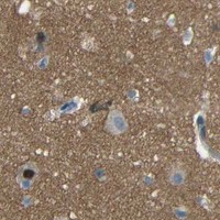
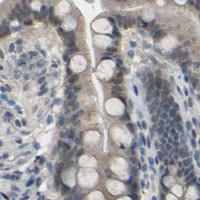

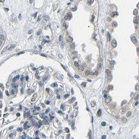
Comprehensive Human Protein Atlas IHC scoring per tissue (reliability: Uncertain — Low consistency between antibody staining and RNA expression data. Presumed off target binding observed and disregarded.). Rows are taken directly from the HPA tissue chart — click any row's HPA link to view the source.
| Tissue | Cell type | Level | Evidence | Source |
|---|---|---|---|---|
| Cerebral cortex | Neuronal cells | High | Protein (IHC) | HPA → |
| Cerebellum | Cells in granular layer | Medium | Protein (IHC) | HPA → |
| Hippocampus | Neuronal cells | Medium | Protein (IHC) | HPA → |
| Tissue | Cell type | Level | Evidence | Source |
|---|---|---|---|---|
| Adipose tissue | Adipocytes | Not detected | Protein (IHC) | HPA → |
| Adrenal gland | Glandular cells | Not detected | Protein (IHC) | HPA → |
| Appendix | Glandular cells | Not detected | Protein (IHC) | HPA → |
| Bone marrow | Hematopoietic cells | Not detected | Protein (IHC) | HPA → |
| Breast | Adipocytes | Not detected | Protein (IHC) | HPA → |
Troubleshoot GPR158 staining in paraffin sections by checking retrieval, compartment, controls and scoring before interpreting chromogenic signal.
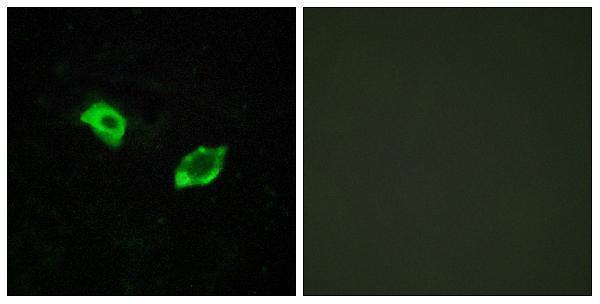
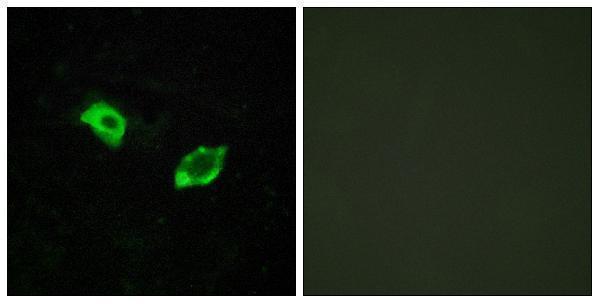
Both antibodies show paraffin-embedded human brain IHC with peptide-blocking images (catalog IHC captions) and HUVEC IF images (catalog IF captions); both list Human and Mouse reactivity (catalog reactivity).
A11699 shows paraffin-embedded human brain IHC with peptide blocking and HUVEC IF (A11699 image captions); its listed applications include IHC and IF (A11699 applications). A30819 shows the same sample types in its own images (A30819 image captions) and also lists ICC (A30819 applications).
Which to pick: For tissue IHC, either SKU has its own paraffin-embedded human brain image with peptide blocking (A11699 IHC caption; A30819 IHC caption); the fixative is unreported in both captions. For IF, both have HUVEC images (A11699 IF caption; A30819 IF caption); choose A30819 when ICC is required because only it lists ICC (A30819 applications; A11699 applications). For Mouse samples, both list Mouse reactivity and are rabbit polyclonals (catalog reactivity and dilution data), but the supplied IHC images show human brain (A11699 IHC caption; A30819 IHC caption).