GSTA1 / Glutathione S-transferase A1 · IHC design guide

Design Immunohistochemistry for GSTA1

Plan paraffin-section GSTA1 IHC around cytoplasmic staining in hepatocytes and kidney tubule cells (HPA tissue IHC). This guide covers fixation consistency, antibody specificity, chromogenic detection and tissue-based scoring.

Evidence assembled Oct 2026 · For research use; verify linked source records and product datasheet before use
Immunohistochemistry protocol sheet for GSTA1 (IHC for GSTA1): expected localisation Cytoplasmic tissue staining (HPA tissue IHC), antibody A01462-1, validated IHC image, and IHC protocol steps
Printable GSTA1 IHC protocol sheet — expected localisation Cytoplasmic tissue staining (HPA tissue IHC), antibody A01462-1, controls and protocol steps. Open the full GSTA1 IHC guide →

GSTA1 Immunohistochemistry Experimental Design Guide

Expected localisation, validated protocols, controls and antibodies — the at-a-glance facts below, then the full design guide.

Must know before staining
Expected localisation Cytoplasmic tissue staining (HPA tissue IHC)
Staining pattern Cytoplasmic signal in hepatocytes and kidney tubule cells (HPA tissue IHC)
Antigen retrieval Citrate pH 6 HIER, heat-mediated (datasheet A01462-1)
Positive control ⓘ Adrenal gland+4 more · see all
Negative control ⓘ Adipose tissue+4 more · see all
Important caveats
Reasons your staining may differ from the expected pattern.
Fixation Keep fixation consistent across sections. (standard IHC practice; not target-specific)
Caveat GSTA-family recognition can confound GSTA1 scoring (datasheet A01462-1)
Regulation Tissue-dependent expression (HPA tissue IHC)
Isoform / epitope No annotated isoforms; N-terminal processing may affect epitopes (UniProt)
Section 1

Recommended GSTA1 IHC & IF Protocols

The catalog antibody’s IHC-P protocol is complemented by 3 published GSTA1 IHC protocols (PMC4476686; PMC12997922; PMC6930411).

Recommended immunohistochemistry (IHC-P) protocol parameters
SampleParaffin-embedded human lung cancer tissues; fixative not specified (datasheet A01462-1)
FixationImage fixative and duration unreported (datasheet A01462-1); verify before use.
Sectioning4–5 µm sections on charged slides (standard)
DeparaffinisationXylene, graded ethanol series to water (standard)
Antigen retrievalHeat retrieval: Citrate pH 6, 20 min (datasheet A01462-1)
Peroxidase block3% H2O2, 10 min, room temperature (standard)
Blocking10% goat serum (datasheet A01462-1)
Primary antibodyRabbit anti-GSTA1, 1μg/ml (datasheet A01462-1)
Primary incubationOvernight at 4 °C (datasheet A01462-1)
DetectionStreptavidin-biotin complex (SABC), DAB chromogen (datasheet A01462-1)
CounterstainHematoxylin, blue, dehydrate and mount (standard)
Expected resultGSTA1-positive staining in glandular cells of adrenal gland (HPA tissue IHC: High). HPA tissue profile: Distinct cytoplasmic expression in several tissues including liver, kidney and adrenal gland. No signal in the no-primary control.
💡Decision noteStart with heat-mediated citrate retrieval at pH 6 (datasheet A01462-1); consider EDTA at pH 9 when optimizing retrieval (PMC12997922).
Section 2

What Is the Expected GSTA1 Staining Pattern?

GSTA1 is a cytoplasmic protein with no transmembrane segment (UniProt P08263). In paraffin-section IHC, expect cytoplasmic staining in hepatocytes, kidney tubular cells, and adrenal glandular cells, each reported as High by HPA (HPA tissue IHC). HPA rates the tissue pattern Supported, with medium agreement between staining and RNA data, and cautions that the antibodies may target proteins from more than one gene (HPA tissue IHC).

What am I looking at on my slide?
Strong cytoplasmic stain in hepatocytes, kidney tubular cells, or adrenal glandular cells.This matches the reported compartment and High staining in those cell populations (UniProt P08263; HPA tissue IHC). Compare cell-by-cell distribution with the counterstain; intensity alone cannot establish that every stained protein is GSTA1 because HPA flags possible multi-gene antibody targeting (HPA tissue IHC).
Predominantly nuclear or membrane-rim staining, with little cytoplasmic signal.This conflicts with the cytoplasmic assignment and absence of a transmembrane segment (UniProt P08263). Treat it as a suspect pattern: review morphology, controls, and detection conditions before assigning it to GSTA1. A different compartment by itself does not identify the source of the stain (standard IHC practice).
Strong staining in adipocytes or bone-marrow hematopoietic cells.HPA reports GSTA1 as Not detected in those cell types (HPA tissue IHC). Recheck cell identity and controls; antibody recognition of another protein or endogenous detection activity are possibilities, especially given HPA's multi-gene caution (HPA tissue IHC; standard IHC practice).
Brown deposit spread across the section without clear cell boundaries.A diffuse deposit is hard to score as cytoplasmic GSTA1 because the reported pattern is cell-associated (HPA tissue IHC; UniProt P08263). Review a no-primary control, blocking, washing, and chromogen development to distinguish background from interpretable cellular staining (standard IHC practice).
No visible stain in a section with identifiable hepatocytes.This disagrees with the High hepatocyte staining reported by HPA (HPA tissue IHC), but a negative slide alone cannot establish absent GSTA1. Confirm that a known-positive section and the detection controls work, then assess tissue preservation and assay conditions using general IHC practice.
💡Expected GSTA1 appearanceA convincing positive is clear, predominantly cytoplasmic staining in hepatocytes, kidney tubular cells, or adrenal glandular cells at the HPA-reported High level; dominant nuclear or membrane-rim staining is suspect (HPA tissue IHC; UniProt P08263).
How each factor affects the staining
Cell compartment and topologyUniProt places GSTA1 in the cytoplasm and reports no transmembrane segment (UniProt P08263). Score signal within the cell body against the counterstain; a crisp membrane outline or isolated nuclear signal does not match that assignment (standard IHC interpretation).
Tissue and cell-type distributionHPA reports High staining in hepatocytes, kidney tubular cells, adrenal glandular cells, duodenal endocrine cells, gallbladder glandular cells, and testicular Leydig cells (HPA tissue IHC). Its Not detected calls apply to specified cell types, not necessarily every cell in those tissues (HPA tissue IHC).
Antibody interpretationThe HPA tissue profile is Supported, yet HPA describes only medium staining–RNA consistency and cautions that antibodies may target proteins from more than one gene (HPA tissue IHC). Agreement with the expected pattern strengthens interpretation but does not establish gene-exclusive recognition.
Processing and protein formsUniProt records full-length residues 1–222 and an N-terminally processed 2–222 chain, with N-terminal acetylation among the listed modifications; it lists no isoforms (UniProt P08263). These annotations do not specify an IHC epitope or demonstrate altered staining after tissue processing.
Why is my staining missing, weak or wrong?
SituationLikely causeNext action
A liver positive control is weak or blank.The result conflicts with HPA's High hepatocyte staining, but the failed step is undetermined (HPA tissue IHC).Verify hepatocyte morphology, primary-antibody application, detection reagents, and counterstain; compare with a working control section before interpreting the specimen (standard IHC practice).
Staining is mainly nuclear or outlines cell membranes.That distribution conflicts with cytoplasmic GSTA1 and its lack of a transmembrane segment (UniProt P08263).Check a no-primary control and inspect whether precipitate or overlapping cells create the apparent compartment; score only well-resolved cellular signal (standard IHC practice).
Adipocytes or marrow hematopoietic cells appear strongly positive.HPA reports Not detected in these populations; non-target recognition or endogenous detection activity is possible (HPA tissue IHC; standard IHC practice).Confirm the cell type, inspect no-primary and positive controls, and review endogenous-enzyme blocking if using enzyme-based chromogenic detection (standard IHC practice).
Diffuse brown background obscures cell boundaries.Background from incomplete blocking, washing, or excess chromogen development is possible (standard IHC practice).Compare a no-primary control and review blocking, washes, and development time; interpret only signal whose cellular compartment remains distinguishable (standard IHC practice).
Some cells stain while nearby cells in the same section do not.HPA reports staining by cell type, so mixed cell populations need cell-specific interpretation; staining intensity alone cannot establish identity (HPA tissue IHC).Use morphology and the counterstain to score the specified cell population, then compare it with a relevant positive control (standard IHC practice).
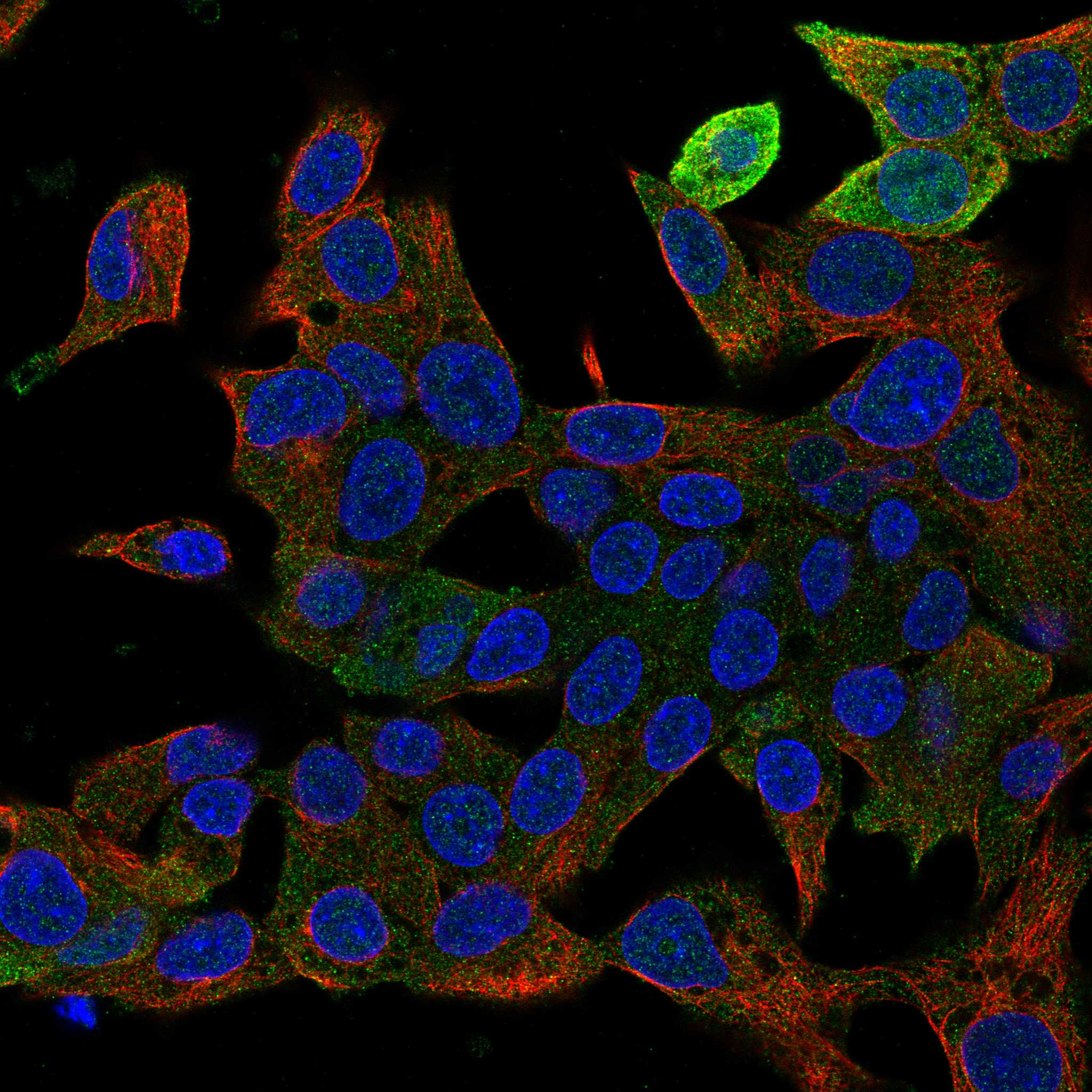
What pattern should IF/ICC show?HPA reports supported cytosolic localization in ICC-IF, while cautioning that the antibodies may target proteins from multiple genes (HPA subcellular ICC-IF).Expect cytosolic fluorescence and assess cell boundaries against an appropriate counterstain; use the separate IF/ICC guide for assay setup (HPA subcellular ICC-IF; standard IF practice).

Sample controls for GSTA1 IHC & IF

🧪Run liver first: hepatocytes should stain (HPA: High in hepatocytes). Run adipose tissue as a negative comparator (HPA: Not detected in adipocytes); on the liver slide, unstained non-hepatocyte areas can show background, but the supplied HPA rows do not establish a specific internal cell type as GSTA1-negative.
Positive control tissue: Adrenal gland (Glandular cells, HPA High)
Negative control tissue: Adipose tissue (HPA Not detected)
ICC-IF cell lines (HPA subcellular resource): HPA ICC-IF images show GSTA1 in Hep-G2, U2OS, with annotated localisation: Cytosol (supported) (HPA subcellular).
Technical controls: Include a no-primary (secondary-only) control, an isotype-matched rabbit IgG control, and a GSTA1 knockout or peptide-block control (caption: rabbit primary; standard IHC control practice). In liver IHC, check endogenous peroxidase and biotin background; for IF, check tissue autofluorescence (standard IHC/IF practice).
⚠️Feasibility: A target-specific fixation window or fixation effect is unreported, and the selected A01462-1 tissue-IHC caption does not state a fixative (selected-SKU caption). The caption reports citrate retrieval at pH 6 for 20 minutes, but does not establish that retrieval is required for GSTA1; the supplied evidence does not establish that frozen sections or IF are easier (selected-SKU caption). Interpret staining cautiously because the catalog antibody is labeled GSTA1/A2/A3/A4/A5, while the HPA cytosol assignment warns that its ICC-IF evidence uses antibodies targeting proteins from multiple genes (selected-SKU caption; HPA: Cytosol, supported).

HPA tissue IHC evidence for GSTA1

Comprehensive Human Protein Atlas IHC scoring per tissue (reliability: Supported — Medium consistency between antibody staining and RNA expression data. Caution, targets protein from more than one gene.). Rows are taken directly from the HPA tissue chart — click any row's HPA link to view the source.

Positive expression · recommended positive controls

TissueCell typeLevelEvidenceSource
Adrenal gland Glandular cells High Protein (IHC) HPA →
Duodenum Endocrine cells High Protein (IHC) HPA →
Gallbladder Glandular cells High Protein (IHC) HPA →
Kidney Cells in tubules High Protein (IHC) HPA →
Liver Hepatocytes High Protein (IHC) HPA →

Undetected expression · recommended negative controls

TissueCell typeLevelEvidenceSource
Adipose tissue Adipocytes Not detected Protein (IHC) HPA →
Appendix Endocrine cells Not detected Protein (IHC) HPA →
Bone marrow Hematopoietic cells Not detected Protein (IHC) HPA →
Caudate Glial cells Not detected Protein (IHC) HPA →
Cerebellum Cells in granular layer Not detected Protein (IHC) HPA →
Section 3

Advanced GSTA1 IHC Tips

Troubleshoot GSTA1 staining by checking retrieval, cytoplasmic localisation, cell identity and antibody specificity before comparing signal across sections.

What retrieval should I use when GSTA1 staining is weak in paraffin sections?
Start with heat-mediated retrieval in citrate buffer at pH 6 (datasheet A01462-1). The selected antibody’s paraffin-section image used 20 minutes of this retrieval, followed by overnight primary incubation at 4°C (caption A01462-1). If staining remains weak, compare retrieval durations on adjacent sections while keeping antibody concentration and detection constant, and check whether tissue morphology deteriorates (standard IHC practice). Use a liver section with hepatocytes as a positive reference, but remember that the HPA tissue profile cautions that its staining may include proteins from more than one gene (HPA: high in hepatocytes; HPA: multi-gene antibody caution).
How should I troubleshoot fixation-related loss of GSTA1 staining?
Target-specific fixation sensitivity is unknown: the selected paraffin-section caption does not state a fixative, so its staining cannot establish how GSTA1 responds to fixation (caption A01462-1). Record each block’s fixative and fixation interval, then compare sections processed with the same citrate pH 6 retrieval and detection settings (datasheet A01462-1; standard IHC practice). If a suspected fixation effect persists, compare matched specimens with documented processing histories and assess tissue preservation alongside signal (standard IHC practice). Use hepatocyte cytoplasmic staining as a reference pattern, while treating a negative result in a poorly preserved block as inconclusive (HPA: high in hepatocytes; UniProt P08263: cytoplasm; standard IHC practice).
Where should convincing GSTA1 staining appear within positive cells?
Expect predominantly cytoplasmic staining, because GSTA1 is annotated in the cytoplasm and has no transmembrane segment (UniProt P08263: localisation and topology). The HPA subcellular summary supports cytosolic localisation, while cautioning that its antibodies can recognise proteins from multiple genes (HPA: cytosol supported; HPA: multi-gene antibody caution). In chromogenic sections, compare signal within intact cells against the nuclear counterstain and nearby unstained structures, using the same exposure or scanning settings across controls (standard IHC practice). Predominantly nuclear, membrane-restricted or extracellular deposits warrant checks of morphology, secondary-only staining and antibody specificity before they are assigned to GSTA1 (UniProt P08263: cytoplasm; standard IHC practice).
Can this antibody distinguish GSTA1 from related alpha GST proteins?
Do not assign staining from A01462-1 uniquely to GSTA1: its selected image explicitly describes recognition of GSTA1/A2/A3/A4/A5 (caption A01462-1). GSTA1 has 0 listed isoforms, can form a homodimer or a heterodimer with GSTA2, and has GST N-terminal and C-terminal domains at residues 3–83 and 85–207 (UniProt P08263). Its listed N-terminal processing and modifications make epitope position relevant when comparing antibodies, but the supplied caption does not locate this antibody’s epitope (UniProt P08263; caption A01462-1). For gene-specific conclusions, establish antibody cross-reactivity experimentally with appropriate loss-of-target or orthogonal evidence before interpreting a brown cell as GSTA1-positive (standard IHC practice).
How can IF help check a GSTA1 pattern seen by chromogenic IHC?
Use IF as a separate localisation check, pairing GSTA1 staining with a marker that identifies the expected cell type, such as hepatocytes in liver, and assess overlap at the cell level (HPA: high in hepatocytes; standard IF practice). Choose fluorophores after inspecting unstained tissue for autofluorescence, and include single-colour controls when multiplexing to distinguish bleed-through from overlap (standard IF practice). Because GSTA1 is cytosolic and lacks a transmembrane segment, permeabilisation must allow antibody access to the inside of cells (UniProt P08263: localisation and topology; standard IF practice). The HPA reports cytosolic IF localisation but cautions that its antibodies target proteins from multiple genes; that observation alone does not establish A01462-1 specificity in IF (HPA: subcellular summary; caption A01462-1).
How do I reduce diffuse brown background without losing cytoplasmic signal?
First inspect a section lacking primary antibody to separate detection-system deposits from primary-antibody staining, and compare it with an identically developed test section (standard IHC practice). The selected image used 10% goat serum blocking, 1 μg/ml primary antibody, a biotinylated secondary, streptavidin-biotin detection and DAB, providing specific conditions to review if background rises (caption A01462-1). Check endogenous peroxidase blocking and, with this detection chemistry, endogenous biotin controls as general chromogenic IHC safeguards (standard IHC practice). Adjust primary concentration, wash stringency or DAB development one variable at a time, preserving the expected cytoplasmic signal in hepatocytes as a reference (HPA: high in hepatocytes; UniProt P08263: cytoplasm; standard IHC practice).
How should I score GSTA1 staining across paraffin sections? ⚠ ANSWER MARKED FOR VERIFICATION
Define the cell population and region before scoring; for liver, hepatocytes provide a documented high-staining population (HPA: high in hepatocytes; standard IHC practice). Score cytoplasmic intensity and the percentage of positive target cells, or combine intensity categories into an H-score, while using the same threshold and imaging conditions for every section (UniProt P08263: cytoplasm; standard IHC practice). Normalise positive counts to the number of eligible cells, or positive area to evaluable tissue area, excluding folds, necrosis and blank space consistently (standard IHC practice). Report the antibody and detection method with the score, because the selected antibody recognises several alpha GST proteins and a measured signal is not necessarily GSTA1-specific (caption A01462-1).
When is a positive stain convincing evidence of GSTA1 expression?
A convincing pattern is cytoplasmic signal in intact expected cells, such as hepatocytes or kidney tubular cells, that exceeds the matched negative-control signal (UniProt P08263: cytoplasm; HPA: high in hepatocytes and kidney tubules; standard IHC practice). Question nuclear-only or membrane-restricted staining because GSTA1 is cytoplasmic and lacks a transmembrane segment (UniProt P08263: localisation and topology). Also inspect section edges, necrotic areas and secondary-only controls for staining caused by processing or endogenous enzyme activity (standard IHC practice). Even a plausible pattern cannot by itself identify GSTA1 specifically: the selected antibody is labelled for GSTA1/A2/A3/A4/A5, and HPA warns that its tissue staining may represent proteins from more than one gene (caption A01462-1; HPA: multi-gene antibody caution).
Boster reagents

Best GSTA1 / Glutathione S-transferase A1 IHC Antibodies

IHC and IF figures show paraffin sections of human lung and liver cancer and mouse and rat liver (A01462-1/PB9627 captions); catalog reactivity spans human, mouse and rat (catalog).

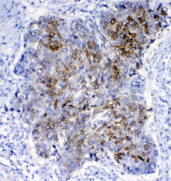
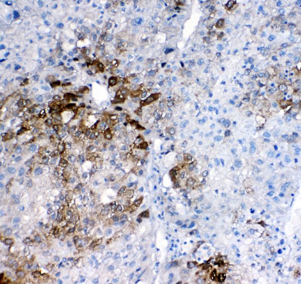
Real IHC data IHC analysis of GSTA1/A2/A3/A4/A5 using anti-GSTA1/A2/A3/A4/A5 antibody (A01462-1). GSTA1/A2/A3/A4/A5 was detected in paraffin-embedded section of human lung cancer tissues. Heat mediated antigen retrieval was performed in citrate buffer (pH6, epitope retrieval solution) for 20 mins. The tissue section was blocked with 10% goat serum. The tissue section was then incubated with 1μg/ml rabbit anti-GSTA1/A2/A3/A4/A5 Antibody (A01462-1) overnight at 4°C. Biotinylated goat anti-rabbit IgG was used as secondary antibody and incubated for 30 minutes at 37°C. The tissue section was developed using Strepavidin-Biotin-Complex (SABC)(Catalog # SA1022) with DAB as the chromogen.
Anti-GSTA1/A2/A3/A4/A5 Antibody ®
Cat # A01462-1
Real IF data IF analysis of GSTA1/A2/A3/A4/A5 using anti-GSTA1/A2/A3/A4/A5 antibody (PB9627) GSTA1/A2/A3/A4/A5 was detected in paraffin-embedded section of mouse liver tissues. Heat mediated antigen retrieval was performed in citrate buffer (pH6, epitope retrieval solution ) for 20 mins. The tissue section was blocked with 10% goat serum. The tissue section was then incubated with 1μg/mL rabbit anti-GSTA1/A2/A3/A4/A5 Antibody (PB9627) overnight at 4°C. DyLight®488 Conjugated Goat Anti-Rabbit IgG (BA1127) was used as secondary antibody at 1:100 dilution and incubated for 30 minutes at 37°C. The section was counterstained with DAPI. Visualize using a fluorescence microscope and filter sets appropriate for the label used.
Anti-GSTA1/GSTA2/GSTA3/GSTA4/GSTA5 Antibody ®
Cat # PB9627

A01462-1 renders with a human lung cancer IHC figure; its other IHC captions document human liver cancer (A01462-1 captions). PB9627 renders with a mouse liver IF figure, and its captions also document rat liver IF (PB9627 captions).

Which to pick: For chromogenic tissue IHC, choose A01462-1: its own paraffin-section caption documents citrate pH 6 retrieval and DAB detection; the fixative is unreported (A01462-1 IHC caption). For tissue IF, choose PB9627 because its own captions show mouse and rat liver sections; for ICC, A01462-3 lists that application but has no supplied figure (PB9627 IF captions; A01462-3 catalog). All three list human, mouse and rat reactivity, but PB9627’s stated IHC-P dilution applies to mouse and rat, so A01462-1 has the stronger supplied evidence for human tissue IHC (catalog; A01462-1 IHC captions).

Each figure is that product's own IHC / IF validation image from its datasheet.

References

  1. UniProt Consortium. UniProt entry P08263 (GSTA1_HUMAN, Glutathione S-transferase A1).
  2. Human Protein Atlas. GSTA1 tissue IHC expression (reliability: Supported).
  3. Human Protein Atlas. GSTA1 subcellular location (ICC-IF): Localized to the cytosol. Caution: Based on antibodies targeting proteins from multiple genes..
  4. Human Protein Atlas. GSTA1 antibody validation summary (3 antibodies).
  5. Oviductal Transcriptome Is Modified after Insemination during Spontaneous Ovulation in the Sow. PloS one 2015 — PMC4476686.
  6. Integrated analysis reveals GSTA1 as a prognostic biomarker in stage I lung adenocarcinoma. BMC cancer 2026 — PMC12997922.
  7. WGCNA-based identification of potential targets and pathways in response to treatment in locally advanced breast cancer patients. Open medicine (Warsaw, Poland) 2023 — PMC9990777.
  8. Glutathione S-transferase A1 suppresses tumor progression and indicates better prognosis of human primary hepatocellular carcinoma. Journal of Cancer 2020 — PMC6930411.
  9. PubMed PMID:3800996 — UniProt-cited evidence.
  10. PubMed PMID:3036131 — UniProt-cited evidence.
  11. PubMed PMID:3678589 — UniProt-cited evidence.