This website uses cookies to ensure you get the best experience on our website.


- Table of Contents
Plan chromogenic GUSB IHC in paraffin sections using liver hepatocytes as a high-staining reference (HPA tissue IHC). Expect cytoplasmic tissue staining while interpreting GUSB as a lysosomal protein (HPA tissue IHC; UniProt).

Expected localisation, validated protocols, controls and antibodies — the at-a-glance facts below, then the full design guide.
| Expected localisation | Cytoplasmic staining (HPA tissue IHC); lysosomal protein (UniProt) | |
| Staining pattern | Cytoplasmic staining in most tissues (HPA tissue IHC) | |
| Antigen retrieval | EDTA pH 9 HIER, heat-mediated (datasheet A02234) | |
| Positive control | Adrenal gland+4 more · see all | |
| Negative control | Adipose tissue+4 more · see all |
| Fixation | Formaldehyde-fixed sections are documented (selected-SKU IHC image A02234); keep fixation consistent (standard IHC practice; not target-specific) | |
| Caveat | Endogenous peroxidase can mimic HRP/DAB staining (standard IHC practice) | |
| Regulation | No expression regulator annotated (UniProt) | |
| Isoform / epitope | 3 isoforms; mature chain begins at residue 23 (UniProt) |
The catalog antibody uses EDTA pH 9 heat retrieval (datasheet A02234). The published mouse colon protocol below uses citrate retrieval (PMC9140600).
| Sample | Formaldehyde-fixed, paraffin-embedded Human liver tissue (datasheet A02234) |
| Fixation | Image formalin-fixed; duration unreported (datasheet A02234); verify before use. |
| Sectioning | 4–5 µm sections on charged slides (standard) |
| Deparaffinisation | Xylene, graded ethanol series to water (standard) |
| Antigen retrieval | Heat retrieval: EDTA pH 9 (datasheet A02234); 20 min, 95–100 °C (standard) |
| Peroxidase block | 3% H2O2, 10 min, room temperature (standard) |
| Blocking | 10% normal serum of the secondary host, 30 min, room temperature (standard) |
| Primary antibody | Rabbit anti-GUSB, 1:100 (datasheet A02234) |
| Primary incubation | 1 hour at room temperature (datasheet A02234) |
| Detection | HRP-conjugated secondary, DAB chromogen (datasheet A02234) |
| Counterstain | Hematoxylin, blue, dehydrate and mount (standard) |
| Expected result | GUSB-positive staining in glandular cells of adrenal gland (HPA tissue IHC: High). HPA tissue profile: Cytoplasmic expression in most tissues. No signal in the no-primary control. |
GUSB is a lysosomal enzyme without a transmembrane segment (UniProt P08236: location, topology). In paraffin IHC, expect cytoplasmic staining in most tissues, especially glandular cells in adrenal gland, endometrium, epididymis and gallbladder, and hepatocytes in liver (HPA: tissue IHC). HPA rates its tissue IHC profile Approved, with medium consistency between antibody staining and RNA expression (HPA: reliability).
| Cytoplasmic staining in adrenal gland, endometrial, epididymal or gallbladder glandular cells, or liver hepatocytes. | This fits HPA's High staining in those cells and its broader cytoplasmic profile (HPA: tissue IHC). A chromogenic slide need not resolve individual lysosomes to be consistent with lysosomal GUSB (UniProt P08236: location). |
| Predominantly nuclear, membranous or extracellular signal, with little cytoplasmic staining. | That distribution conflicts with the reported cytoplasmic tissue pattern and lysosomal location (HPA: tissue IHC; UniProt P08236: location). Check staining specificity and detection background before calling it GUSB; tissue IHC alone cannot identify the source of the signal. |
| Strong staining in a cell population HPA lists as Not detected, such as adipocytes or skeletal myocytes. | Treat it as unexpected for that cell type (HPA: adipocytes and myocytes, Not detected). Consider nonspecific antibody binding or endogenous detection activity (general IHC practice). An HPA negative is a reference pattern, not proof that every specimen must be negative. |
| Widespread, fairly uniform chromogen obscures cell borders and expected tissue differences. | The diffuse background makes the reported cell specific pattern difficult to judge (HPA: tissue IHC). Nonspecific binding or endogenous detection activity can produce background (general IHC practice); evaluate controls before assigning it to GUSB. |
| No convincing cytoplasmic signal in a well preserved liver section or another HPA High tissue. | This is inconsistent with the HPA reference pattern for hepatocytes or the specified glandular cells (HPA: High in liver, adrenal gland, endometrium, epididymis and gallbladder). Review the IHC run and controls before interpreting the sample as GUSB negative (general IHC practice). |
| Compartment and resolution | UniProt places GUSB in lysosomes, while HPA tissue IHC describes cytoplasmic expression (UniProt P08236: location; HPA: tissue IHC). Judge chromogenic sections by a cytoplasmic pattern; discrete vesicles are documented in HPA ICC-IF, where they can be resolved more readily (HPA: subcellular). |
| Choice of reference tissue and cell | Adrenal gland, endometrium, epididymis and gallbladder glandular cells, plus liver hepatocytes, are HPA High examples (HPA: tissue IHC). Breast glandular cells and caudate glia are Medium; several other cell types are Low or Not detected, so score the specified cell population rather than the whole section (HPA: tissue IHC). |
| Strength of tissue evidence | HPA labels the tissue IHC profile Approved and describes medium consistency with RNA data; RNA tissue specificity is Low (HPA: reliability, RNA specificity). Use the listed staining levels as reference observations, with that stated validation limit, rather than absolute thresholds for a new specimen. |
| Processing, glycosylation and isoforms | GUSB has a signal peptide at residues 1–22, a mature chain at 23–651, four annotated glycosylation sites and three isoforms (UniProt P08236: processing, glycosylation, isoforms). Neither source supplies the antibody epitope or isoform coverage, so these annotations cannot predict staining intensity or retrieval needs. |
| Topology and protocol limits | No transmembrane segment is annotated (UniProt P08236: topology). That supports checking an apparent membrane only pattern, but it does not specify antigen retrieval or fixation sensitivity; no target specific fixation effect is supplied (UniProt P08236: topology; HPA: tissue IHC scope). |
| Situation | Likely cause | Next action |
|---|---|---|
| Known HPA High tissue shows no staining. | The result conflicts with the reference cell pattern; the supplied sources do not establish which workflow step failed (HPA: High in liver and listed glandular cells). | Confirm that the intended cell population is present and that run controls worked. Review retrieval, primary antibody use and detection against the selected IHC-P protocol (general IHC practice); no dilution or retrieval setting is supplied here. |
| Signal is mainly nuclear or outlines cell membranes. | This conflicts with cytoplasmic tissue staining and lysosomal localization (HPA: tissue IHC; UniProt P08236: location). | Compare a HPA High reference tissue and the negative control in the same run. Reassess specificity and background before scoring the unexpected compartment as positive (general IHC practice). |
| Adipocytes, skeletal myocytes or rectal glandular cells stain strongly. | HPA reports those cell types as Not detected (HPA: tissue IHC); nonspecific binding or endogenous detection activity is possible (general IHC practice). | Check the exact cell identity, then examine a negative control and detection controls. Report the disagreement with HPA rather than treating one reference negative as an absolute biological rule. |
| Chromogen is diffuse across cells or the entire section. | Diffuse background can conceal the cell pattern; nonspecific binding or endogenous detection activity are general IHC possibilities (general IHC practice). | Review blocking, washes, primary antibody concentration and detection controls using the chosen IHC-P workflow (general IHC practice). Reassess the HPA expected cell pattern only after background is controlled (HPA: tissue IHC). |
| Staining differs between HPA High and Low tissues less than expected. | HPA levels describe observations in specified cell populations; its Approved profile has medium consistency with RNA data (HPA: tissue IHC, reliability). | Score the named cell types separately and compare sections processed in the same run (general IHC practice). Record the observed difference without converting HPA categories into a quantitative cutoff. |
| IF/ICC Q: Must a separate fluorescence protocol be inferred from the IHC result? | HPA reports vesicular ICC-IF localization and lists HPA036322 as ICC Supported (HPA: subcellular, antibodies); its tissue IHC profile is cytoplasmic (HPA: tissue IHC). | A: Use vesicular localization as the IF/ICC pattern check (HPA: subcellular). Consult the separate IF/ICC guide for that application; the IHC tissue pattern does not specify an IF/ICC protocol. |
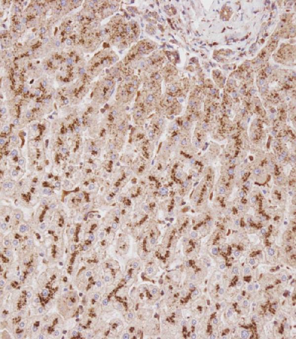
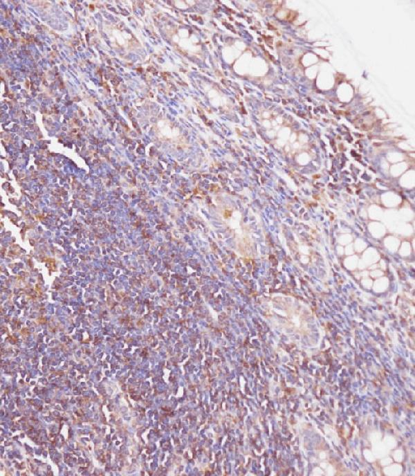
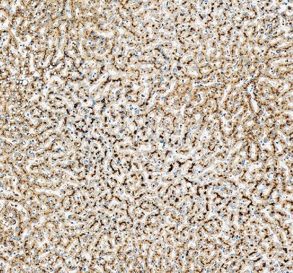
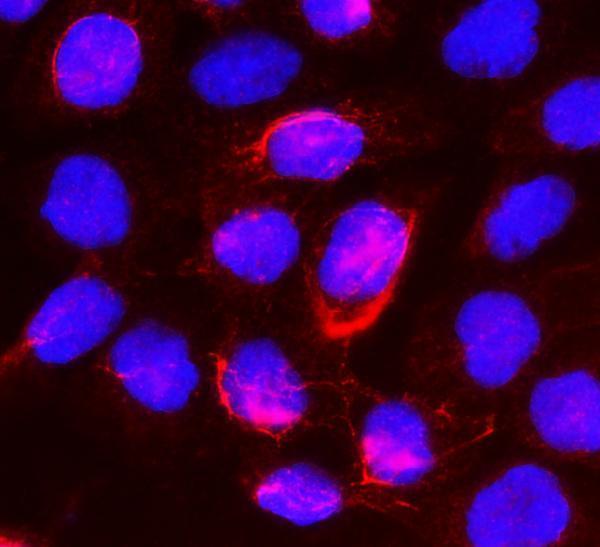
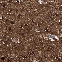


Comprehensive Human Protein Atlas IHC scoring per tissue (reliability: Approved — Medium consistency between antibody staining and RNA expression data.). Rows are taken directly from the HPA tissue chart — click any row's HPA link to view the source.
| Tissue | Cell type | Level | Evidence | Source |
|---|---|---|---|---|
| Adrenal gland | Glandular cells | High | Protein (IHC) | HPA → |
| Endometrium | Glandular cells | High | Protein (IHC) | HPA → |
| Epididymis | Glandular cells | High | Protein (IHC) | HPA → |
| Gallbladder | Glandular cells | High | Protein (IHC) | HPA → |
| Liver | Hepatocytes | High | Protein (IHC) | HPA → |
| Tissue | Cell type | Level | Evidence | Source |
|---|---|---|---|---|
| Adipose tissue | Adipocytes | Not detected | Protein (IHC) | HPA → |
| Nasopharynx | Respiratory epithelial cells | Not detected | Protein (IHC) | HPA → |
| Ovary | Follicle cells | Not detected | Protein (IHC) | HPA → |
| Rectum | Glandular cells | Not detected | Protein (IHC) | HPA → |
| Seminal vesicle | Glandular cells | Not detected | Protein (IHC) | HPA → |
Troubleshoot GUSB staining in paraffin section IHC using the catalog antibody’s documented retrieval conditions, expected lysosomal localisation, and tissue staining controls.
Two anti-GUSB antibodies have human paraffin-section IHC images (catalog IHC captions); A02234-1 also has an A431-cell IF image (catalog IF caption). A02234 lists mouse reactivity (catalog reactivity).
A02234 has IHC images from formaldehyde-fixed, paraffin-embedded human liver and colon carcinoma, using EDTA pH 9 retrieval and 1:100 primary antibody (A02234 IHC captions). A02234-1 has an IHC image from paraffin-embedded human liver cancer using EDTA pH 8 retrieval and 2 μg/ml primary antibody, plus an IF image from A431 cells (A02234-1 IHC and IF captions).
Which to pick: For tissue IHC, choose A02234 to match its formaldehyde-fixed human liver or colon carcinoma examples (A02234 IHC captions), or A02234-1 to match its human liver cancer example; the latter caption does not report a fixative (A02234-1 IHC caption). For IF/ICC, choose A02234-1 because both applications are listed and its A431-cell IF image uses 2 μg/ml (A02234-1 applications; IF caption). For mouse samples, A02234 is the listed human/mouse-reactive polyclonal option with IHC-P listed, though its IHC images show human tissue (A02234 reactivity; dilution listing; applications; IHC captions).