GYPA / Glycophorin-A · IHC design guide

Design Immunohistochemistry for GYPA

Plan chromogenic paraffin IHC around selective erythrocyte staining (HPA tissue IHC) and expected membrane localization (UniProt). This guide covers fixation consistency, epitope placement, and interpretation of kidney distal tubule staining (HPA tissue IHC).

Evidence assembled Oct 2026 · For research use; verify linked source records and product datasheet before use
Immunohistochemistry protocol sheet for GYPA (IHC for GYPA): expected localisation Erythrocyte membrane expected (UniProt), antibody A02184-2, validated IHC image, and IHC protocol steps
Printable GYPA IHC protocol sheet — expected localisation Erythrocyte membrane expected (UniProt), antibody A02184-2, controls and protocol steps. Open the full GYPA IHC guide →

GYPA Immunohistochemistry Experimental Design Guide

Expected localisation, validated protocols, controls and antibodies — the at-a-glance facts below, then the full design guide.

Must know before staining
Expected localisation Erythrocyte membrane expected (UniProt)
Staining pattern Selective erythrocyte staining; compartment unresolved (HPA tissue IHC)
Antigen retrieval Citrate pH 6 HIER, heat-mediated (datasheet A02184-2)
Positive control ⓘ Bone marrow+1 more · see all
Negative control ⓘ Adipose tissue+4 more · see all
Important caveats
Reasons your staining may differ from the expected pattern.
Fixation Keep formalin fixation consistent across paraffin sections. (standard IHC practice; not target-specific)
Caveat Kidney distal tubules also show medium staining (HPA tissue IHC)
Regulation Expression regulation not specified (UniProt)
Isoform / epitope 3 isoforms; check extracellular vs cytoplasmic epitope (UniProt)
Section 1

Recommended GYPA IHC & IF Protocols

The catalog antibody’s IHC-P protocol is followed by published GYPA staining methods for placenta, mouse choroid plexus, fetal tissues, and hepatocellular carcinoma (PMC8036899; PMC3310074; PMC7780123; PMC11106372).

Recommended immunohistochemistry (IHC-P) protocol parameters
SampleParaffin-embedded human liver cancer tissues; fixative not specified (datasheet A02184-2)
FixationImage fixative and duration unreported (datasheet A02184-2); verify before use.
Sectioning4–5 µm sections on charged slides (standard)
DeparaffinisationXylene, graded ethanol series to water (standard)
Antigen retrievalHeat retrieval: Citrate pH 6, 20 min (datasheet A02184-2)
Peroxidase block3% H2O2, 10 min, room temperature (standard)
Blocking10% goat serum (datasheet A02184-2)
Primary antibodyRabbit anti-GYPA, 1μg/ml (datasheet A02184-2)
Primary incubationOvernight at 4 °C (datasheet A02184-2)
DetectionStreptavidin-biotin complex (SABC), DAB chromogen (datasheet A02184-2)
CounterstainHematoxylin, blue, dehydrate and mount (standard)
Expected resultGYPA-positive staining in hematopoietic cells of bone marrow (HPA tissue IHC: Medium). HPA tissue profile: Selective expression in erythrocytes. No signal in the no-primary control.
💡Decision noteStart with heat-mediated citrate retrieval at pH 6 (datasheet: A02184-2); published placental and fetal-tissue methods also use pH 6 retrieval (PMC8036899; PMC7780123).
Section 2

What Is the Expected GYPA Staining Pattern?

GYPA is an erythrocyte membrane protein with an extracellular segment at residues 20–91 and a transmembrane segment at 92–114 (UniProt P02724 topology/function). In paraffin-section IHC, expect staining associated with erythrocytes; HPA describes the tissue profile as selective for erythrocytes and rates its IHC evidence Enhanced (HPA tissue IHC). Interpret other stained structures against HPA's cell-level observations, including marrow hematopoietic cells and kidney distal tubules (HPA tissue IHC).

What am I looking at on my slide?
Crisp staining outlines erythrocytes, with little signal in surrounding cells.This fits GYPA's role as a major erythrocyte membrane protein and its cell-membrane location (UniProt P02724 function/subcellular). HPA calls the tissue profile selective for erythrocytes (HPA tissue IHC). Judge the result by cell identity and membrane association as well as chromogen intensity; a dark deposit alone does not identify its source (general IHC practice).
Medium staining appears among bone-marrow hematopoietic cells, or in kidney distal tubules.Both are recorded observations: bone-marrow hematopoietic cells and kidney distal tubules are listed at Medium staining (HPA tissue IHC). HPA's overall erythrocyte-selective profile does not erase the distal-tubule entry. Review the image and local morphology before treating kidney staining as either confirmed GYPA expression or an artefact (HPA tissue IHC; general IHC practice).
Nuclear or diffuse cytoplasmic staining dominates the tissue section.That distribution is discordant with UniProt's cell-membrane assignment and the erythrocyte-selective tissue profile (UniProt P02724 subcellular; HPA tissue IHC). HPA separately reports nucleoplasm and cytosol in ICC-IF, so those cell-line observations should not be used to validate a dominant nuclear IHC pattern in tissue (HPA subcellular ICC-IF). Check controls and morphology before calling it GYPA (general IHC practice).
Strong stain appears in unrelated cells or across an HPA-listed negative compartment.Cross-reactivity or endogenous detection activity is possible when signal does not follow the expected cell and compartment pattern (general IHC practice). HPA lists adipocytes in adipose tissue and respiratory epithelial cells in bronchus as Not detected; these are compartment-level comparisons, not claims that every cell in those sections must be unstained (HPA tissue IHC).
The known-positive area is blank, or faint colour covers nearly the whole slide.A blank erythrocyte-rich area leaves the staining run uninterpretable until a positive control works (UniProt P02724 function; general IHC practice). Broad colour without cell boundaries suggests background rather than the selective tissue pattern described by HPA (HPA tissue IHC; general IHC practice). Evaluate the positive and negative controls, tissue preservation and detection run before assigning biological absence (general IHC practice).
💡Expected GYPA appearanceCall an IHC result positive when erythrocytes show clear membrane-associated staining consistent with HPA's selective erythrocyte profile; HPA records Medium staining in marrow hematopoietic cells, while dominant unrelated-cell or nuclear staining calls for review (UniProt P02724 subcellular; HPA tissue IHC; general IHC practice).
How each factor affects the staining
Epitope locationThe mature chain spans residues 20–150, including an extracellular region at 20–91 and cytoplasmic region at 115–150 (UniProt P02724 processing/topology). Interpret staining and any retrieval comparison in light of the antibody's documented epitope, if available; the supplied record does not identify that epitope (general IHC practice).
Extracellular glycosylationUniProt annotates 17 glycosylation sites within residues 21–69 (UniProt P02724 glycosylation/topology). This locates many modifications on the extracellular segment, but the record does not show whether they alter this antibody's IHC recognition; avoid predicting a staining gain or loss from glycosylation alone (UniProt P02724 glycosylation; general IHC practice).
Tissue-specific interpretationHPA rates tissue IHC evidence Enhanced and describes selective erythrocyte expression, yet also lists Medium staining in kidney distal tubules and marrow hematopoietic cells (HPA tissue IHC). Keep those observations distinct when selecting a positive area or investigating an unexpected cell type; HPA also flags stained structures that were not annotated (HPA tissue IHC).
Assay and antibody contextThe listed mouse monoclonal has Enhanced IHC validation; the rabbit polyclonal has Supported IHC validation (HPA antibodies). HPA's ICC-IF summary additionally includes plasma membrane, nucleoplasm and cytosol, with HEL among imaged lines (HPA subcellular ICC-IF). Those IF locations describe a different assay context and do not define an IHC-P protocol (HPA subcellular ICC-IF; general IHC practice).
Isoforms and retrieval evidenceUniProt lists 3 isoforms but supplies no isoform-specific staining pattern here (UniProt P02724 isoforms). The supplied sources give no target-specific fixation sensitivity or antigen-retrieval result, so choose and assess IHC-P retrieval using the antibody's validated procedure and run controls, without attributing an observed change to GYPA chemistry (general IHC practice).
Why is my staining missing, weak or wrong?
SituationLikely causeNext action
No erythrocyte staining in a section expected to contain them.A failed stain or unsuitable positive area is possible; GYPA is a major erythrocyte membrane protein (UniProt P02724 function; general IHC practice).Confirm erythrocytes on the counterstained section, run a known-positive control, and review the antibody's IHC-P procedure and detection reagents before interpreting a negative result (general IHC practice).
Signal is predominantly nuclear in tissue IHC.Nuclear dominance conflicts with the UniProt membrane assignment, although HPA reports nucleoplasm in ICC-IF (UniProt P02724 subcellular; HPA subcellular ICC-IF).Check whether nuclear colour persists in a no-primary control, review morphology, and compare a positive erythrocyte area; keep the ICC-IF observation separate from the IHC call (general IHC practice; HPA tissue IHC).
Diffuse stain obscures cell boundaries.Excess detection signal, nonspecific binding or endogenous activity can produce broad background (general IHC practice).Inspect no-primary and negative-compartment controls; review blocking, antibody dilution, washes and chromogen development against the validated IHC-P procedure (general IHC practice).
Kidney distal tubules stain despite an erythrocyte-selective summary.HPA explicitly records Medium distal-tubule staining alongside the selective erythrocyte profile (HPA tissue IHC).Record the cell type and staining distribution, examine HPA's tissue image, and compare an erythrocyte-positive area and negative controls before classifying the finding (HPA tissue IHC; general IHC practice).
Adipocytes or bronchial respiratory epithelium stain strongly.Those specific cell compartments are listed as Not detected, so cross-reactivity or endogenous detection activity warrants investigation (HPA tissue IHC; general IHC practice).Verify the stained cells by morphology, assess the no-primary control, and repeat with an independently validated IHC antibody if needed; distinguish stained erythrocytes in the section from the named negative cell compartment (general IHC practice; HPA tissue IHC).
IF/ICC shows cytosolic or nucleoplasmic signal alongside plasma-membrane signal.HPA reports all three locations in ICC-IF, whereas UniProt assigns GYPA to the cell membrane (HPA subcellular ICC-IF; UniProt P02724 subcellular).Evaluate IF/ICC with its own controls and localisation criteria; use the separate IF/ICC guide for assay design rather than treating these locations as an IHC-P protocol option (general IHC practice; HPA subcellular ICC-IF).

Sample controls for GYPA IHC & IF

🧪Run bone marrow first and assess its hematopoietic cells for GYPA staining (HPA: Medium in bone marrow hematopoietic cells). Use adipose tissue as the negative comparator, scoring adipocytes, which are listed as not detected (HPA: Not detected in adipocytes); on the marrow slide, use cells without specific membrane staining as internal negative comparators (UniProt P02724: cell membrane localization).
Positive control tissue: Bone marrow (Hematopoietic cells, HPA Medium)
Negative control tissue: Adipose tissue (HPA Not detected)
ICC-IF cell lines (HPA subcellular resource): HPA ICC-IF images show GYPA in HEL, with annotated localisation: Nucleoplasm (approved), Plasma membrane (supported), Cytosol (approved) (HPA subcellular).
Technical controls: Include a no-primary, secondary-only control and a host- and class-matched rabbit IgG isotype control (selected-SKU caption: rabbit primary antibody). Use a GYPA knockout specimen for biological specificity where available; in bone marrow, block endogenous peroxidase and assess endogenous biotin when using the caption’s biotin-based DAB detection (selected-SKU caption: biotinylated secondary, SABC and DAB).
⚠️Feasibility: A target-specific fixation window or fixation effect is unreported, and the exact selected-SKU tissue-IHC caption does not state its fixative (selected-SKU caption: fixative not stated). The caption uses heat retrieval in citrate buffer at pH 6 for 20 minutes on a paraffin section, but whether retrieval is required for bone marrow is unreported (selected-SKU caption: liver cancer tissue-IHC conditions). The supplied evidence does not establish that frozen sections or IF are easier; assess marrow background carefully because GYPA is an erythrocyte membrane protein (UniProt P02724: function).

HPA tissue IHC evidence for GYPA

Comprehensive Human Protein Atlas IHC scoring per tissue (reliability: Enhanced — High consistency between antibody staining and RNA expression data. Antibody staining in cells/structures not annotated, view images.). Rows are taken directly from the HPA tissue chart — click any row's HPA link to view the source.

Positive expression · recommended positive controls

TissueCell typeLevelEvidenceSource
Bone marrow Hematopoietic cells Medium Protein (IHC) HPA →
Kidney Distal tubules Medium Protein (IHC) HPA →

Undetected expression · recommended negative controls

TissueCell typeLevelEvidenceSource
Adipose tissue Adipocytes Not detected Protein (IHC) HPA →
Adrenal gland Glandular cells Not detected Protein (IHC) HPA →
Appendix Glandular cells Not detected Protein (IHC) HPA →
Breast Adipocytes Not detected Protein (IHC) HPA →
Bronchus Respiratory epithelial cells Not detected Protein (IHC) HPA →
Section 3

Advanced GYPA IHC Tips

Use membrane localisation, erythroid context and matched controls to troubleshoot GYPA staining in paraffin section chromogenic IHC (UniProt P02724; HPA tissue IHC).

How should I adjust retrieval when GYPA staining is weak?
Start with heat mediated retrieval in citrate buffer at pH 6 for 20 minutes (datasheet A02184-2). Keep section thickness, heating and cooling consistent across the test and control slides so changes in staining can be attributed to retrieval (standard IHC practice). The catalog antibody's paraffin section image used 1 μg/mL primary antibody overnight at 4°C; match those conditions before changing retrieval (datasheet A02184-2). If signal remains weak, compare a modest change in heating time on adjacent sections while checking for tissue damage and nonspecific staining (standard IHC practice). Judge improvement by erythrocyte membrane staining, the expected GYPA location (UniProt P02724 topology; HPA tissue IHC).
Could fixation explain weak or uneven GYPA staining?
Target specific fixation sensitivity is unknown because the selected paraffin section caption does not state a fixative (datasheet A02184-2). Record the fixative, fixation duration and processing history for each specimen before comparing staining intensity across cases (standard IHC practice). Compare adjacent sections processed together using the same citrate pH 6, 20-minute retrieval and 1 μg/mL antibody conditions from the catalog example (datasheet A02184-2). Uneven signal can also reflect section handling or reagent coverage, so inspect the whole slide and repeat affected sections with uniform reagent application (standard IHC practice). Do not assign a GYPA specific fixation mechanism without a controlled comparison (standard IHC practice).
Where should convincing GYPA staining appear in tissue sections?
Look first for membrane associated staining of erythrocytes, consistent with GYPA's membrane assignment and its role as a major erythrocyte membrane protein (UniProt P02724). Its extracellular region spans residues 20–91 and its transmembrane segment spans 92–114, so the expected chromogenic pattern follows cell outlines (UniProt P02724 topology). HPA describes selective expression in erythrocytes but also reports medium staining in bone marrow hematopoietic cells and kidney distal tubules (HPA tissue IHC). Resolve those observations by recording the stained cell or structure rather than calling every brown area erythrocyte staining (standard IHC practice; HPA tissue IHC). Diffuse nuclear staining needs independent validation before assignment to erythrocyte membrane GYPA (UniProt P02724; standard IHC practice).
How can epitope location affect GYPA staining?
GYPA has 3 annotated isoforms, but the supplied caption does not identify the catalog antibody's epitope or establish isoform specificity (UniProt P02724; datasheet A02184-2). The extracellular segment spans residues 20–91 and contains 17 annotated glycosylation sites, while the cytoplasmic segment spans 115–150 (UniProt P02724 topology and glycosylation). Treat changes after retrieval as evidence of altered staining performance, not proof that a particular glycan or isoform was detected (standard IHC practice). Check the antibody's documented immunogen before comparing reagents aimed at different regions, and keep retrieval and detection conditions matched during that comparison (standard IHC practice). Report isoform selective staining only if an independent validation supports that assignment (standard IHC practice).
How should I plan a complementary GYPA immunofluorescence experiment?
For a separate IF experiment, pair GYPA with an independently validated marker of the expected erythroid cell population and assess whether their cell assignments agree (HPA tissue IHC; standard IF practice). Choose spectrally separated fluorophores, testing a far red channel if specimen autofluorescence interferes with shorter wavelength channels (standard IF practice). Plan permeabilisation around the antibody's documented epitope: extracellular residues 20–91 can be assessed without membrane permeabilisation, whereas a cytoplasmic epitope at 115–150 requires access to the cell interior (UniProt P02724 topology; standard IF practice). The supplied caption does not map this antibody's epitope, so confirm that information before choosing a permeabilisation condition (datasheet A02184-2). Include single channel and secondary only controls when interpreting multiplex signal (standard IF practice).
What causes brown background around blood rich areas?
GYPA is a major erythrocyte membrane protein, so staining associated with erythrocytes can represent genuine target signal even when it dominates a tissue section (UniProt P02724). Inspect whether DAB follows individual cell outlines or forms diffuse deposits over vessels, damaged regions or section edges (UniProt P02724 topology; standard IHC practice). Use a peroxide block before chromogenic detection and compare a no primary control to assess endogenous peroxidase activity and detection background (standard IHC practice). The catalog example used 10% goat serum blocking, biotinylated secondary antibody, a streptavidin biotin complex and DAB (datasheet A02184-2). If background persists, optimise blocking and antibody concentration while retaining a matched positive control (standard IHC practice).
How should I quantify GYPA staining across sections? ⚠ ANSWER MARKED FOR VERIFICATION
Define the compartment and cell population before scoring, because GYPA is associated with the erythrocyte membrane and HPA also records staining in other annotated structures (UniProt P02724; HPA tissue IHC). For cell based analysis, report the percentage of membrane positive cells and, if intensity is reproducible, an H-score using a prespecified intensity scale (standard IHC practice). For spatial analysis, report positive cell density per mm² of evaluable tissue and distinguish cells within vascular spaces from those elsewhere (standard IHC practice). Normalise counts to the same eligible cell population or tissue area across sections, excluding folds and necrotic regions consistently (standard IHC practice). Keep exposure, staining batch and threshold rules matched, and review automated calls against morphology (standard IHC practice).
How can I separate true GYPA signal from artefact?
A convincing result aligns membrane staining with erythrocyte morphology and the expected cell context, since GYPA is an erythrocyte membrane protein (UniProt P02724; HPA tissue IHC). Check brown signal in unexpected nuclei or cell populations against adjacent morphology and controls; HPA's broader subcellular summary alone does not establish those tissue IHC assignments (HPA subcellular; standard IHC practice). Edge accentuation, staining over necrosis and deposits that ignore cell boundaries warrant review as possible processing or chromogen artefacts (standard IHC practice). A no primary control helps identify endogenous enzyme or detection signal, especially in blood rich regions (standard IHC practice). Reproduce questionable staining on an adjacent section before assigning it to GYPA (standard IHC practice).
Boster reagents

Best GYPA / Glycophorin-A IHC Antibodies

Two anti-GYPA antibodies have IHC images from human paraffin-embedded tumor sections (A02184-2 and M02184-1 IHC image captions). Neither has IF image data (catalog IF image fields).

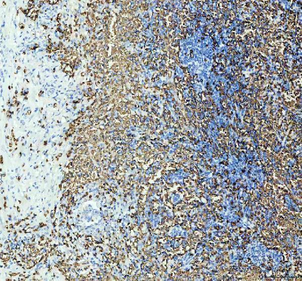
Real IHC data IHC analysis of GYPA using anti-GYPA antibody (A02184-2). GYPA was detected in paraffin-embedded section of human liver cancer tissues. Heat mediated antigen retrieval was performed in citrate buffer (pH6, epitope retrieval solution) for 20 mins. The tissue section was blocked with 10% goat serum. The tissue section was then incubated with 1μg/ml rabbit anti-GYPA Antibody (A02184-2) overnight at 4°C. Biotinylated goat anti-rabbit IgG was used as secondary antibody and incubated for 30 minutes at 37°C. The tissue section was developed using Strepavidin-Biotin-Complex (SABC)(Catalog # SA1022) with DAB as the chromogen.
Anti-Glycophorin A/GYPA Antibody ®
Cat # A02184-2
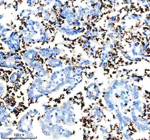
Real IHC data IHC analysis of Glycophorin A/GYPA using anti-Glycophorin A/GYPA antibody (M02184-1). Glycophorin A/GYPA was detected in a paraffin-embedded section of human lung cancer tissue. Heat mediated antigen retrieval was performed in EDTA buffer (pH 8.0, epitope retrieval solution). The tissue section was blocked with 10% goat serum. The tissue section was then incubated with 1:100 rabbit anti-Glycophorin A/GYPA Antibody (M02184-1) overnight at 4°C. Peroxidase Conjugated Goat Anti-rabbit IgG was used as secondary antibody and incubated for 30 minutes at 37°C. The tissue section was developed using HRP Conjugated Rabbit IgG Super Vision Assay Kit (Catalog # SV0002) with DAB as the chromogen.
Anti-Glycophorin A (CD235a) GYPA Monoclonal Antibody
Cat # M02184-1

A02184-2 has IHC images from human liver and renal cancer paraffin sections (A02184-2 IHC image captions). M02184-1 has IHC images from human lung and colon cancer paraffin sections (M02184-1 IHC image captions).

Which to pick: For tissue IHC, choose A02184-2 for the documented citrate pH 6 retrieval or M02184-1 for the documented EDTA pH 8 retrieval; both captions show paraffin sections, but neither reports the fixative (A02184-2 and M02184-1 IHC image captions). Neither SKU lists IF/ICC as a tested application or supplies an IF image, so neither has documented IF/ICC validation here (catalog applications and IF image fields). Neither has documented cross-species reactivity beyond human (catalog reactivity fields).

Each figure is that product's own IHC / IF validation image from its datasheet.

References

  1. UniProt Consortium. UniProt entry P02724 (GLPA_HUMAN, Glycophorin-A).
  2. Human Protein Atlas. GYPA tissue IHC expression (reliability: Enhanced).
  3. Human Protein Atlas. GYPA subcellular location (ICC-IF): Localized to the nucleoplasm, plasma membrane and cytosol..
  4. Human Protein Atlas. GYPA antibody validation summary (2 antibodies).
  5. Hypoxia-Induced Alpha-Globin Expression in Syncytiotrophoblasts Mimics the Pattern Observed in Preeclamptic Placentas. International journal of molecular sciences 2021 — PMC8036899.
  6. Molecular characterisation of transport mechanisms at the developing mouse blood-CSF interface: a transcriptome approach. PloS one 2012 — PMC3310074.
  7. A human cell atlas of fetal gene expression. Science (New York, N.Y.) 2020 — PMC7780123.
  8. Constructing immune and prognostic features associated with ADCP in hepatocellular carcinoma and pan-cancer based on scRNA-seq and bulk RNA-seq. Frontiers in immunology 2024 — PMC11106372.
  9. PubMed PMID:3456608 — UniProt-cited evidence.
  10. PubMed PMID:3196288 — UniProt-cited evidence.
  11. PubMed PMID:2734312 — UniProt-cited evidence.