This website uses cookies to ensure you get the best experience on our website.


- Table of Contents
Plan chromogenic GZMA IHC-P around the cytoplasmic tissue pattern (HPA tissue IHC), using human tonsil as a positive control (HPA tissue IHC; datasheet: PA1588 IHC-P image). Start the catalog antibody at 0.5–1 μg/mL (datasheet: PA1588) and compare staining with matched controls.

Expected localisation, validated protocols, controls and antibodies — the at-a-glance facts below, then the full design guide.
| Expected localisation | Cytoplasmic staining in tissue (HPA tissue IHC) | |
| Staining pattern | Cytoplasmic signal in several tissues, including immune cells (HPA tissue IHC) | |
| Antigen retrieval | Citrate pH 6.0 HIER, 95–98 °C, 20 min (rule: cytoplasmic / membrane antigen) | |
| Positive control | Appendix+4 more · see all | |
| Negative control | Adipose tissue+4 more · see all |
| Fixation | Matched tissue-IHC evidence does not establish the fixation claim. Validate the specimen-specific method before use. (selected-SKU IHC image PA1588) | |
| Caveat | Secreted GZMA may stain away from its RNA source (HPA tissue IHC) | |
| Regulation | Cytotoxic T/NK cell content affects signal (UniProt) | |
| Isoform / epitope | 2 isoforms; check epitope against mature chain 29–262 (UniProt) |
The catalog antibody’s IHC-P protocol is accompanied by published GZMA IHC protocols for salivary gland and premotor cortex sections (PMC10710186; PMC11555691).
| Sample | Tissue sections; selected-image fixative not specified (standard IHC workflow) |
| Fixation | Image fixative and duration unreported (datasheet PA1588); verify before use. |
| Sectioning | 4–5 µm sections on charged slides (standard) |
| Deparaffinisation | Xylene, graded ethanol series to water (standard) |
| Antigen retrieval | Heat-induced epitope retrieval in citrate buffer, pH 6.0, 20 min at 95–98 °C (standard rule: cytoplasmic / membrane antigen) |
| Peroxidase block | 3% H2O2, 10 min, room temperature (standard) |
| Blocking | 10% normal serum of the secondary host, 30 min, room temperature (standard) |
| Primary antibody | Rabbit anti-GZMA, 0.5-1μg/ml (datasheet PA1588) |
| Primary incubation | Overnight at 4 °C (standard) |
| Detection | HRP-polymer secondary, DAB chromogen 5–10 min (standard) |
| Counterstain | Hematoxylin, blue, dehydrate and mount (standard) |
| Expected result | GZMA-positive staining in lymphoid tissue of appendix (HPA tissue IHC: Medium). HPA tissue profile: Cytoplasmic expression in several tissues, including immune cells. No signal in the no-primary control. |
GZMA is a secreted protein stored in cytoplasmic granules of cytotoxic T cells and NK cells; it has no transmembrane segment (UniProt P12544). In tissue IHC, expect cytoplasmic staining among immune cells, with Medium staining reported in selected lymphoid and hematopoietic compartments (HPA tissue IHC). HPA rates the tissue staining reliability Enhanced, while noting low agreement between antibody staining and RNA expression because secreted protein can appear away from its site of production (HPA tissue IHC).
| Discrete cytoplasmic staining in immune cells of appendix lymphoid tissue, bone marrow hematopoietic cells, lymph node or tonsil germinal center cells, or splenic red pulp cells. | This matches the compartments where HPA reports Medium tissue staining (HPA tissue IHC). A compatible cell and compartment support the expected pattern, but morphology alone cannot establish that each stained cell is a cytotoxic T cell or NK cell (UniProt P12544; standard IHC interpretation). |
| Predominantly nuclear staining, or a crisp membrane outline instead of cytoplasmic staining. | Treat this as a pattern mismatch: HPA describes cytoplasmic tissue expression, and UniProt places GZMA in cytoplasmic granules or outside the cell, with no transmembrane segment (HPA tissue IHC; UniProt P12544). Check whether counterstain, precipitate, or nonspecific detection could explain the apparent compartment (standard IHC practice). |
| Strong signal in adipocytes or bronchial respiratory epithelial cells while the expected immune-cell pattern is absent. | HPA reports GZMA as Not detected in those listed cell types (HPA tissue IHC). Consider antibody cross-reactivity or endogenous chromogenic detection activity before assigning the signal to GZMA; compare the staining with a no-primary control (standard IHC practice). |
| Diffuse color across tissue, including cells expected to be negative, with little cell-specific contrast. | A widespread haze does not resemble HPA's cytoplasmic pattern in several tissues, including immune cells (HPA tissue IHC). Possible technical sources include insufficient blocking, excessive antibody concentration, or detection background; these are general IHC possibilities, not documented GZMA-specific effects (standard IHC practice). |
| No cytoplasmic signal in a section containing one of HPA's Medium-staining compartments. | A negative result there is inconclusive until section quality and detection are checked: HPA reports staining in the specified compartments, but does not establish that every specimen or cell will stain (HPA tissue IHC). Verify the anatomical compartment and assess a positive-control section and the antibody's IHC suitability (standard IHC practice; HPA antibodies). |
| Subcellular distribution | GZMA is stored in cytoplasmic granules, is secreted, and can be delivered into target cells by perforin (UniProt P12544). Interpret cell-associated cytoplasmic signal in its tissue context; extracellular signal alone cannot identify the producing cell (UniProt P12544; standard IHC interpretation). |
| Choice of tissue and compartment | HPA reports Medium staining in specified appendix, bone marrow, lymph node, spleen, and tonsil compartments, but Not detected staining in listed adipocytes and bronchial epithelium (HPA tissue IHC). Score the annotated cells within each tissue instead of treating the whole section as uniformly positive or negative (standard IHC practice). |
| Antibody validation | HPA054134 is IHC Approved; HPA076751 is IHC Enhanced, meaning its pattern is supported by at least two independent antibodies or orthogonal data (HPA antibodies). These labels inform confidence in an observed pattern; they do not prove that every signal in a new specimen is specific (HPA antibodies; standard IHC interpretation). |
| Processing and epitope uncertainty | UniProt lists a signal peptide at residues 1–26, a propeptide at 27–28, a mature chain at 29–262, and one glycosylation site at residue 170 (UniProt P12544). The payload gives no antibody epitope, so it cannot establish which processed form an IHC antibody recognizes (UniProt P12544; HPA antibodies). |
| Retrieval and detection | Follow the chosen IHC-validated antibody's documented paraffin-section workflow when available; optimize retrieval, antibody concentration, blocking, and chromogenic detection against controls as general IHC practice. Neither HPA tissue staining nor UniProt topology establishes a GZMA-specific fixation or retrieval effect (HPA tissue IHC; UniProt P12544; standard IHC practice). |
| IF/ICC Q: Can this IHC pattern define an IF/ICC protocol? | No ICC-IF images or main cellular location are available in the supplied HPA subcellular record, and neither listed HPA antibody has an ICC validation entry (HPA subcellular; HPA antibodies). The IHC tissue pattern supports an expectation of cytoplasmic signal, but does not establish IF/ICC conditions or validate an IF/ICC result (HPA tissue IHC; standard assay interpretation). |
| Situation | Likely cause | Next action |
|---|---|---|
| A proposed positive control has no staining. | The sampled area may lack the specific HPA-reported positive compartment, or the staining run may have failed; a negative section alone cannot distinguish these explanations (HPA tissue IHC; standard IHC practice). | Confirm that the section contains an HPA-listed Medium compartment, then review section integrity, the IHC-validated antibody instructions, and a working positive-control section (HPA tissue IHC; HPA antibodies; standard IHC practice). |
| Nuclei dominate the positive signal. | Predominantly nuclear staining conflicts with the reported cytoplasmic tissue pattern and cytoplasmic granule location (HPA tissue IHC; UniProt P12544). Counterstain or nonspecific detection may complicate interpretation (standard IHC practice). | Inspect the signal before and after counterstaining where feasible, compare with a no-primary control, and repeat using the chosen antibody's IHC conditions if the mismatch persists (standard IHC practice; HPA antibodies). |
| Adipocytes or bronchial respiratory epithelium stain strongly. | Those cell types are Not detected in the supplied HPA tissue record; cross-reactivity or endogenous chromogenic activity is possible (HPA tissue IHC; standard IHC practice). | Compare a no-primary control and a positive-compartment section; address endogenous detection activity and reassess antibody concentration if controls implicate technical staining (HPA tissue IHC; standard IHC practice). |
| The entire section shows weak, diffuse color. | Background from blocking, antibody concentration, washing, or chromogenic detection can obscure a cell-specific pattern (standard IHC practice). HPA's reported cytoplasmic staining does not establish which technical step caused the haze (HPA tissue IHC). | Check a no-primary control, then optimize blocking, washing, antibody concentration, and detection while preserving a positive tissue control for comparison (standard IHC practice; HPA tissue IHC). |
| Protein staining and local RNA expectations disagree. | HPA explicitly notes low consistency between antibody staining and RNA expression data because secreted protein may be located away from the tissue site of its RNA (HPA tissue IHC). | Record the stained cell type and compartment, compare them with HPA's tissue observations, and avoid calling the IHC result false solely from local RNA abundance (HPA tissue IHC; standard IHC interpretation). |
| Granular cytoplasmic staining is hard to distinguish from nearby extracellular color. | UniProt describes both cytoplasmic granule storage and secretion, so location alone may not identify the cell producing the protein (UniProt P12544). Chromogenic spread or background can further blur boundaries (standard IHC practice). | Use the counterstain to identify cell borders, compare negative and no-primary controls, and score convincing cell-associated cytoplasmic staining separately from extracellular color (UniProt P12544; standard IHC practice). |



Comprehensive Human Protein Atlas IHC scoring per tissue (reliability: Enhanced — Low consistency between antibody staining and RNA expression data. Secreted protein, tissue location of RNA and protein is expected to differ.). Rows are taken directly from the HPA tissue chart — click any row's HPA link to view the source.
| Tissue | Cell type | Level | Evidence | Source |
|---|---|---|---|---|
| Appendix | Lymphoid tissue | Medium | Protein (IHC) | HPA → |
| Bone marrow | Hematopoietic cells | Medium | Protein (IHC) | HPA → |
| Lymph node | Germinal center cells | Medium | Protein (IHC) | HPA → |
| Spleen | Cells in red pulp | Medium | Protein (IHC) | HPA → |
| Tonsil | Germinal center cells | Medium | Protein (IHC) | HPA → |
| Tissue | Cell type | Level | Evidence | Source |
|---|---|---|---|---|
| Adipose tissue | Adipocytes | Not detected | Protein (IHC) | HPA → |
| Breast | Adipocytes | Not detected | Protein (IHC) | HPA → |
| Bronchus | Respiratory epithelial cells | Not detected | Protein (IHC) | HPA → |
| Caudate | Glial cells | Not detected | Protein (IHC) | HPA → |
| Cerebellum | Cells in granular layer | Not detected | Protein (IHC) | HPA → |
Use compartment, cell identity, and section controls together when troubleshooting GZMA staining in chromogenic paraffin-section IHC.
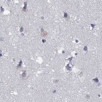
PA1588 is an anti-GZMA antibody with real human tonsil paraffin-section IHC data (PA1588 image caption: human tonsil IHC(P)); no IF figure is supplied (catalog: IF image alts empty).
PA1588 will render with a human tonsil IHC(P) figure (PA1588 image caption: human tonsil IHC(P)). Its listed applications are IHC and WB, and its listed reactivity is Human (catalog: PA1588 applications and reactivity).
Which to pick: Choose PA1588 for human paraffin-section IHC because its own figure shows human tonsil IHC(P); the fixative is unreported (PA1588 image caption: human tonsil IHC(P)). No listed SKU has IF/ICC validation or nonhuman reactivity (catalog: PA1588 applications IHC/WB; reactivity Human). Clonality is unreported, so it cannot guide selection.