This website uses cookies to ensure you get the best experience on our website.


- Table of Contents
Plan HAVCR2 IHC around cytoplasmic staining in a subset of lymphoid cells (HPA tissue IHC) and its expected membrane location (UniProt). This guide covers fixation, controls, and interpretation of paraffin-section staining.

Expected localisation, validated protocols, controls and antibodies — the at-a-glance facts below, then the full design guide.
| Expected localisation | Cytoplasm observed in some lymphoid cells (HPA tissue IHC); cell membrane expected (UniProt) | |
| Staining pattern | Subset of lymphoid cells with cytoplasmic staining (HPA tissue IHC) | |
| Antigen retrieval | Citrate pH 6.0 HIER, 95–98 °C, 20 min (rule: cytoplasmic / membrane antigen) | |
| Positive control | Kidney+3 more · see all | |
| Negative control | Adipose tissue+4 more · see all |
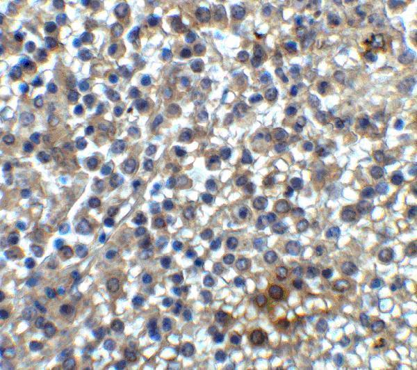
| Fixation | Keep formalin fixation consistent across sections (standard IHC practice; not target-specific); Matched tissue-IHC evidence does not establish the fixation claim. Validate the specimen-specific method before use. (selected-SKU IHC image A00657) | |
| Caveat | Presumed off-target staining may mimic positive cells (HPA tissue IHC) | |
| Regulation | Treg expression follows TCR stimulation (UniProt) | |
| Isoform / epitope | 2 isoforms; check epitope against extracellular/cytoplasmic topology (UniProt) |
The catalog antibody protocol is accompanied by published IHC methods for paraffin-embedded tissues (PMC9751455) and human breast cancer FFPE tissue (PMC13397360).
| Sample | Tissue sections; selected-image fixative not specified (standard IHC workflow) |
| Fixation | Image fixative and duration unreported (datasheet A00657); verify before use. |
| Sectioning | 4–5 µm sections on charged slides (standard) |
| Deparaffinisation | Xylene, graded ethanol series to water (standard) |
| Antigen retrieval | Heat-induced epitope retrieval in citrate buffer, pH 6.0, 20 min at 95–98 °C (standard rule: cytoplasmic / membrane antigen) |
| Peroxidase block | 3% H2O2, 10 min, room temperature (standard) |
| Blocking | 10% normal serum of the secondary host, 30 min, room temperature (standard) |
| Primary antibody | Rabbit anti-HAVCR2, 2 μg/mL (datasheet A00657) |
| Primary incubation | Overnight at 4 °C (standard) |
| Detection | HRP-polymer secondary, DAB chromogen 5–10 min (standard) |
| Counterstain | Hematoxylin, blue, dehydrate and mount (standard) |
| Expected result | HAVCR2-positive staining in proximal tubules (microvilli) of kidney (HPA tissue IHC: High). HPA tissue profile: Cytoplasmic expression in a subset of lymphoid cells. No signal in the no-primary control. |
HAVCR2 is a cell membrane protein with an extracellular region at residues 22–202 and a cytoplasmic region at 224–301 (UniProt Q8TDQ0 topology). In IHC, expect strong proximal-tubule microvillus staining in kidney and staining in a subset of lymphoid cells; HPA also describes cytoplasmic staining in those lymphoid cells (HPA tissue IHC). HPA rates its IHC evidence Enhanced, while noting medium agreement with RNA data and disregarded presumed off-target staining (HPA tissue IHC).
| Strong staining along kidney proximal-tubule microvilli, with scattered positive non-germinal-center lymphoid cells. | This fits HPA's High kidney microvillus signal and Medium lymph-node and tonsil signal (HPA tissue IHC). Assess each named cell population separately: the lymphoid pattern is a subset, so a positive section need not show every lymphoid cell stained (HPA tissue IHC). |
| Predominantly nuclear staining, especially where expected microvillus or lymphoid staining is absent. | A nuclear-dominant pattern conflicts with the reported membrane location and transmembrane topology (UniProt Q8TDQ0 topology; HPA subcellular). Review localisation against a positive kidney section and a no-primary control before scoring it as HAVCR2 (general IHC practice). HPA's reported cytoplasmic lymphoid staining should not be mistaken for nuclear staining (HPA tissue IHC). |
| Convincing staining in adipocytes or other cells HPA lists as not detected. | HPA reports adipocytes as Not detected in adipose tissue and breast (HPA tissue IHC). Check cell identification and control staining; cross-reactivity or detection-system activity can mimic a positive result (general IHC practice). HPA also reports presumed off-target binding in its overall reliability assessment (HPA tissue IHC). |
| Diffuse chromogen covers cells and extracellular areas without a readable cell pattern. | A uniform haze cannot establish the reported proximal-tubule or subset-of-lymphoid-cells pattern (HPA tissue IHC). Inspect a no-primary section for detection background and review blocking, washes and chromogen development before interpreting individual cells (general IHC practice). |
| No signal in an adequately sampled kidney section, including proximal-tubule microvilli. | This conflicts with HPA's High proximal-tubule microvillus staining (HPA tissue IHC). First assess section quality, tissue identification and assay controls, then review the antibody dilution, retrieval and detection steps against the laboratory's validated IHC procedure (general IHC practice). |
| Membrane topology and compartment | HAVCR2 spans residues 203–223, with extracellular and cytoplasmic portions on opposite sides of the membrane (UniProt Q8TDQ0 topology). The catalog antibody's epitope is not supplied here, so topology alone does not establish which portion it recognizes or which retrieval condition works. |
| Cell population and tissue choice | Kidney proximal-tubule microvilli are High; duodenal Paneth cells and lymph-node and tonsil non-germinal-center cells are Medium (HPA tissue IHC). Lung macrophages, small-intestinal Paneth cells and appendix non-germinal-center cells are listed as Low, making a faint result less decisive there (HPA tissue IHC). |
| Evidence and antibody validation | HPA calls the tissue IHC result Enhanced and lists CAB026003 as IHC Enhanced (HPA tissue IHC; HPA antibodies). Its reliability note also reports medium staining–RNA consistency and presumed off-target binding that was disregarded; unusual positives therefore need control review (HPA tissue IHC; general IHC practice). |
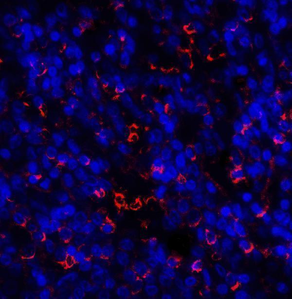
| IF/ICC Q&A: Where should signal appear? | HPA's subcellular summary is Membrane, and UniProt lists cell membrane and cell junction, including the immunological synapse between CD8+ T cells and targets (HPA subcellular; UniProt Q8TDQ0 subcellular). HPA supplies no ICC-IF cell-line images here, so this record cannot confirm a cell-line-specific IF pattern (HPA subcellular). |
| Protein forms and modifications | UniProt lists two isoforms, a cleaved signal peptide at residues 1–21, and glycosylation sites at 145 and 172 (UniProt Q8TDQ0). Without epitope information, these annotations do not establish isoform discrimination, staining intensity or a HAVCR2-specific retrieval effect. |
| Situation | Likely cause | Next action |
|---|---|---|
| Kidney proximal tubules are blank. | The result disagrees with HPA's High microvillus staining; the specific failed assay step cannot be inferred from the image alone (HPA tissue IHC; general IHC practice). | Confirm proximal tubules and tissue integrity, then compare a positive control and review antibody dilution, retrieval, incubation and detection against the validated IHC procedure (general IHC practice). No HAVCR2-specific retrieval response is supplied. |
| Lymph node or tonsil looks negative at low magnification. | HPA places the Medium signal in non-germinal-center cells and describes expression in a subset of lymphoid cells, so sparse positives can be missed (HPA tissue IHC). | Inspect representative non-germinal-center areas at higher magnification and score cells individually; use the kidney microvillus pattern as a separate reference for assay performance (HPA tissue IHC; general IHC practice). |
| Nuclei carry the strongest signal. | Nuclear-dominant staining does not fit the membrane localisation or topology reported for HAVCR2 (HPA subcellular; UniProt Q8TDQ0 topology). Background or nonspecific detection is a possible assay explanation (general IHC practice). | Compare with a no-primary section, reassess counterstain versus chromogen, and verify whether the expected kidney or lymphoid compartments stain (general IHC practice; HPA tissue IHC). |
| Adipocytes stain strongly. | HPA reports adipocytes as Not detected in adipose tissue and breast; its reliability note also flags presumed off-target binding (HPA tissue IHC). Endogenous detection activity is another possible explanation (general IHC practice). | Verify the stained cell type, examine a no-primary control and review blocking and detection controls before assigning HAVCR2 positivity (general IHC practice). |
| Colour spreads across most of the section. | A widespread, poorly localised signal does not match HPA's specified microvillus and subset-of-lymphoid-cells observations (HPA tissue IHC). Excess detection background or endogenous activity can produce this appearance (general IHC practice). | Check a no-primary control, review endogenous-enzyme blocking where applicable, and assess washes and chromogen development; interpret target staining only after cellular boundaries are readable (general IHC practice). |


Comprehensive Human Protein Atlas IHC scoring per tissue (reliability: Enhanced — Medium consistency between antibody staining and RNA expression data. Presumed off target binding observed and disregarded.). Rows are taken directly from the HPA tissue chart — click any row's HPA link to view the source.
| Tissue | Cell type | Level | Evidence | Source |
|---|---|---|---|---|
| Kidney | Proximal tubules (microvilli) | High | Protein (IHC) | HPA → |
| Duodenum | Paneth cells | Medium | Protein (IHC) | HPA → |
| Lymph node | Non-germinal center cells | Medium | Protein (IHC) | HPA → |
| Tonsil | Non-germinal center cells | Medium | Protein (IHC) | HPA → |
| Tissue | Cell type | Level | Evidence | Source |
|---|---|---|---|---|
| Adipose tissue | Adipocytes | Not detected | Protein (IHC) | HPA → |
| Adrenal gland | Glandular cells | Not detected | Protein (IHC) | HPA → |
| Bone marrow | Hematopoietic cells | Not detected | Protein (IHC) | HPA → |
| Breast | Adipocytes | Not detected | Protein (IHC) | HPA → |
| Bronchus | Respiratory epithelial cells | Not detected | Protein (IHC) | HPA → |
Troubleshoot HAVCR2 chromogenic IHC in paraffin sections by checking retrieval, cell identity, staining compartment, and assay controls before interpreting signal.
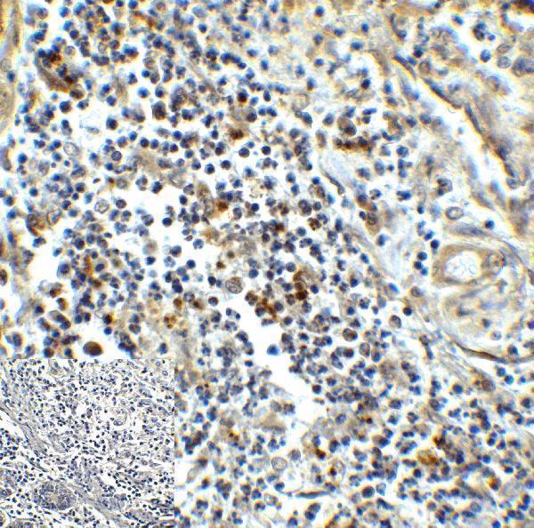
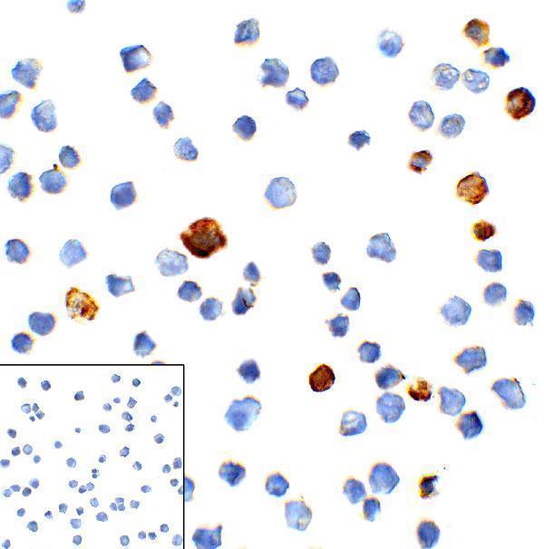
The catalog includes human-reactive anti-HAVCR2 antibodies with IHC images from spleen and colon carcinoma tissue and IF images from spleen tissue and transfected HEK293 cells (catalog reactivity and image captions).
A00657 has an IHC-P image from human spleen at 2 μg/mL and an IF image from human spleen (catalog applications and image captions). M00657 has an IHC-P image from human colon carcinoma at 5 μg/mL, plus IF and ICC images from transfected HEK293 cells (catalog applications and image captions).
Which to pick: For paraffin-section IHC, choose A00657 for human spleen or M00657 for human colon carcinoma: each has its own IHC-P listing and tissue image, and neither image caption reports the fixative (catalog applications and respective IHC image captions). For IF or ICC, M00657 is the mouse monoclonal with both applications listed; A00657 is a rabbit antibody with IF listed and a human spleen IF image (catalog host, clone, applications and IF image caption). For work spanning human, mouse and rat, consider A00657-5, a rabbit polyclonal listed for IHC and IF in those species; the payload provides no IHC or IF image for it (catalog reactivity, applications, dilution_raw and image captions).