This website uses cookies to ensure you get the best experience on our website.


- Table of Contents
Use bone marrow erythrocyte staining as a reference for chromogenic HBA1 IHC in paraffin sections (HPA tissue IHC). The guide covers staining controls and interpretation where the catalog antibody also recognizes HBA2 (catalog A00233-1).

Expected localisation, validated protocols, controls and antibodies — the at-a-glance facts below, then the full design guide.
| Expected localisation | Erythrocyte positivity; intracellular compartment unassigned (HPA tissue IHC) | |
| Staining pattern | Distinct erythrocyte staining in bone marrow (HPA tissue IHC) | |
| Antigen retrieval | EDTA pH 8.0 HIER, heat-mediated (datasheet A00233-1) | |
| Positive control | Bone marrow+1 more · see all | |
| Negative control | Adipose tissue+4 more · see all |
| Fixation | Keep fixation consistent across sections (standard IHC practice; not target-specific); target-specific effects are unknown. | |
| Caveat | HBA2 recognition may confound HBA1 attribution (catalog A00233-1; HPA tissue IHC) | |
| Regulation | Staining intensity reflects erythrocyte content (UniProt; HPA tissue IHC) | |
| Isoform / epitope | 0 isoforms reported; mature chain spans residues 2–142 (UniProt) |
Compare the catalog antibody’s IHC-P protocol (datasheet: A00233-1) with two published chromogenic IHC protocols (PMC7753232; PMC3547883).
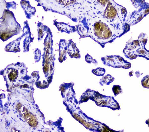
| Sample | Paraffin-embedded human placenta tissue; fixative not specified (datasheet A00233-1) |
| Fixation | Image fixative and duration unreported (datasheet A00233-1); verify before use. |
| Sectioning | 4–5 µm sections on charged slides (standard) |
| Deparaffinisation | Xylene, graded ethanol series to water (standard) |
| Antigen retrieval | Heat retrieval: EDTA pH 8.0 (datasheet A00233-1); 20 min, 95–100 °C (standard) |
| Peroxidase block | 3% H2O2, 10 min, room temperature (standard) |
| Blocking | 10% goat serum (datasheet A00233-1) |
| Primary antibody | Rabbit anti-HBA1, 2-5μg/ml (datasheet A00233-1) |
| Primary incubation | Overnight at 4 °C (datasheet A00233-1) |
| Detection | HRP-conjugated secondary, DAB chromogen (datasheet A00233-1) |
| Counterstain | Hematoxylin, blue, dehydrate and mount (standard) |
| Expected result | HBA1-positive staining in hematopoietic cells of bone marrow (HPA tissue IHC: High). HPA tissue profile: Selective expression in bone marrow with distinct positivity in erythrocytes. No signal in the no-primary control. |
HBA1 is associated with red blood cells (UniProt P69905: tissue specificity). In paraffin sections, expect intracellular staining of erythrocytes, particularly in bone marrow, and staining in splenic red pulp (HPA: distinct erythrocyte positivity in bone marrow; High in red pulp cells). UniProt does not annotate a subcellular location, but reports no transmembrane segment (UniProt P69905: topology). HPA rates the tissue pattern Enhanced while cautioning that staining may reflect protein from more than one gene (HPA: reliability description).
| Strong staining outlines the contents of erythrocytes in bone marrow; nearby hematopoietic cells may also stain. | This fits the reported marrow pattern: High staining in hematopoietic cells with distinct erythrocyte positivity (HPA: bone marrow tissue IHC). Score the cell population and distribution together; color intensity alone cannot establish which alpha-globin gene supplied the protein (HPA: reliability description). |
| Cells in splenic red pulp stain strongly, while adipocytes in adipose tissue lack comparable staining. | The contrast agrees with High staining in splenic red pulp cells and Not detected staining in adipocytes (HPA: spleen and adipose tissue IHC). Red pulp staining is supportive context, but its cell identity should be checked morphologically before assigning a result to erythrocytes (HPA: red pulp cell designation; general IHC practice). |
| A crisp nuclear, membrane-only, or extracellular signal dominates where erythrocytes are present. | Treat the compartment assignment as suspect: hemoglobin alpha is a red-blood-cell protein with no annotated membrane-spanning segment or secretory signal (UniProt P69905: tissue specificity, topology, processing). Review morphology and controls before reporting localization; UniProt does not provide a formal subcellular annotation for HBA1 (UniProt P69905: subcellular location). |
| Adrenal glandular cells, bronchial respiratory epithelium, or adipocytes stain prominently. | Those cell types are reported as Not detected (HPA: adrenal gland, bronchus, adipose tissue IHC). Check for antibody cross-reactivity and detection-system activity with appropriate controls (general IHC practice). Do not call such staining HBA1-specific on appearance alone, especially given HPA's more-than-one-gene caution (HPA: reliability description). |
| Brown color spreads across the section, or bone marrow has no identifiable positive erythrocytes. | Widespread color obscures the reported cell pattern; an absent marrow signal conflicts with its High staining and distinct erythrocyte positivity (HPA: bone marrow tissue IHC). Interpret either result only after checking section quality, detection controls, and whether erythrocytes are actually present in the sampled area (general IHC practice). |
| Tissue and cell selection | Bone marrow offers distinct erythrocyte positivity, and spleen offers High red pulp staining (HPA: tissue IHC). Adipocytes and several listed glandular or epithelial cell types are Not detected comparators (HPA: negative tissues). Compare identified cells within each section rather than treating all tissue-wide color as target signal (general IHC practice). |
| Location and protein processing | The annotated chain spans residues 2–142, with no signal peptide, propeptide, or transmembrane segment (UniProt P69905: processing, topology). Those annotations support examining intracellular erythrocyte contents; they do not supply a validated microscopic subcellular location or establish how fixation affects staining (UniProt P69905: subcellular location; HPA: tissue IHC). |
| Antibody interpretation | HPA lists CAB038417 as IHC Enhanced and HPA043780 as IHC Supported (HPA: antibody validation). HPA also cautions that its tissue staining targets protein from more than one gene (HPA: reliability description). A matching pattern supports an alpha-globin-associated result, but morphology and controls cannot by themselves prove gene-specific HBA1 detection (HPA: reliability description; general IHC practice). |
| Molecular variation | UniProt lists 0 isoforms and multiple modified or glycosylated sites (UniProt P69905: isoforms, modifications, glycosylation). No antibody epitope or target-specific fixation study is supplied, so these annotations cannot predict a particular retrieval requirement, staining loss, or alternate compartment (UniProt P69905: annotations; HPA: supplied tissue and antibody records). |
| Chromogenic detection background | Erythrocyte-rich areas require careful review of chromogen controls because endogenous activity or background color can complicate peroxidase-based IHC interpretation (general IHC practice). Evaluate staining against a no-primary control and the expected cell distribution; the supplied HPA tissue pattern does not establish the source of background in an individual section (HPA: tissue IHC). |
| Situation | Likely cause | Next action |
|---|---|---|
| Bone marrow is blank despite visible erythrocytes. | The observed result conflicts with HPA's distinct, High marrow erythrocyte-associated pattern (HPA: bone marrow tissue IHC); the cause is not determined by that record. | Confirm the section contains intact erythrocytes, then check the catalog antibody's stated IHC-P conditions, detection reagents, and a positive control (general IHC practice). Do not infer an HBA1-specific fixation defect without evidence. |
| Splenic red pulp is positive, but the stained cells are unclear. | HPA reports High staining in red pulp cells without assigning every stained cell to a named lineage (HPA: spleen tissue IHC). | Identify cells from morphology and counterstain before scoring them; compare the distribution with a marrow section showing distinct erythrocyte positivity (HPA: bone marrow tissue IHC; general IHC practice). |
| Nuclei or tissue edges carry the strongest color. | That pattern does not match an intracellular erythrocyte-centered reading of the reported tissue distribution (HPA: bone marrow tissue IHC; UniProt P69905: red-blood-cell specificity). | Inspect a no-primary control and section edges, then adjust detection or blocking using ordinary IHC controls if background persists (general IHC practice). Avoid assigning HBA1 a nuclear location from this result. |
| Adipocytes or respiratory epithelial cells stain strongly. | Those cells are Not detected in the supplied HPA tissue IHC data (HPA: adipose tissue and bronchus). Cross-reactivity or detection background is possible (general IHC practice). | Compare matched negative tissue and no-primary controls, then reassess antibody specificity. HPA warns that the tissue stain may recognize protein from more than one gene (HPA: reliability description). |
| A broad brown haze prevents cell-level scoring. | Diffuse color cannot be matched reliably to the distinct marrow erythrocyte pattern (HPA: bone marrow tissue IHC); its source needs experimental controls (general IHC practice). | Check no-primary and detection-only controls, blocking, washing, and chromogen development, then score only resolvable cells (general IHC practice). Do not interpret haze as evidence of widespread HBA1 expression. |
| Can this IHC-P pattern be used as an ICC/IF localization result? | HPA supplies no ICC-IF images or main subcellular location for HBA1, and neither listed antibody has an ICC validation entry (HPA: subcellular and antibody records). | Use the separate IF/ICC guide for that application. Treat the paraffin-section tissue pattern as IHC evidence only; it does not establish an ICC/IF compartment or an IF protocol (HPA: tissue, subcellular, and antibody records). |


Comprehensive Human Protein Atlas IHC scoring per tissue (reliability: Enhanced — High consistency between antibody staining and RNA expression data. Caution, targets protein from more than one gene.). Rows are taken directly from the HPA tissue chart — click any row's HPA link to view the source.
| Tissue | Cell type | Level | Evidence | Source |
|---|---|---|---|---|
| Bone marrow | Hematopoietic cells | High | Protein (IHC) | HPA → |
| Spleen | Cells in red pulp | High | Protein (IHC) | HPA → |
| Tissue | Cell type | Level | Evidence | Source |
|---|---|---|---|---|
| Adipose tissue | Adipocytes | Not detected | Protein (IHC) | HPA → |
| Adrenal gland | Glandular cells | Not detected | Protein (IHC) | HPA → |
| Appendix | Glandular cells | Not detected | Protein (IHC) | HPA → |
| Breast | Adipocytes | Not detected | Protein (IHC) | HPA → |
| Bronchus | Respiratory epithelial cells | Not detected | Protein (IHC) | HPA → |
Troubleshoot HBA1 staining in paraffin sections by checking retrieval, cell identity, assay controls, and the specificity limits of hemoglobin antibodies.
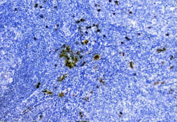
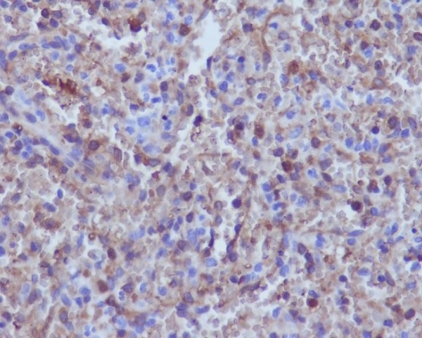
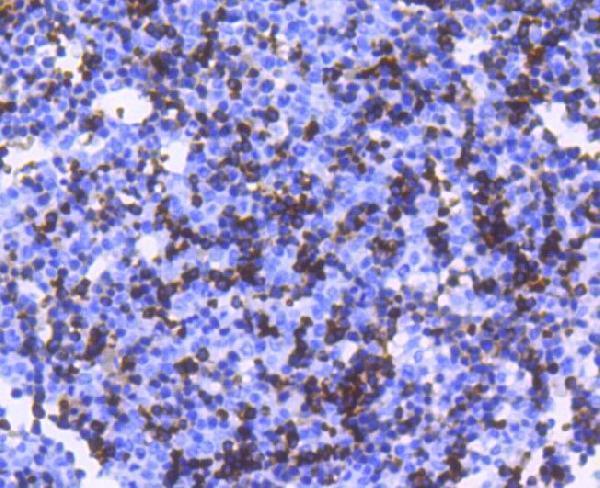
IHC images show paraffin sections of human placenta and tonsil for A00233-1, and human spleen and mouse embryo for M00233 (catalog IHC captions). Neither has IF imagery (catalog IF image fields).
A00233-1 lists human IHC and shows staining in paraffin sections of human placenta and tonsil (catalog applications, reactivity and IHC captions). M00233 lists IHC with Human, Mouse and Rat reactivity, and shows staining in paraffin sections of human spleen and mouse embryo (catalog applications, reactivity and IHC captions).
Which to pick: For human paraffin-section IHC, choose A00233-1 when its polyclonal Hemoglobin/HBA1/HBA2 target scope suits the experiment; its images show human placenta and tonsil, and the fixative is unreported (catalog title and dilution_raw; A00233-1 IHC captions). For HBA1-focused or cross-species tissue IHC, choose monoclonal M00233, clone EGI-8: it lists Human, Mouse and Rat reactivity, while its paraffin-section images show human spleen and mouse embryo only; the fixative is unreported (catalog title, clone, reactivity and M00233 IHC captions). For IF/ICC, neither SKU has a listed IF/ICC application or IF image, so neither is supported for that use by this catalog evidence (catalog applications and IF image fields).