This website uses cookies to ensure you get the best experience on our website.
- Table of Contents
2 Citations 6 Q&As
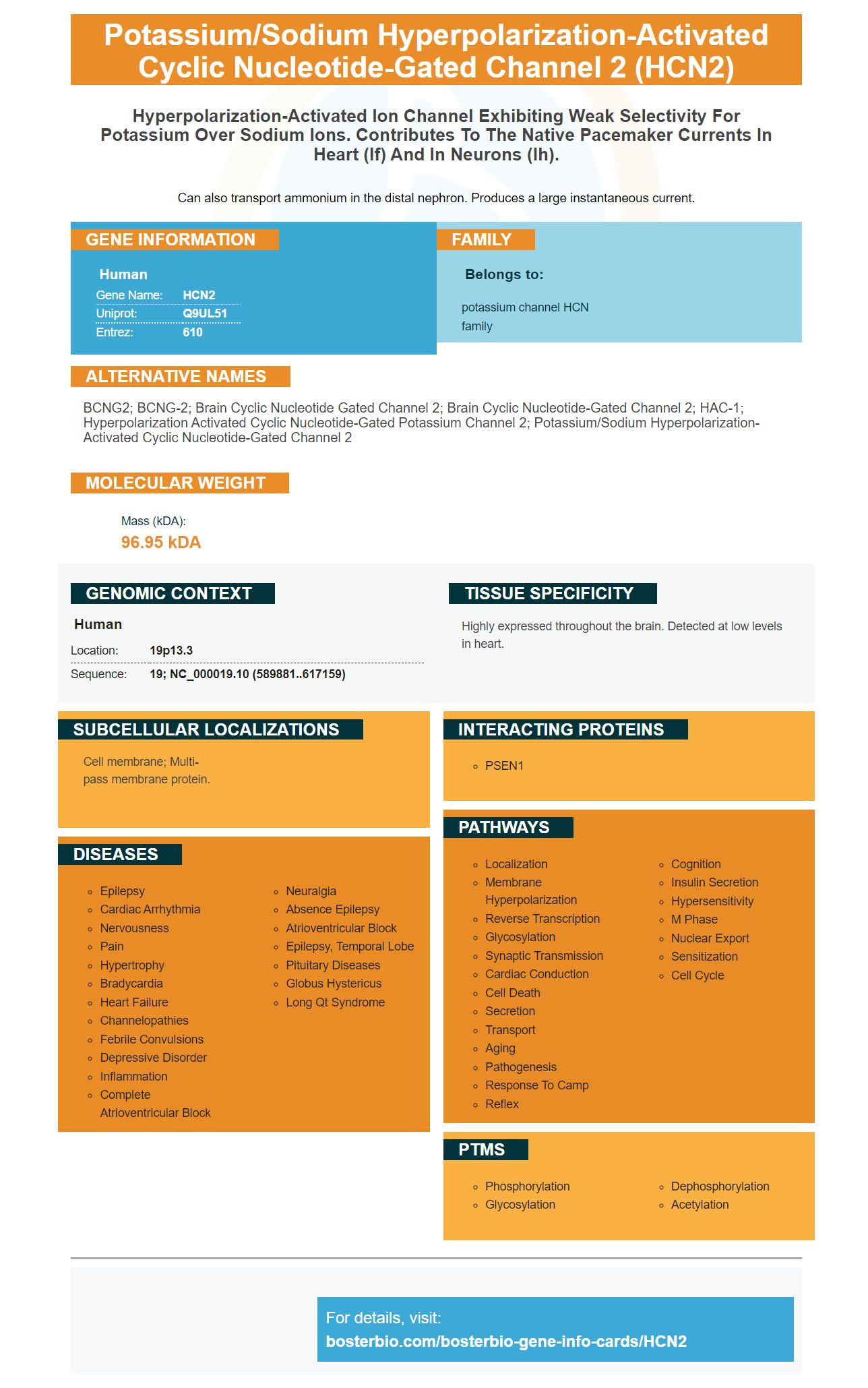
Facts about Potassium/sodium hyperpolarization-activated cyclic nucleotide-gated channel 2.
Can also transport ammonium in the distal nephron. Produces a large instantaneous current.
Human | |
---|---|
Gene Name: | HCN2 |
Uniprot: | Q9UL51 |
Entrez: | 610 |
Belongs to: |
---|
potassium channel HCN family |
BCNG2; BCNG-2; brain cyclic nucleotide gated channel 2; brain cyclic nucleotide-gated channel 2; HAC-1; hyperpolarization activated cyclic nucleotide-gated potassium channel 2; potassium/sodium hyperpolarization-activated cyclic nucleotide-gated channel 2
Mass (kDA):
96.95 kDA
Human | |
---|---|
Location: | 19p13.3 |
Sequence: | 19; NC_000019.10 (589881..617159) |
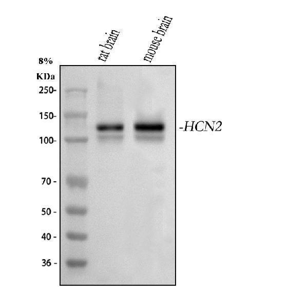
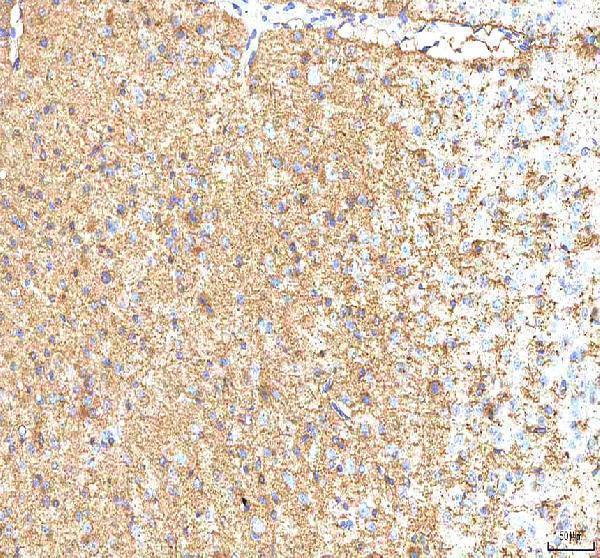
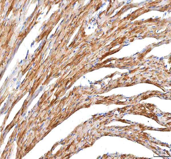
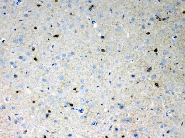
Highly expressed throughout the brain. Detected at low levels in heart.
Cell membrane; Multi-pass membrane protein.
If you are researching the best primary antibodies against HCN2, you'll be interested in Boster Bio. Their antibody catalog is filled with high-affinity antibodies that have been validated and have been cited in numerous publications over the past 25 years. Boster's antibodies have been validated for use in Western Blotting, Immunohistochemistry, and ELISA. Learn more about Boster's products to learn more about its use in research and its applications in the biological sciences.
High-affinity primary antibodies are highly effective tools for detecting protein targets in tissues and cells. There are a number of protocols for preparing tissue samples for antigenicity testing. The fixation process may alter epitope accessibility or nature, and thus should be performed in an in-vitro setting. Additionally, KO animal tissues are particularly valuable for binding assays.
The N423 project produced a series of candidate mAbs with varying IgG concentrations. Many of these candidates showed monospecific immunoreactivity against the target band. Based on other assays, N423/75 was chosen as the NeuroMab. Despite the difficulties associated with designing such antibodies, Boster Bio is confident of their high-affinity primary antibodies against the HCN2 marker.
In addition to its role in sensory processing, HCN2 may also contribute to the modulation of the membrane excitability of SP-containing primary afferent terminals. In this regard, it may play a role in the fine-tuning of pain transmission. Currently, there are no therapeutic drugs targeting this HCN2 receptor. However, new studies may shed light on this issue. This study provides new insights into the mechanism of action of HCN2 in the central nervous system.
The peripheral expression of HCN2 is critical for regulating pain and mechanical hypersensitivity in diabetes. Immunohistochemistry studies of lumbar DRG sections have revealed that this marker colocalizes with b3-tubulin in the majority of neurons. This finding suggests that HCN2 is expressed in both small and large diameter neurons. Furthermore, colocalization of this marker with membrane ring staining has been suggested to reveal its function in peripheral neurons.
Furthermore, the HCN2 marker may also be useful in identifying glia with a low number of HCN channels. Similarly, HCN channels are implicated in the regulation of intracellular calcium concentration. In glial cells, the HCN channel is associated with neuroinflammation. In addition, HCN channels play an important role in the secretion of cytokines and reactive oxygen species. Several researches have shown that selective HCN blockers like ivabradine can significantly improve the recovery of patients following brain injury.
The HCN2 marker is localized to the basolateral membranes of principal and intercalated cells that secrete acid. It also appears in other tissues, such as the kidney and the spleen, and has a variety of functions. However, its location in the cell is not well understood. The presence of this marker in these tissues could suggest possible biological functions. Therefore, it is crucial to investigate the sources of this marker to better understand its biological roles.
The HCN2 marker is expressed in various parts of the brain, including the hippocampal region. The levels of immunolabeling were highest in the ventricular zone, while the subunits of HCN1 and HCN2 were found in different cell types. HCN2 expression in pyramidal cells is correlated with functional integration of dendritic fibers. Moreover, the HCN subunit immunolabeling is cell type-specific, suggesting that the HCN channel subunit is expressed specifically in the brain.
Although the HCN1 isoform is more abundant in human atria than in the SAN, HCN2 is more abundant in SAN tissue. The relative abundance of HCN4 in human SAN tissue is 4-6 times greater than in the surrounding right atrial myocardium. However, recent immunolabeling studies suggest that HCN4 cannot be used as an exclusive marker for mapping the human SAN.
PMID: 10524219 by Vaccari T., et al. The human gene coding for HCN2, a pacemaker channel of the heart.
PMID: 10228147 by Ludwig A., et al. Two pacemaker channels from human heart with profoundly different activation kinetics.
*More publications can be found for each product on its corresponding product page