HDAC1 / Histone deacetylase 1 · Western blot design guide

Design a Western Blot for HDAC1

Real validated HDAC1 Western blot protocols, expected-band and isoform facts, troubleshooting for weak or shifted signal, and recommended anti-HDAC1 WB antibodies. Everything you need to plan the experiment before you commit precious samples.

Evidence assembled September 2026 · For research use; verify linked source records and product datasheet before use
Western blot protocol sheet for HDAC1: expected band ~55.1 kDa, hero antibody A00256-4, catalog values and labelled standard workflow; separate PMC comparisons on the guide
Printable HDAC1 Western blot protocol sheet — expected band ~55.1 kDa, antibody A00256-4, controls and PMC citations. Open the full HDAC1 WB guide →

HDAC1 Western Blot Experimental Design Guide

Expected bands, validated protocols, controls and antibodies — the at-a-glance facts below, then the full design guide.

Must know before running
Expected band ~55.1 kDa
Observed band ~65 kDa
Gel 5–20% (catalog A00256-4)
Positive control ⓘ Appendix (IHC candidate; verify WB) +4 more
Negative control ⓘ Adrenal gland (IHC candidate; verify WB)
Important caveats
Reasons your observed band may differ from the expected size.
PTM Phosphorylated + Acetylated
Caveat Modification-state controls
Gene-set association MSigDB Hallmark membership
Isoform 1 isoform(s)
Section 1

Real Curated HDAC1 Western Blot Protocols

The A00256-4 protocol combines labelled catalog values with standard starting conditions. Published comparisons retain their own sample, reagent and detection scope.

Recommended Western blot protocol parameters
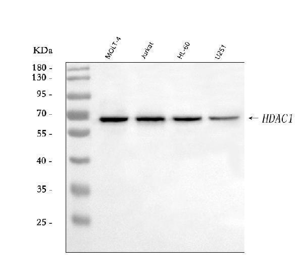
Sample / lysatehuman MOLT-4, human Jurkat, human HL-60 (catalog A00256-4)
Gel %5–20% (catalog A00256-4)
Load30 ug; reducing conditions (catalog A00256-4)
Transfera nitrocellulose membrane at 150 mA for 50-90 minutes (catalog A00256-4)
Membranenitrocellulose membrane (catalog A00256-4)
Blocking5% non-fat milk/TBS for 1.5 hour at RT (catalog A00256-4)
Primary antibodyA00256-4 · 0.5 μg/mL (catalog A00256-4)
Primary incubationovernight at 4°C (catalog A00256-4)
Secondary antibodygoat anti-rabbit IgG-HRP, 1:5000 (catalog A00256-4)
Secondary incubation1.5 hour at RT (catalog A00256-4)
WashTBS-0.1%Tween 3 times with 5 minutes each (catalog A00256-4)
DetectionECL (catalog A00256-4)
Section 2

What Is the Expected HDAC1 Western Blot Band Size?

HDAC1 has a predicted mass of 55.1 kDa and an observed band near 65 kDa; the cause of the difference is not established.

What am I looking at on my blot?
Band near 65 kDaEmpirical HDAC1 band in reducing whole-cell lysates; identity should be confirmed with controls
Band near 55.1 kDaNear the UniProt predicted mass of HDAC1; identity requires confirmation
Little or no band in whole-cell lysateNuclear HDAC1 may be poorly recovered or underrepresented in this preparation
Additional nearby bandsIdentity and any contribution from documented modifications require testing
💡Expected HDAC1 appearanceUniProt predicts 55.1 kDa for HDAC1, while antibody QC shows a band near 65 kDa in reducing whole-cell lysates; confirm band identity with appropriate controls because the cause of the difference is unestablished.
How each factor affects band size
UniProt predicted mass55.1 kDa for the 482-residue HDAC1 sequence; the empirical band is near 65 kDa
Phosphoserines at residues 393, 406, 409, and 421Documented modifications, but their effect on migration is not established
N6-acetyllysines at residues 74 and 220Documented modifications, but their effect on migration is not established
S-nitrosocysteines at residues 261 and 273Documented modifications, but their effect on migration is not established
Why is my band missing or off?
SituationLikely causeNext action
No band in lysateNuclear HDAC1 may be poorly recoveredCheck nuclear extraction and a nuclear loading marker
Band higher than expectedThe observed HDAC1 band is near 65 kDa versus a predicted 55.1 kDa; the cause is unestablishedCompare with the antibody QC band and verify identity by HDAC1 depletion
Band lower than expectedBand identity is uncertain; no cleavage feature is suppliedVerify specificity by HDAC1 depletion and compare with the 65 kDa QC band
Multiple bandsAdditional bands cannot be assigned to isoforms or modifications from these dataUse HDAC1 depletion to identify the specific band
Weak or no signalLow HDAC1 recovery or insufficient detectionCheck nuclear protein recovery, loading, and antibody performance
Fragments below expected sizePossible sample degradation, with no documented HDAC1 cleavage product suppliedCheck sample integrity and test whether fragments track with HDAC1 depletion

Sample controls for HDAC1 Western blot

🧪HPA-IHC candidate guidance (verify in WB): For positive controls for HDAC1 in Western blot, you can use appendix tissue, which has high HPA expression.
Positive control: Appendix (IHC candidate; verify WB)
Negative control: Adrenal gland (IHC candidate; verify WB)
Loading controls: Run GAPDH, β-actin, and a total-protein stain such as Ponceau alongside the samples.
⚠️Feasibility: HDAC1 is nuclear, so a nuclear extract may improve detection; HPA lists adrenal gland as a potential negative tissue.

HPA tissue expression evidence for HDAC1

Comprehensive Human Protein Atlas IHC scoring per tissue. Rows are taken directly from the HPA tissue chart — click any row's HPA link to view the source.

Higher expression tissues · candidate positive controls from IHC

TissueCell typeLevelEvidenceSource
Appendix glandular cells High Protein (IHC) HPA →
Bone marrow hematopoietic cells High Protein (IHC) HPA →
Bronchus respiratory epithelial cells High Protein (IHC) HPA →
Cervix squamous epithelial cells High Protein (IHC) HPA →
Colon glandular cells High Protein (IHC) HPA →

Lower expression tissues · IHC evidence, not confirmed WB-negative controls

TissueCell typeLevelEvidenceSource
Adrenal gland glandular cells Not detected Protein (IHC) HPA →
Cerebellum cells in granular layer Not detected Protein (IHC) HPA →
Hippocampus glial cells Not detected Protein (IHC) HPA →
Skeletal muscle myocytes Not detected Protein (IHC) HPA →
Adipose tissue adipocytes Low Protein (IHC) HPA →
Section 3

Advanced HDAC1 Western Blot Tips

Deeper troubleshooting and optimisation questions for HDAC1, answered from its protein features.

How should HDAC1 band migration be interpreted?
Band shift · Use the separately labelled calculated mass and catalog-observed evidence above. A sequence annotation does not establish an observed migration shift. Verify target identity with orthogonal controls.
Could HDAC1 isoforms explain multiple bands?
Isoforms · The supplied UniProt record lists one isoform and no alternative sequence. It therefore provides no isoform-based explanation for multiple bands; verify any additional bands independently.
Which HDAC1 phosphorylation sites matter when interpreting bands?
PTM · UniProt lists phosphoserines at positions 393, 406, 409, 421 and 423; CK2 is specified for 421 and 423. These are UniProt coordinates, which may differ from antibody or paper numbering. Their annotation does not establish a visible band shift.

UniProt lists acetyllysine at 74 (alternate) and 220, S-nitrosocysteine at 261 and 273, and EHMT2-mediated methyllysine at 432. Use UniProt coordinates when comparing site-specific reagents; numbering in papers or antibody descriptions may differ. These annotations alone do not identify an unexpected band.

The supplied record lists no glycosylation sites, signal peptide or propeptide. It provides no basis to attribute an unexpected band to glycosylation or precursor processing.
Does this guide establish induction of HDAC1?
Induction · No general induction response is established by this guide. A pathway or gene-set association is not evidence of induction in a particular specimen. Verify the relevant treatment and control in a target-specific experiment.
How should transfer be checked for HDAC1?
Transfer · Standard workflow guidance: verify transfer efficiency for the intended target size before interpreting a weak signal. Use total-protein assessment and optimize transfer for the membrane, gel and apparatus; the labelled catalog values take precedence.
How should blocking be optimized?
Blocking · Standard workflow guidance: follow the A00256-4 datasheet where specified. Otherwise compare 5% milk or 5% BSA in TBST; for a phospho-specific assay start with BSA. Optimize background and specific signal with matched controls.
How should I quantify HDAC1 in cell fractions?
Quantitation · HDAC1 is annotated in the nucleus. Quantify the band in a consistently prepared nuclear fraction or clearly define the fraction used across samples. The record does not establish that a band at any particular apparent mass is specific to HDAC1.
Why does HDAC1 appear near 65 kDa instead of 55.1 kDa?
Interpretation · The supplied observed band is approximately 65 kDa, while the predicted mass is 55.1 kDa. UniProt lists several modified residues, but their presence alone does not establish the cause of this difference. Compare the band with an appropriate HDAC1 control before assigning it.

HDAC1 belongs to a core complex with HDAC2, RBBP4 and RBBP7 and associates with the SIN3 complex. Complex membership identifies possible biological partners but does not, by itself, assign extra Western-blot bands to HDAC1 or its partners.
Boster reagents

HDAC1 Western Blot Antibodies

Catalog antibodies with Western blot application and product-specific WB images. Evaluate suitability with the reported sample, controls and experimental conditions.

Real WB data Western blot analysis of HDAC1 using anti-HDAC1 antibody (A00256-4). Electrophoresis was performed on a 5-20% SDS-PAGE gel at 70V (Stacking gel) / 90V (Resolving gel) for 2-3 hours. The sample well of each lane was loaded with 30 ug of sample under reducing conditions. Lane 1: human MOLT-4 whole cell lysates, Lane 2: human Jurkat whole cell lysates, Lane 3: human HL-60 whole cell lysates, Lane 4: human U251 whole cell lysates. After electrophoresis, proteins were transferred to a nitrocellulose membrane at 150 mA for 50-90 minutes. Blocked the membrane with 5% non-fat milk/TBS for 1.5 hour at RT. The membrane was incubated with rabbit anti-HDAC1 antigen affinity purified polyclonal antibody (Catalog # A00256-4) at 0.5 μg/mL overnight at 4°C, then washed with TBS-0.1%Tween 3 times with 5 minutes each and probed with a goat anti-rabbit IgG-HRP secondary antibody at a dilution of 1:5000 for 1.5 hour at RT. The signal is developed using an Enhanced Chemiluminescent detection (ECL) kit (Catalog # EK1002) with Tanon 5200 system. A specific band was detected for HDAC1 at approximately 65 kDa. The expected band size for HDAC1 is at 65 kDa.
Anti-HDAC1 Antibody Picoband®
Cat # A00256-4
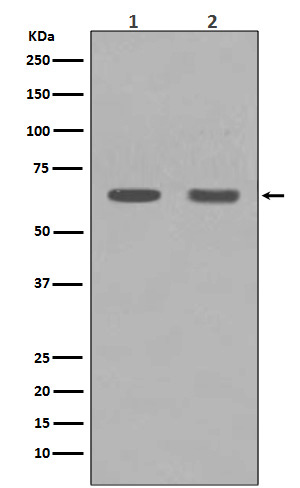
Real WB data Western blot analysis of HDAC1 expression in (1) C6 cell lysate; (2) NIH/3T3 cell lysate.
Anti-HDAC1 Rabbit Monoclonal Antibody
Cat # M00256

Two catalogued anti-HDAC1 antibodies, A00256-4 and M00256, have Western blot images. The supplied evidence does not establish comparative performance or broader validation.

Which to pick: A00256-4 shows WB in human MOLT-4, Jurkat, HL-60, and U251 whole cell lysates; M00256 shows WB in C6 and NIH/3T3 cell lysates. Choose the image with samples closest to your context.

Source: BosterBio HDAC1 gene-info card — filtered to Western-blot-capable antibodies; each card shows that product's actual WB validation figure.