This website uses cookies to ensure you get the best experience on our website.


- Table of Contents
Plan HDAC5 staining in paraffin sections using its predominantly cytoplasmic tissue pattern, with nuclear staining in some tissues (HPA tissue IHC). Compare compartments as well as intensity because HDAC5 shuttles between nucleus and cytoplasm (UniProt).

Expected localisation, validated protocols, controls and antibodies — the at-a-glance facts below, then the full design guide.
| Expected localisation | Cytoplasmic in most tissues; nuclear in several (HPA tissue IHC) | |
| Staining pattern | Mostly cytoplasmic staining, with some nuclear staining (HPA tissue IHC) | |
| Antigen retrieval | EDTA pH 8.0 HIER, heat-mediated (datasheet A01230-6) | |
| Positive control | Appendix+4 more · see all | |
| Negative control | None in HPA (detected in all 44 tissues); use no-primary + isotype controls |
| Fixation | Keep formalin fixation consistent across sections. (standard IHC practice; not target-specific) | |
| Caveat | Nuclear–cytoplasmic shuttling can change compartment scores (UniProt) | |
| Regulation | Differentiation promotes cytoplasmic export (UniProt) | |
| Isoform / epitope | 3 isoforms; check antibody epitope coverage (UniProt) |
The catalog antibody uses EDTA pH 8.0 retrieval (datasheet A01230-6). Two published IHC protocols describe HDAC5 staining in kidney and scar tissue (PMC7791055; PMC9576526).
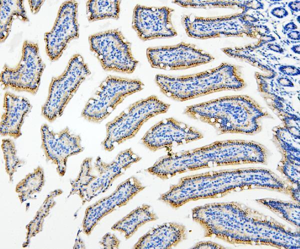
| Sample | Paraffin-embedded mouse small intestine tissue; fixative not specified (datasheet A01230-6) |
| Fixation | Image fixative and duration unreported (datasheet A01230-6); verify before use. |
| Sectioning | 4–5 µm sections on charged slides (standard) |
| Deparaffinisation | Xylene, graded ethanol series to water (standard) |
| Antigen retrieval | Heat retrieval: EDTA pH 8.0 (datasheet A01230-6); 20 min, 95–100 °C (standard) |
| Peroxidase block | 3% H2O2, 10 min, room temperature (standard) |
| Blocking | 10% goat serum (datasheet A01230-6) |
| Primary antibody | Rabbit anti-HDAC5, 2-5μg/ml (datasheet A01230-6) |
| Primary incubation | Overnight at 4 °C (datasheet A01230-6) |
| Detection | Streptavidin-biotin complex (SABC), DAB chromogen (datasheet A01230-6) |
| Counterstain | Hematoxylin, blue, dehydrate and mount (standard) |
| Expected result | HDAC5-positive staining in glandular cells of appendix (HPA tissue IHC: High). HPA tissue profile: Cytoplasmic expression in most tissues. Additional nuclear expression in several tissues. No signal in the no-primary control. |
HDAC5 staining is predominantly cytoplasmic across tissues, with additional nuclear staining in several tissues (HPA tissue IHC). Glandular cells in appendix, colon, duodenum, salivary gland and thyroid are among the high staining examples (HPA tissue IHC). Nuclear and cytoplasmic localisation is consistent with HDAC5 shuttling between those compartments (UniProt Q9UQL6); HDAC5 has no transmembrane segment (UniProt Q9UQL6 topology). HPA rates its tissue staining Approved, with medium consistency against RNA expression (HPA tissue IHC).
| Glandular cells show clear cytoplasmic staining, with nuclear staining in some cells. | This fits the reported tissue pattern: cytoplasmic expression in most tissues and additional nuclear expression in several (HPA tissue IHC). Interpret nuclear staining alongside the tissue and cell type; HDAC5 can shuttle between nucleus and cytoplasm (UniProt Q9UQL6). |
| Signal outlines cell membranes or fills gland lumens, without convincing intracellular staining. | A membrane or luminal pattern alone does not match the reported cytoplasmic and nuclear localisation (HPA tissue IHC; UniProt Q9UQL6 topology). Check morphology and controls before treating it as HDAC5; edge staining or deposited chromogen can mislead localisation (general IHC practice). |
| Strong staining appears mainly in a cell population expected to have low staining. | For example, HPA reports low staining in cardiomyocytes, skeletal myocytes and glial cells in caudate (HPA tissue IHC). Unexpected strong signal raises the possibility of cross-reactivity or endogenous detection activity (general IHC practice); low reported staining does not mean every cell must be negative. |
| A faint, even tint covers cells and surrounding tissue. | Diffuse colour without cell boundaries or a discernible compartment is difficult to score as HDAC5 (general IHC practice). Compare with a negative reagent control and inspect washing, blocking and chromogen development before calling weak cytoplasmic staining (general IHC practice). |
| No convincing staining appears in a tissue with an HPA high staining example. | Appendix or colon glandular cells and tonsil germinal center cells are reported High (HPA tissue IHC). Absence of signal there suggests reviewing antibody and detection performance, section quality and control results (general IHC practice); HPA staining levels alone do not establish a cause. |
| Tissue and cell choice | HPA reports High staining in glandular cells of appendix, colon, duodenum, salivary gland and thyroid, as well as bone marrow hematopoietic cells, skin Langerhans cells and tonsil germinal center cells (HPA tissue IHC). These are useful comparison patterns, not guarantees for every section. |
| Compartment and cell state | Nuclear and cytoplasmic localisation is reported for HDAC5 (UniProt Q9UQL6). In muscle cells, differentiation can drive cytoplasmic shuttling after phosphorylation at Ser-259 and Ser-498 (UniProt Q9UQL6). Interpret a compartment shift in its biological context rather than requiring one fixed nuclear-to-cytoplasmic ratio. |
| Antibody evidence | HPA rates its tissue IHC profile Approved, describing medium consistency between antibody staining and RNA expression (HPA tissue IHC). HPA030991, HPA076420 and CAB019400 each have IHC Approved status (HPA antibodies). Approved status supports use as a reference but does not make an unexpected pattern specific by itself. |
| Protein forms and topology | UniProt lists three HDAC5 isoforms, one chain spanning residues 1–1122, no signal peptide and no transmembrane segment (UniProt Q9UQL6). Those annotations support an intracellular interpretation; they do not identify which isoform a particular antibody recognises or establish an antigen retrieval condition. |
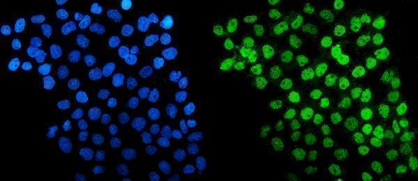
| What should IF/ICC show? | HPA reports mainly nuclear speckles, with additional Golgi apparatus and cytosol localisation in ICC-IF (HPA subcellular). That finer pattern can inform localisation comparisons, while the tissue IHC summary remains predominantly cytoplasmic with nuclear expression in several tissues (HPA tissue IHC). |
| Target-specific fixation sensitivity | No target-specific fixation effect is established by the supplied UniProt or HPA localisation and staining records. Choose and assess routine paraffin-section preparation with appropriate controls (general IHC practice); do not infer an HDAC5 antigen retrieval requirement from tissue staining intensity. |
| Situation | Likely cause | Next action |
|---|---|---|
| High staining example gives no signal. | A failed antibody or detection step, damaged section, or unsuitable working conditions are possibilities (general IHC practice); the HPA High label does not diagnose the failure (HPA tissue IHC). | Check section morphology and a known working staining control, then verify antibody dilution, detection reagents and chromogen development (general IHC practice). |
| Signal is strong on membranes or in lumens. | The pattern conflicts with HDAC5's reported nuclear and cytoplasmic localisation and lack of a transmembrane segment (UniProt Q9UQL6; HPA tissue IHC). Artefactual deposition is possible (general IHC practice). | Inspect cell boundaries at higher magnification and compare negative reagent controls; score only convincing intracellular signal (general IHC practice). |
| Unexpected cells stain strongly. | Cell type differences may matter: HPA reports low staining in cardiomyocytes and skeletal myocytes (HPA tissue IHC). Cross-reactivity or endogenous detection activity is also possible (general IHC practice). | Compare a high staining reference cell population and an appropriate negative reagent control; assess endogenous enzyme blocking if using enzyme-based detection (general IHC practice). |
| Nuclei stain but cytoplasm appears weak. | Nuclear expression occurs in several tissues, and HDAC5 shuttles between nucleus and cytoplasm (HPA tissue IHC; UniProt Q9UQL6). Nuclear signal alone therefore does not prove a technical failure. | Confirm nuclear boundaries with the counterstain and assess the expected cell type, morphology and controls before assigning localisation (general IHC practice). |
| Colour is diffuse across the section. | Nonspecific background or excessive chromogen development can obscure intracellular boundaries (general IHC practice). HPA's cytoplasmic profile requires a cellular pattern for comparison (HPA tissue IHC). | Compare negative reagent controls; review blocking, washing, antibody concentration and chromogen development under the laboratory's routine IHC workflow (general IHC practice). |
| Muscle staining differs from a reference image. | HPA reports low staining in heart cardiomyocytes and skeletal myocytes, while UniProt describes differentiation-linked cytoplasmic shuttling in muscle cells (HPA tissue IHC; UniProt Q9UQL6). | Compare the same tissue and cell type, assess nuclear and cytoplasmic compartments separately, and verify controls before attributing a difference to biology or staining conditions (general IHC practice). |


Comprehensive Human Protein Atlas IHC scoring per tissue (reliability: Approved — Medium consistency between antibody staining and RNA expression data.). Rows are taken directly from the HPA tissue chart — click any row's HPA link to view the source.
| Tissue | Cell type | Level | Evidence | Source |
|---|---|---|---|---|
| Appendix | Glandular cells | High | Protein (IHC) | HPA → |
| Bone marrow | Hematopoietic cells | High | Protein (IHC) | HPA → |
| Colon | Glandular cells | High | Protein (IHC) | HPA → |
| Duodenum | Glandular cells | High | Protein (IHC) | HPA → |
| Salivary gland | Glandular cells | High | Protein (IHC) | HPA → |
| Tissue | Cell type | Level | Evidence | Source |
|---|---|---|---|---|
| None in HPA: HDAC5 is detected in all 44 scored tissues. Use a no-primary (secondary-only) and an isotype control instead. | ||||
Troubleshoot HDAC5 staining in paraffin sections by checking retrieval, compartment patterns, detection background, and cell-specific scoring.
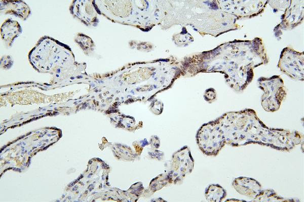
Two anti-HDAC5 antibodies have paraffin-section IHC images from human and mouse samples (catalog IHC captions); one also has IF images from A431 and U20S cells (A01230-6 IF captions).
A01230-5 has IHC images from human placenta and mouse and rat small intestine paraffin sections (A01230-5 IHC captions). A01230-6 has IHC images from mouse small intestine, human placenta, esophageal squamous carcinoma and lung cancer paraffin sections, plus IF images from A431 and U20S cells (A01230-6 IHC and IF captions).
Which to pick: For tissue IHC across human, mouse and rat samples, choose A01230-5: its paraffin-section images cover all three species (A01230-5 IHC captions); start at 0.5–1 μg/ml (catalog IHC dilution). For IF/ICC, choose A01230-6: those applications are listed and its cell images use 5 μg/ml (catalog applications; A01230-6 IF captions). A01230-6 also has human and mouse paraffin-section IHC images at 2 μg/ml (A01230-6 IHC captions); clonality and the tissue fixative are unreported (catalog clone field; IHC captions).