HDAC9 / Histone deacetylase 9 · IHC design guide

Design Immunohistochemistry for HDAC9

Plan chromogenic HDAC9 IHC in paraffin sections around a mainly nuclear tissue pattern (HPA tissue IHC). This guide covers consistent fixation (standard IHC practice), controls for uncertain tissue staining (HPA tissue IHC), and interpretation across 11 isoforms (UniProt).

Evidence assembled Oct 2026 · For research use; verify linked source records and product datasheet before use
Immunohistochemistry protocol sheet for HDAC9 (IHC for HDAC9): expected localisation Mainly nuclear tissue staining; reliability uncertain (HPA tissue IHC), antibody M02177, validated IHC image, and IHC protocol steps
Printable HDAC9 IHC protocol sheet — expected localisation Mainly nuclear tissue staining; reliability uncertain (HPA tissue IHC), antibody M02177, controls and protocol steps. Open the full HDAC9 IHC guide →

HDAC9 Immunohistochemistry Experimental Design Guide

Expected localisation, validated protocols, controls and antibodies — the at-a-glance facts below, then the full design guide.

Must know before staining
Expected localisation Mainly nuclear tissue staining; reliability uncertain (HPA tissue IHC)
Staining pattern Mainly nuclear staining across diverse cell types (HPA tissue IHC)
Antigen retrieval Tris-EDTA pH 9.0 HIER, 95–98 °C, 20 min (rule: nuclear antigen)
Positive control ⓘ Adrenal gland+4 more · see all
Negative control ⓘ Hippocampus+1 more · see all
Important caveats
Reasons your staining may differ from the expected pattern.
Fixation Keep paraffin-section fixation consistent across samples (standard IHC practice; not target-specific)
Caveat Tissue staining has uncertain reliability (HPA tissue IHC)
Regulation Expression varies by tissue (UniProt)
Isoform / epitope 11 isoforms; check antibody epitope coverage (UniProt)
Section 1

Recommended HDAC9 IHC & IF Protocols

The catalog antibody’s IHC-P protocol is accompanied by published HDAC9 protocols for arteries, endometrial carcinoma, and lung tumors (PMC4206941; PMC8884513; PMC7304847).

Recommended immunohistochemistry (IHC-P) protocol parameters
SampleParaffin-embedded human brain tissue; fixative not specified (datasheet M02177)
FixationImage fixative and duration unreported (datasheet M02177); verify before use.
Sectioning4–5 µm sections on charged slides (standard)
DeparaffinisationXylene, graded ethanol series to water (standard)
Antigen retrievalHeat-induced epitope retrieval in Tris-EDTA buffer, pH 9.0, 20 min at 95–98 °C (standard rule: nuclear antigen)
Peroxidase block3% H2O2, 10 min, room temperature (standard)
Blocking10% normal serum of the secondary host, 30 min, room temperature (standard)
Primary antibodyRabbit monoclonal (clone HEF-8) anti-HDAC9, 1:50-1:200 (datasheet M02177)
Primary incubationOvernight at 4 °C (standard)
DetectionHRP-polymer secondary, DAB chromogen 5–10 min (standard)
CounterstainHematoxylin, blue, dehydrate and mount (standard)
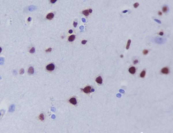
Expected resultHDAC9-positive staining in glandular cells of adrenal gland (HPA tissue IHC: High). HPA tissue profile: Mainly general nuclear expression. No signal in the no-primary control.
💡Decision noteStart with Tris-EDTA pH 9.0 retrieval at 95–98 °C for 20 min (page retrieval rule); compare the published conditions if staining needs optimization.
Section 2

What Is the Expected HDAC9 Staining Pattern?

HDAC9 should appear mainly in nuclei in IHC sections (UniProt Q9UKV0: nucleus; HPA tissue IHC: mainly general nuclear expression). HPA reports high staining in several epithelial, glandular and hematopoietic cell populations (HPA tissue IHC), but rates the tissue IHC profile Uncertain, with medium agreement between staining and RNA data and pending external verification (HPA tissue IHC). HDAC9 has no transmembrane segment (UniProt Q9UKV0: topology).

What am I looking at on my slide?
Nuclear staining in breast or colon glandular cells, bronchial respiratory epithelial cells, or bone marrow hematopoietic cells.This fits the cell types and High levels reported for those tissues (HPA tissue IHC). Compare staining within the named cell population, since a tissue-wide impression can hide which cells carry the signal (general IHC practice). Treat agreement as supportive rather than definitive because the tissue IHC profile is Uncertain (HPA tissue IHC).
Predominantly membranous or extracellular deposit, with little nuclear staining.This conflicts with the reported nuclear location and absence of a transmembrane segment (UniProt Q9UKV0: subcellular location and topology; HPA tissue IHC: nuclear profile). Review morphology and detection controls before calling it HDAC9; a misplaced deposit alone does not identify the source of the artefact (general IHC practice).
Strong staining in hippocampal glial cells or parathyroid glandular cells.Those specific populations are reported Not detected (HPA tissue IHC). Consider nonspecific binding, cross-reactivity or endogenous chromogenic activity, and check controls (general IHC practice). The HPA result is an uncertain IHC observation, so an unexpected positive warrants investigation rather than automatic rejection (HPA tissue IHC: Uncertain).
Diffuse color across nuclei, cytoplasm and empty spaces, obscuring cell boundaries.This is harder to reconcile with mainly nuclear HDAC9 (UniProt Q9UKV0: nucleus; HPA tissue IHC: mainly general nuclear expression). Excess background or detection chemistry may be involved; inspect the no-primary control and review blocking, antibody concentration and washing as general IHC checks (general IHC practice).
No nuclear signal in a section expected to contain HPA-reported High cells.An absent result in breast glandular or bone marrow hematopoietic cells conflicts with the reported High staining (HPA tissue IHC). First assess whether the relevant cells are present, then check positive-control performance and the antibody's IHC-P instructions (general IHC practice). HPA's Uncertain rating limits how strongly one negative section can be interpreted (HPA tissue IHC).
💡Expected HDAC9 appearanceA convincing positive is predominantly nuclear color in the named HPA-reported High cell populations, while strong membrane or extracellular color or equally strong staining in HPA-reported Not detected cells is a result to investigate (UniProt Q9UKV0: nucleus and topology; HPA tissue IHC: High, Not detected and Uncertain).
How each factor affects the staining
Tissue and cell selectionUse the cell-specific HPA observations to choose comparisons: breast glandular and bronchial respiratory epithelial cells are High, whereas parathyroid glandular cells are Not detected (HPA tissue IHC). These are reported staining levels, not guaranteed positive and negative controls, because HPA rates tissue IHC Uncertain (HPA tissue IHC).
Isoforms and antibody epitopeUniProt lists 11 HDAC9 isoforms (UniProt Q9UKV0: isoforms). The supplied record does not locate the IHC-validated antibody's epitope or establish which isoforms it detects; check that information before interpreting a missing signal as absence of all HDAC9 forms (UniProt Q9UKV0: isoforms; supplied antibody record: no epitope specified).
Phosphorylation and localizationUniProt annotates five modified residues, including phosphoserines, and an interaction between the phosphorylated form and 14-3-3 by similarity (UniProt Q9UKV0: modified residues and subunit). Those entries do not predict a different staining compartment in a particular section; use the observed nuclear IHC profile as the comparison (HPA tissue IHC).
IHC evidence and retrievalThe listed antibody HPA028926 has Uncertain IHC validation, and the overall tissue IHC profile is Uncertain (HPA antibodies; HPA tissue IHC). No target-specific retrieval or fixation sensitivity is supplied; follow the IHC-P instructions for the antibody in use and assess controls when optimizing retrieval (general IHC practice).
IF/ICC Q&A: where should HDAC9 appear?Mainly in the nucleoplasm, with additional mitotic chromosome localization (HPA subcellular ICC-IF: Approved); this is the IF/ICC localization answer. HPA028926 is Approved for ICC but Uncertain for IHC, so its ICC status does not validate a paraffin-section result (HPA antibodies).
Why is my staining missing, weak or wrong?
SituationLikely causeNext action
A HPA-reported High cell population shows no nuclear color.The expected cells may be absent from the sampled area, or the IHC detection workflow may have failed (HPA tissue IHC: cell-specific High reports; general IHC practice).Confirm the named cells on the counterstained section; run an appropriate positive control and check the IHC-validated antibody's prescribed IHC-P conditions (general IHC practice). Do not infer an HDAC9-specific fixation effect from this result (supplied sources: no fixation sensitivity reported).
Color is mainly at membranes or outside cells.That distribution conflicts with nuclear HDAC9 and its lack of a transmembrane segment (UniProt Q9UKV0: subcellular location and topology). Its precise cause cannot be assigned from location alone (general IHC practice).Review tissue morphology, the no-primary control and wash quality; score nuclear staining separately from surface or extracellular deposits (general IHC practice).
Unexpected strong color appears in hippocampal glia or parathyroid glandular cells.HPA reports those cell populations as Not detected, although its tissue IHC reliability is Uncertain (HPA tissue IHC). Nonspecific binding or endogenous detection activity remains possible (general IHC practice).Compare matched controls, including a no-primary section, and inspect the cell identity and nuclear compartment before scoring the result (general IHC practice). Replicate unexpected findings before relying on the HPA comparison (HPA tissue IHC: Uncertain).
Diffuse chromogen obscures nuclei and cell borders.Background can arise from antibody concentration, insufficient blocking or washing, or endogenous detection activity (general IHC practice); the supplied sources do not establish which applies to HDAC9.Inspect no-primary controls, review blocking and washes, and adjust antibody concentration within the antibody's IHC-P instructions (general IHC practice). Reassess whether discrete nuclear staining becomes visible (UniProt Q9UKV0: nucleus).
A low-level population is scored as a definite negative or strong positive.HPA reports Low staining in heart cardiomyocytes, thyroid glandular cells, caudate glia and splenic red-pulp cells (HPA tissue IHC); faint color is especially vulnerable to scoring differences (general IHC practice).Record intensity and the stained cell population separately, compare sections processed together, and avoid turning a Low HPA observation into a binary control (HPA tissue IHC: Low and Uncertain; general IHC practice).
IF/ICC nucleoplasmic staining agrees with HPA, but paraffin IHC remains ambiguous.The antibody's ICC status is Approved while its IHC status is Uncertain (HPA antibodies: HPA028926); agreement in ICC does not resolve ambiguous section staining (general assay interpretation).Interpret the paraffin section using its nuclear morphology, cell-specific HPA IHC comparisons and IHC controls (HPA tissue IHC; general IHC practice). Keep the IF/ICC result as supporting localization evidence (HPA subcellular ICC-IF: Approved).

Sample controls for HDAC9 IHC & IF

🧪Run breast first: glandular cells should stain (HPA: High in breast glandular cells). Run parathyroid gland as the tissue negative (HPA: Not detected in parathyroid glandular cells); on the breast slide, cells without nuclear chromogen may provide an internal background reference, but their HDAC9-negative status needs validation (UniProt Q9UKV0: nucleus; standard IHC practice).
Positive control tissue: Adrenal gland (Glandular cells, HPA High)
Negative control tissue: Hippocampus (HPA Not detected)
ICC-IF cell lines (HPA subcellular resource): HPA ICC-IF images show HDAC9 in A-431, U-251MG, U2OS, KOLF2.1J, with annotated localisation: Nucleoplasm (approved) (HPA subcellular).
Technical controls: Include a no-primary (secondary-only) control, an isotype-matched control for a monoclonal antibody or host-matched nonimmune IgG for a polyclonal antibody, and identically processed HDAC9 knockout material if available (standard IHC practice). For breast sections, check background after endogenous peroxidase blocking if using HRP detection, and block endogenous biotin if using a biotin-based detector (standard IHC practice).
⚠️Feasibility: A target-specific fixation window is unreported, and the selected M02177 paraffin-section caption does not state a fixative (selected-SKU tissue-IHC caption). Retrieval dependency is unreported; optimize antigen retrieval against the positive and negative tissues (standard IHC practice; HPA: High in breast glandular cells; HPA: Not detected in parathyroid glandular cells). These data do not establish whether frozen sections or IF are easier; breast adipose tissue can autofluoresce in IF, so assess an unstained section (standard IF practice).

HPA tissue IHC evidence for HDAC9

Comprehensive Human Protein Atlas IHC scoring per tissue (reliability: Uncertain — Medium consistency between antibody staining and RNA expression data. Pending external verification.). Rows are taken directly from the HPA tissue chart — click any row's HPA link to view the source.

Positive expression · recommended positive controls

TissueCell typeLevelEvidenceSource
Adrenal gland Glandular cells High Protein (IHC) HPA →
Bone marrow Hematopoietic cells High Protein (IHC) HPA →
Breast Glandular cells High Protein (IHC) HPA →
Bronchus Respiratory epithelial cells High Protein (IHC) HPA →
Cervix Glandular cells High Protein (IHC) HPA →

Undetected expression · recommended negative controls

TissueCell typeLevelEvidenceSource
Hippocampus Glial cells Not detected Protein (IHC) HPA →
Parathyroid gland Glandular cells Not detected Protein (IHC) HPA →
Section 3

Advanced HDAC9 IHC Tips

Use nuclear staining, matched controls and cell-specific scoring to troubleshoot HDAC9 in paraffin-section chromogenic IHC; interpret tissue patterns cautiously (UniProt Q9UKV0; HPA tissue IHC).

How should I retrieve HDAC9 when nuclear staining is weak?
Start with Tris-EDTA at pH 9.0 for heat-induced epitope retrieval at 95–98 °C for 20 min (page retrieval specification). Keep section thickness, cooling, primary-antibody incubation and chromogen development consistent while comparing retrieved sections with a no-primary control (standard IHC practice). If staining remains weak, compare a shorter retrieval time or citrate buffer at pH 6.0 on adjacent sections, checking whether either change improves nuclear signal without increasing background (standard IHC practice). Score signal in intact nuclei rather than treating diffuse cytoplasmic color as rescued staining, because HDAC9 is nuclear and HPA describes mainly general nuclear expression (UniProt Q9UKV0 subcellular location; HPA tissue IHC).
Could fixation explain weak or patchy HDAC9 staining?
Target-specific fixation sensitivity is unknown: the selected M02177 image identifies paraffin-embedded human brain but does not report its fixative (selected M02177 tissue-IHC caption). Record fixative, fixation duration and processing history for each specimen, then compare similarly processed sections before changing antibody conditions (standard IHC practice). If the sample is known to be formalin fixed, compare a controlled retrieval series on adjacent sections and inspect morphology for underfixation or excessive heat damage (standard IHC practice). Do not assign a fixation effect to HDAC9 from its phosphoserine annotations or tissue staining patterns; those sources document modifications and expression, respectively, without measuring fixation sensitivity (UniProt Q9UKV0 modified residues; HPA tissue IHC).
What staining compartment should count as HDAC9 positive?
Prioritise nuclear chromogen in morphologically intact cells, because HDAC9 is annotated as nuclear and HPA reports mainly general nuclear tissue staining (UniProt Q9UKV0 subcellular location; HPA tissue IHC). The approved cellular imaging location is nucleoplasm, with an additional mitotic-chromosome location, so distinguish chromatin-associated mitotic signal from diffuse cytoplasmic color during review (HPA subcellular). Compare nuclear and cytoplasmic signal separately on matched sections, including a no-primary control, before calling an unexpected compartment positive (standard IHC practice). The protein has no annotated transmembrane segment, so a membrane-only pattern warrants checks for nonspecific binding, section edges and chromogen precipitate (UniProt Q9UKV0 topology; standard IHC practice).
Could the antibody miss particular HDAC9 isoforms or modified epitopes?
HDAC9 has 11 annotated isoforms, so first obtain the catalog antibody's immunogen or epitope information before assuming that every isoform is detected (UniProt Q9UKV0 isoforms; standard antibody-validation practice). Map that epitope against isoform sequences, then compare nuclear staining with an independently validated antibody directed to a different region when available (standard antibody-validation practice). Five modified residues are listed, including phosphoserines at 22, 220, 240, 451 and 554, but their effect on this antibody's binding is unreported (UniProt Q9UKV0 modified residues). Record retrieval conditions and compare adjacent sections before interpreting a negative region as isoform absence or phosphorylation-dependent epitope loss (standard IHC practice).
How can IF help investigate ambiguous HDAC9 IHC staining?
In a separate IF experiment, pair HDAC9 with an epithelial marker when examining bronchus, where respiratory epithelial cells are listed as high in tissue IHC (HPA tissue IHC; standard multiplex IF practice). Choose a far-red fluorophore for the weaker signal and inspect an unstained section for tissue autofluorescence before assigning overlapping fluorescence to the same cell (standard IF practice). Because HDAC9 is nuclear and has no transmembrane segment, use a controlled permeabilisation step that permits access across the plasma and nuclear membranes, then check nuclear morphology (UniProt Q9UKV0 subcellular location and topology; standard IF practice). Include single-label and no-primary controls, and treat IF colocalisation as supporting evidence rather than proof that chromogenic IHC signal is specific (standard IF practice).
How do I reduce diffuse brown staining without losing nuclear signal?
Inspect a no-primary section for endogenous peroxidase activity and a reagent-control section for nonspecific detection before changing the HDAC9 antibody incubation (standard chromogenic IHC practice). Apply a peroxidase block and an appropriate protein block, then keep DAB development consistent across test and reference sections (standard chromogenic IHC practice). If diffuse color persists, compare antibody incubation conditions on adjacent sections while retaining the same retrieval, wash and detection steps (standard IHC practice). Judge improvement by preserved nuclear contrast in morphologically intact cells, since HDAC9 is nuclear and the tissue profile is mainly general nuclear expression, with HPA tissue-IHC reliability rated uncertain (UniProt Q9UKV0 subcellular location; HPA tissue IHC).
What is a defensible way to quantify HDAC9 IHC? ⚠ ANSWER MARKED FOR VERIFICATION
Define the cell population and tissue region before scoring, then report the percentage of nuclei at each prespecified intensity and calculate an H-score from 0–300 (standard IHC scoring practice). Also report the number of evaluable nuclei or positive-cell density per mm², normalised to viable tissue area rather than the whole section (standard image-analysis practice). Keep retrieval, DAB development, imaging settings and intensity thresholds fixed across the comparison set, with no-primary controls reviewed for background (standard IHC practice). Report cell types separately because HPA lists high staining in several glandular and hematopoietic populations, and flag that its tissue-IHC reliability is uncertain (HPA tissue IHC).
When is an apparent HDAC9-positive region likely to be artefactual?
A credible call combines nuclear staining in intact cells with a matching cell-type pattern and low signal in the no-primary control (UniProt Q9UKV0 subcellular location; HPA tissue IHC; standard IHC practice). Reconsider membrane-only or diffuse cytoplasmic color, since HDAC9 lacks an annotated transmembrane segment and its approved cellular imaging location is mainly nucleoplasm (UniProt Q9UKV0 topology; HPA subcellular). Exclude section edges, folds and necrotic areas from scoring, and test persistent brown deposits against a peroxidase-blocked control for endogenous enzyme activity (standard chromogenic IHC practice). Interpret discordance cautiously: HPA reports uncertain tissue-IHC reliability, and the selected M02177 caption documents staining in paraffin-embedded human brain without specifying fixation (HPA tissue IHC; selected M02177 tissue-IHC caption).
Boster reagents

Best HDAC9 / Histone deacetylase 9 IHC Antibodies

HDAC9 antibody data include paraffin-section human brain IHC (M02177 image caption) and HeLa-cell IF (A02177-4 image caption); listed reactivity covers human, rat, and mouse (catalog reactivity).

Real IHC data Immunohistochemical analysis of paraffin-embedded human brain, using HDAC9 Antibody.
Anti-HDAC9 Rabbit Monoclonal Antibody
Cat # M02177
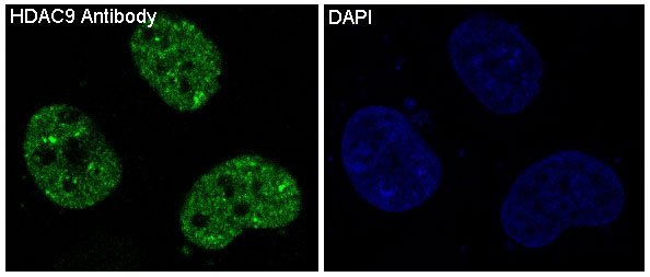
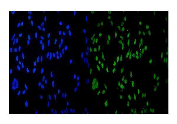
Real IF data IF analysis of HDAC9 using anti-HDAC9 antibody (A02177-4). HDAC9 was detected in immunocytochemical section of Hela cells. Enzyme antigen retrieval was performed using IHC enzyme antigen retrieval reagent (AR0022) for 15 mins. The cells were blocked with 10% goat serum. And then incubated with 4μg/mL rabbit anti-HDAC9 Antibody (A02177-4) overnight at 4°C. DyLight®488 Conjugated Goat Anti-Rabbit IgG (BA1127) was used as secondary antibody at 1:100 dilution and incubated for 30 minutes at 37°C. The section was counterstained with DAPI. Visualize using a fluorescence microscope and filter sets appropriate for the label used.
Anti-HDAC9 Antibody ®
Cat # A02177-4

M02177 is a rabbit monoclonal listed for IHC and IF, with an IHC image of paraffin-embedded human brain (catalog applications; M02177 image caption). A02177-4 is listed for ICC/IF, with an IF image of HeLa cells (catalog applications; A02177-4 image caption).

Which to pick: Choose M02177 for tissue IHC: its own image shows a paraffin-embedded human brain section, and its listed IHC dilution is 1:50–1:200 (M02177 image caption; catalog IHC dilution); the fixative is unreported (M02177 image caption). Choose A02177-4 for IF/ICC: its HeLa-cell IF image uses 4 μg/mL, while IHC is absent from its application list (A02177-4 image caption; catalog applications). For species selection, M02177 lists human and rat reactivity, while A02177-4 lists human, mouse, and rat reactivity; the shown IF example is human HeLa cells (catalog reactivity; A02177-4 image caption).

Each figure is that product's own IHC / IF validation image from its datasheet.

References

  1. UniProt Consortium. UniProt entry Q9UKV0 (HDAC9_HUMAN, Histone deacetylase 9).
  2. Human Protein Atlas. HDAC9 tissue IHC expression (reliability: Uncertain).
  3. Human Protein Atlas. HDAC9 subcellular location (ICC-IF): Mainly localized to the nucleoplasm. In addition localized to the mitotic chromosome..
  4. Human Protein Atlas. HDAC9 antibody validation summary (1 antibodies).
  5. Evidence HDAC9 genetic variant associated with ischemic stroke increases risk via promoting carotid atherosclerosis. Stroke 2013 — PMC4206941.
  6. Nucleocytoplasmic Shuttling of Histone Deacetylase 9 Controls Activity-Dependent Thalamocortical Axon Branching. Scientific reports 2017 — PMC5519695.
  7. Expression of epigenetic pathway related genes in association with PD-L1, ER/PgR and MLH1 in endometrial carcinoma. PloS one 2022 — PMC8884513.
  8. HDAC9 deficiency promotes tumor progression by decreasing the CD8(+) dendritic cell infiltration of the tumor microenvironment. Journal for immunotherapy of cancer 2020 — PMC7304847.
  9. PubMed PMID:11535832 — UniProt-cited evidence.
  10. PubMed PMID:12706107 — UniProt-cited evidence.
  11. PubMed PMID:12590135 — UniProt-cited evidence.