This website uses cookies to ensure you get the best experience on our website.


- Table of Contents
Plan chromogenic paraffin IHC for HECTD3 using the catalog antibody’s documented tissue protocol (datasheet A11560). Compare staining with the cytoplasmic tissue profile and use controls when scoring, as presumed off-target binding was observed (HPA tissue IHC).

Expected localisation, validated protocols, controls and antibodies — the at-a-glance facts below, then the full design guide.
| Expected localisation | Cytoplasmic staining in most tissues (HPA tissue IHC) | |
| Staining pattern | Most tissues: cytoplasmic; breast glandular cells: medium (HPA tissue IHC) | |
| Antigen retrieval | Citrate pH 6 HIER, heat-mediated (datasheet A11560) | |
| Positive control | Adrenal gland+4 more · see all | |
| Negative control | Adipose tissue+1 more · see all |
| Fixation | Keep fixation consistent across sections (standard IHC practice; not target-specific) | |
| Caveat | Presumed off-target binding can confound scoring (HPA tissue IHC) | |
| Regulation | No expression regulator annotated in the record (UniProt) | |
| Isoform / epitope | 2 isoforms; cytoplasmic target, epitope map unspecified (UniProt) |
Use the catalog antibody’s IHC-P protocol as the starting point, then compare the published HECTD3 IHC methods from three PMC articles (PMC8994759; PMC3847339; PMC7904838).
| Sample | Paraffin-embedded human lung cancer tissue; fixative not specified (datasheet A11560) |
| Fixation | Image fixative and duration unreported (datasheet A11560); verify before use. |
| Sectioning | 4–5 µm sections on charged slides (standard) |
| Deparaffinisation | Xylene, graded ethanol series to water (standard) |
| Antigen retrieval | Heat retrieval: Citrate pH 6, 20 min (datasheet A11560) |
| Peroxidase block | 3% H2O2, 10 min, room temperature (standard) |
| Blocking | 10% goat serum (datasheet A11560) |
| Primary antibody | Rabbit anti-HECTD3, 1μg/ml (datasheet A11560) |
| Primary incubation | Overnight at 4 °C (datasheet A11560) |
| Detection | Streptavidin-biotin complex (SABC), DAB chromogen (datasheet A11560) |
| Counterstain | Hematoxylin, blue, dehydrate and mount (standard) |
| Expected result | HECTD3-positive staining in glandular cells of adrenal gland (HPA tissue IHC: Medium). HPA tissue profile: Cytoplasmic expression in most tissues. No signal in the no-primary control. |
HECTD3 is a cytoplasmic protein with a perinuclear location and no transmembrane segment (UniProt Q5T447). In paraffin sections, expect cytoplasmic staining in many tissues, including glandular cells, marrow hematopoietic cells and selected brain cells (HPA: cytoplasmic expression in most tissues; Medium in listed cells). Treat the pattern as a guide: HPA rates tissue IHC Approved, with medium staining–RNA consistency, presumed off-target staining disregarded and external verification pending (HPA: reliability description).
| Cytoplasmic signal, sometimes concentrated near nuclei, in glandular cells or marrow hematopoietic cells. | This fits UniProt’s cytoplasmic, perinuclear location and HPA’s Medium staining in glandular cells of breast, appendix and adrenal gland and in bone marrow hematopoietic cells (UniProt Q5T447; HPA: tissue IHC). Judge the stained cells within their tissue architecture. |
| Predominantly nuclear or sharply cell-surface signal, with little cytoplasmic staining. | That distribution conflicts with the reported cytoplasmic, perinuclear location and lack of a transmembrane segment (UniProt Q5T447). Check morphology and controls before calling it HECTD3; a compartment mismatch alone cannot identify the source of the signal. |
| Prominent staining in adipocytes or splenic red-pulp cells. | HPA reports HECTD3 as Not detected in those cells (HPA: adipocytes; spleen red pulp). Consider cross-reactivity or endogenous detection activity, and compare a no-primary control. A negative HPA observation is a reference pattern, not proof that every specimen must be unstained. |
| A uniform haze covers cells and surrounding tissue, obscuring their boundaries. | A field-wide haze cannot establish the cell-restricted cytoplasmic pattern described by HPA (HPA: cytoplasmic expression in most tissues). Assess background with a no-primary control, then review blocking, washes and detection conditions as general IHC troubleshooting steps. |
| No signal in breast glandular cells or bone marrow hematopoietic cells. | Both are reported at Medium staining in HPA tissue IHC (HPA: breast glandular cells; bone marrow hematopoietic cells). Confirm the expected cells are present, then check assay controls and reagent performance. The HPA rating is a reference, not a guarantee for every section. |
| Cellular location and topology | UniProt places HECTD3 in the cytoplasm and perinuclear region and lists no transmembrane segment (UniProt Q5T447). Use compartment as a pattern check; these annotations do not predict antigen-retrieval conditions or fixation sensitivity. |
| Strength and limits of the tissue reference | HPA describes cytoplasmic expression in most tissues but rates antibody staining–RNA consistency Medium, notes disregarded presumed off-target binding and awaits external verification (HPA: reliability description). Interpret unusual staining with controls. |
| Isoforms and unknown epitope coverage | UniProt lists two HECTD3 isoforms (UniProt Q5T447). The supplied record does not locate the antibody epitope or establish isoform-specific staining, so do not assign a cell pattern to one isoform from IHC alone. |
| IF/ICC: is a validated cell-image pattern available? | No main subcellular location or ICC/IF cell-line images are supplied, and the listed antibody has no ICC validation entry (HPA: subcellular record; HPA027467). UniProt’s cytoplasmic, perinuclear location can guide interpretation, but IF/ICC needs its own validation (UniProt Q5T447). |
| Situation | Likely cause | Next action |
|---|---|---|
| Expected HPA-positive cells are unstained. | The relevant cells may be absent from the section, or the IHC assay may have failed; HPA reports Medium staining, not assured positivity in every specimen (HPA: breast; bone marrow). | Locate the expected cells on the counterstained section and check a suitable positive control. As general IHC practice, review retrieval, primary-antibody conditions and detection reagents without assuming a HECTD3-specific fixation effect. |
| Signal is mainly nuclear or outlines cell borders. | That pattern disagrees with cytoplasmic, perinuclear HECTD3 and its lack of a transmembrane segment (UniProt Q5T447); the underlying cause remains undetermined. | Recheck cell boundaries and compare the positive and no-primary controls. Treat the compartment call as unresolved until reproducible cytoplasmic staining is demonstrated. |
| Adipocytes or splenic red-pulp cells stain prominently. | Those cell groups are Not detected in the HPA tissue reference (HPA: adipocytes; spleen red pulp). Cross-reactivity or endogenous detection activity is possible. | Compare a no-primary control and nearby tissue compartments. If staining persists without primary antibody, investigate endogenous detection activity using controls appropriate to the chromogenic system. |
| The whole section has diffuse color. | Broad background can obscure the cell-specific cytoplasmic pattern reported by HPA (HPA: tissue IHC); excessive detection signal or inadequate washing are general IHC possibilities. | Inspect a no-primary control and review wash and blocking steps. Adjust assay conditions only after confirming that the tissue’s morphology and expected cell populations remain assessable. |
| Only one unexpected cell population shows a strong signal. | HPA reports a broad cytoplasmic profile but also notes presumed off-target binding was observed and disregarded (HPA: profile; reliability description). The new pattern needs independent checking. | Compare tissue morphology and HPA-listed cell populations, then test specificity with an independent antibody or an appropriate orthogonal control when available; do not infer a new tissue restriction from one slide. |
| Perinuclear accentuation is unclear despite visible cytoplasmic signal. | UniProt reports a perinuclear location, while HPA’s tissue-level summary states cytoplasmic expression (UniProt Q5T447; HPA: tissue IHC). Neither source requires a perinuclear ring in every cell. | Score the reproducible cytoplasmic pattern and HPA-listed cell type first. Record whether perinuclear emphasis is visible, without making it a mandatory criterion for positivity. |


Comprehensive Human Protein Atlas IHC scoring per tissue (reliability: Approved — Medium consistency between antibody staining and RNA expression data. Presumed off target binding observed and disregarded. Pending external verification.). Rows are taken directly from the HPA tissue chart — click any row's HPA link to view the source.
| Tissue | Cell type | Level | Evidence | Source |
|---|---|---|---|---|
| Adrenal gland | Glandular cells | Medium | Protein (IHC) | HPA → |
| Appendix | Glandular cells | Medium | Protein (IHC) | HPA → |
| Bone marrow | Hematopoietic cells | Medium | Protein (IHC) | HPA → |
| Breast | Glandular cells | Medium | Protein (IHC) | HPA → |
| Caudate | Glial cells | Medium | Protein (IHC) | HPA → |
Troubleshoot HECTD3 staining in paraffin sections by checking retrieval, cellular localisation, controls and scoring before interpreting differences between specimens.
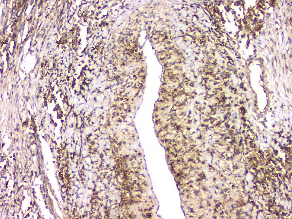
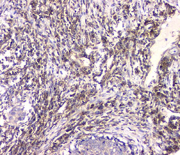
A11560 has paraffin-section IHC images from human lung cancer and mouse gaster tissue (catalog IHC captions). IHC-F and ICC are listed, but no IF image is supplied (catalog applications/images).
A11560 is shown in paraffin-section IHC of human lung cancer and mouse gaster tissue (catalog IHC captions). A11560 lists IHC, IHC-F, and ICC applications and human, mouse, and rat reactivity (catalog applications/reactivity).
Which to pick: Choose A11560 for paraffin-section tissue IHC: its caption documents citrate retrieval at pH 6 for 20 minutes and 1 μg/ml primary antibody overnight at 4°C; the fixative is unreported (A11560 IHC caption). For IF/ICC, A11560 lists IHC-F and ICC, but supplies no IF image or IF dilution (catalog applications/IF images/dilution). For cross-species work, A11560 is polyclonal and lists human, mouse, and rat reactivity, with IHC images from human and mouse tissue (catalog dilution_raw/reactivity; A11560 IHC captions).