This website uses cookies to ensure you get the best experience on our website.


- Table of Contents
Plan HES5 paraffin IHC around nuclear staining in brain and testis (HPA tissue IHC). Use strongly stained neuronal cells in the caudate, cerebral cortex and hippocampus as references, and assess signal carefully because presumed off-target staining has been observed (HPA tissue IHC).

Expected localisation, validated protocols, controls and antibodies — the at-a-glance facts below, then the full design guide.
| Expected localisation | Nuclear staining in brain and testis (HPA tissue IHC) | |
| Staining pattern | Strong nuclear staining in brain neuronal cells (HPA tissue IHC) | |
| Antigen retrieval | EDTA pH 8.0 HIER, heat-mediated (datasheet A05384-1) | |
| Positive control | Caudate+4 more · see all | |
| Negative control | Adipose tissue+4 more · see all |
| Fixation | Keep section fixation consistent. (standard IHC practice; not target-specific) | |
| Caveat | Presumed off-target staining has been observed (HPA tissue IHC) | |
| Regulation | Staining intensity varies by tissue (HPA tissue IHC) | |
| Isoform / epitope | No annotated isoforms; one 1–166 chain (UniProt) |
Compare the catalog antibody’s IHC-P protocol (datasheet: A05384-1) with four published HES5 IHC protocols (PMC5806807; PMC2874490; PMC5879899; PMC13477445).
| Sample | Paraffin-embedded human bladder epithelial carcinoma tissue; fixative not specified (datasheet A05384-1) |
| Fixation | Image fixative and duration unreported (datasheet A05384-1); verify before use. |
| Sectioning | 4–5 µm sections on charged slides (standard) |
| Deparaffinisation | Xylene, graded ethanol series to water (standard) |
| Antigen retrieval | Heat retrieval: EDTA pH 8.0 (datasheet A05384-1); 20 min, 95–100 °C (standard) |
| Peroxidase block | 3% H2O2, 10 min, room temperature (standard) |
| Blocking | 10% goat serum (datasheet A05384-1) |
| Primary antibody | Rabbit anti-HES5, 2-5 μg/ml (datasheet A05384-1) |
| Primary incubation | Overnight at 4 °C (datasheet A05384-1) |
| Detection | Streptavidin-biotin complex (SABC), DAB chromogen (datasheet A05384-1) |
| Counterstain | Hematoxylin, blue, dehydrate and mount (standard) |
| Expected result | HES5-positive staining in neuronal cells of caudate (HPA tissue IHC: High). HPA tissue profile: Nuclear expression in brain and testis. No signal in the no-primary control. |
HES5 is a nuclear transcription factor with no transmembrane segment (UniProt Q5TA89: nucleus; no transmembrane segment). In paraffin-section IHC, expect nuclear staining in neuronal cells of the caudate, cerebral cortex and hippocampus, where HPA reports high staining (HPA: tissue IHC). HPA describes nuclear expression in brain and testis, but reports only medium consistency between staining and RNA data and notes presumed off-target binding (HPA: Enhanced reliability description).
| Distinct nuclear chromogen in neuronal cells of the caudate, cerebral cortex or hippocampus. | This matches the strongest listed tissue observations: neuronal cells stain at a high level in each region (HPA: tissue IHC). Judge the cellular location as well as the intensity; HES5 is assigned to the nucleus (UniProt Q5TA89: subcellular location). |
| Predominantly cytoplasmic, membranous or extracellular chromogen, with little nuclear staining. | Treat this IHC pattern as suspect because UniProt places HES5 in the nucleus and lists no transmembrane segment or signal peptide (UniProt Q5TA89: location; topology; processing). Review the staining distribution and controls before assigning a biological explanation (standard IHC practice). |
| Strong staining in a cell population listed as undetected, such as adipocytes in adipose tissue. | This conflicts with that population's HPA observation (HPA: adipose-tissue adipocytes, not detected). Consider antibody cross-reactivity or endogenous chromogenic activity; HPA explicitly reports presumed off-target binding in its tissue assessment (HPA: reliability description; standard IHC practice). |
| Diffuse chromogen across tissue, including nuclei and surrounding spaces, without clear cell boundaries. | A diffuse field does not support a cell-specific nuclear call (UniProt Q5TA89: nucleus). Assess no-primary and detection controls, blocking and wash conditions to identify nonspecific or detection-related background (standard IHC practice). |
| No nuclear signal in neuronal cells of a caudate, cerebral cortex or hippocampus section. | An absent result in these high-staining HPA reference cells calls for a technical check, but does not by itself prove failed staining: HPA reports medium consistency with RNA data (HPA: tissue IHC; reliability description). Check tissue identity, primary-antibody use and detection controls (standard IHC practice). |
| Antibody evidence | HPA lists HPA076164 as IHC Enhanced; its tissue assessment still reports medium staining–RNA consistency and disregarded presumed off-target binding (HPA: antibody validation; reliability description). Interpret a new cell-type pattern against tissue controls rather than treating the validation label as universal specificity (standard IHC practice). |
| Tissue and cell context | HPA reports high neuronal staining in three listed brain regions, medium staining in several other cell populations, and no detection in specified populations (HPA: tissue IHC). Choose the comparison at the cell-population level: a negative entry for one population does not describe every cell in its tissue (HPA: tissue IHC). |
| Protein location and form | HES5 is a nuclear, 166-aa protein with no annotated signal peptide or transmembrane segment (UniProt Q5TA89: chain; location; topology; processing). These annotations support a nuclear interpretation, not a predicted membrane rim or extracellular deposit; they do not establish fixation sensitivity (UniProt Q5TA89: location; topology). |
| Chromogenic detection | With an enzyme-based chromogen, endogenous activity or nonspecific detection can produce color unrelated to the primary antibody (standard IHC practice). Compare appropriate detection controls before calling unexpected staining HES5; HPA's off-target caution makes that check relevant when the pattern differs from its tissue observations (HPA: reliability description). |
| Situation | Likely cause | Next action |
|---|---|---|
| Expected brain-region nuclei appear blank. | The selected section may lack the listed positive neuronal population, or an IHC workflow step may have failed (HPA: tissue IHC; standard IHC practice). | Confirm the neuronal population and run a documented positive section alongside detection controls; review primary-antibody dilution and retrieval against the chosen antibody's IHC instructions (standard IHC practice). No HES5-specific retrieval setting is established here. |
| Color is widespread outside nuclei. | Nonspecific binding or detection background can obscure a nuclear pattern (standard IHC practice; UniProt Q5TA89: nucleus). | Compare no-primary and detection controls, then review blocking, washes and chromogen development (standard IHC practice). Score HES5 only where cellular nuclear staining can be distinguished. |
| A listed negative cell population stains strongly. | The result disagrees with the relevant HPA cell-level observation; presumed off-target binding is a documented limitation of the tissue dataset (HPA: tissue IHC; reliability description). | Check the exact cell identity, inspect controls and repeat with independently supported IHC evidence if available (standard IHC practice). Report the discrepancy rather than reclassifying the HPA population as positive. |
| Only faint, uneven color appears in a high-staining reference region. | Uneven section handling or detection may reduce interpretability (standard IHC practice); HPA's high level is an observed pattern, not a guaranteed intensity in every run (HPA: tissue IHC). | Compare sections processed together, verify consistent reagent coverage and assess the positive control before changing the scoring threshold (standard IHC practice). |
| Nuclear color appears in bronchial respiratory epithelium or lung macrophages. | These cell populations have medium staining in HPA, so their presence alone is not an unexpected tissue result (HPA: bronchus respiratory epithelial cells; lung macrophages). | Score the named cell population and nuclear location separately from background, then compare with a listed high-staining neuronal reference (HPA: tissue IHC; UniProt Q5TA89: nucleus). |
| IF/ICC Q&A: Is cytosolic fluorescence automatically an artifact? | No. HPA approves nucleoplasm as the main ICC-IF location and also lists nuclear speckles and cytosol (HPA: subcellular ICC-IF). | Interpret that fluorescence using the separate IF/ICC guide and its controls; HPA065458 is ICC Approved, while HPA076164 is IHC Enhanced (HPA: antibody validation; standard IF practice). |
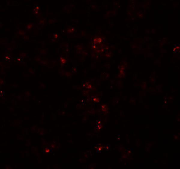

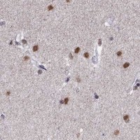


Comprehensive Human Protein Atlas IHC scoring per tissue (reliability: Enhanced — Medium consistency between antibody staining and RNA expression data. Presumed off target binding observed and disregarded.). Rows are taken directly from the HPA tissue chart — click any row's HPA link to view the source.
| Tissue | Cell type | Level | Evidence | Source |
|---|---|---|---|---|
| Caudate | Neuronal cells | High | Protein (IHC) | HPA → |
| Cerebral cortex | Neuronal cells | High | Protein (IHC) | HPA → |
| Hippocampus | Neuronal cells | High | Protein (IHC) | HPA → |
| Bronchus | Respiratory epithelial cells | Medium | Protein (IHC) | HPA → |
| Cerebellum | Cells in molecular layer | Medium | Protein (IHC) | HPA → |
| Tissue | Cell type | Level | Evidence | Source |
|---|---|---|---|---|
| Adipose tissue | Adipocytes | Not detected | Protein (IHC) | HPA → |
| Appendix | Glandular cells | Not detected | Protein (IHC) | HPA → |
| Bone marrow | Hematopoietic cells | Not detected | Protein (IHC) | HPA → |
| Breast | Adipocytes | Not detected | Protein (IHC) | HPA → |
| Cervix | Squamous epithelial cells | Not detected | Protein (IHC) | HPA → |
Troubleshoot HES5 staining by checking nuclear signal, section processing, and cell type against the available tissue evidence (UniProt Q5TA89; HPA tissue IHC).
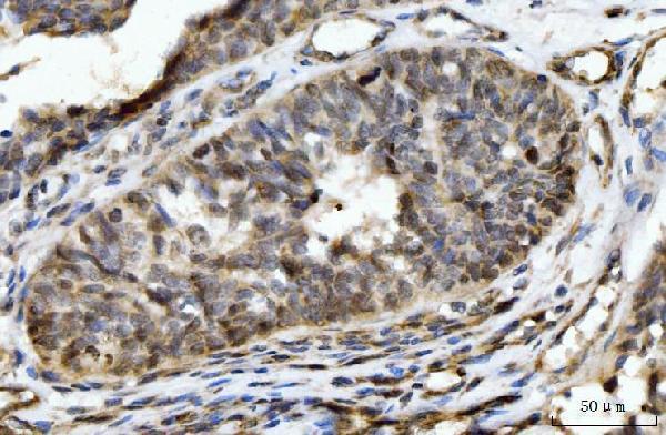
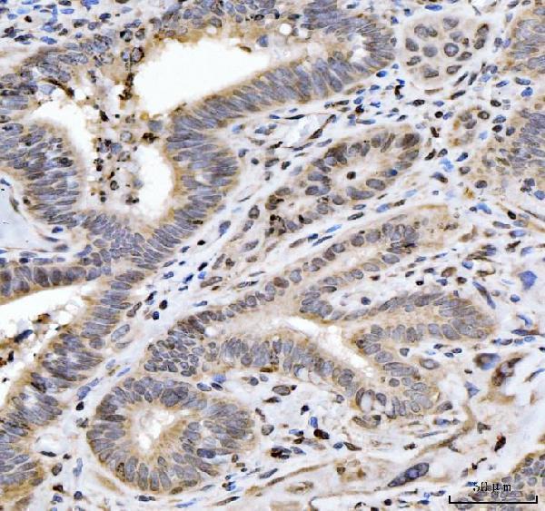
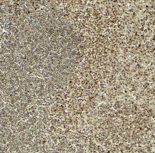
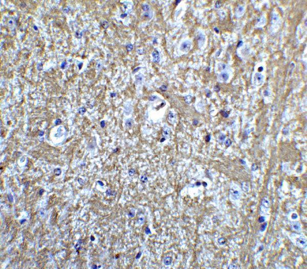
The catalog shows HES5 IHC images in human bladder epithelial carcinoma and spleen and mouse brain, plus IF images in human spleen and mouse brain (catalog image captions).
A05384-1 has IHC images from paraffin sections of human bladder epithelial carcinoma, gall bladder adenosquamous carcinoma and placenta (A05384-1 image captions). A05384 has IHC and IF images from human spleen and mouse brain (A05384 image captions).
Which to pick: For human paraffin-section IHC, choose A05384-1: its own caption documents EDTA retrieval and staining at 2 μg/ml; the fixative is unreported (A05384-1 IHC caption). For cross-species IHC or tissue IF, choose A05384: it lists human, mouse and rat reactivity and IHC-P and IF applications, with human and mouse tissue images; its image captions do not report a fixative (A05384 catalog and image captions). Neither listing specifies a clone, and ICC validation is not listed (catalog: clone and applications fields).