This website uses cookies to ensure you get the best experience on our website.


- Table of Contents
Plan chromogenic HINT1 staining in paraffin sections with the catalog antibody’s 0.5–1 μg/mL IHC range (datasheet PB9632). Use cytoplasmic and occasional nuclear staining as the tissue pattern, with kidney tubule cells as a high-staining reference (HPA tissue IHC).

Expected localisation, validated protocols, controls and antibodies — the at-a-glance facts below, then the full design guide.
| Expected localisation | Mainly cytoplasmic; occasional nuclear staining (HPA tissue IHC) | |
| Staining pattern | Cytoplasmic, sometimes nuclear, in glandular and tubular cells (HPA tissue IHC) | |
| Antigen retrieval | EDTA pH 8.0 HIER, heat-mediated (datasheet PB9632) | |
| Positive control | Epididymis+4 more · see all | |
| Negative control | Heart muscle+4 more · see all |
| Fixation | Matched tissue-IHC evidence does not establish the fixation claim. Validate the specimen-specific method before use. (selected-SKU IHC image PB9632) | |
| Caveat | Antibody staining and RNA show medium consistency (HPA tissue IHC) | |
| Regulation | Intensity regulation unreported (UniProt) | |
| Isoform / epitope | No listed isoforms; one chain spans residues 2–126 (UniProt) |
The catalog antibody’s IHC-P protocol is accompanied by published HINT1 staining methods from four articles (PMC3522093; PMC7922225; PMC7592009; PMC11850129).
| Sample | Paraffin-embedded rat kidney tissue; fixative not specified (datasheet PB9632) |
| Fixation | Image fixative and duration unreported (datasheet PB9632); verify before use. |
| Sectioning | 4–5 µm sections on charged slides (standard) |
| Deparaffinisation | Xylene, graded ethanol series to water (standard) |
| Antigen retrieval | Heat retrieval: EDTA pH 8.0 (datasheet PB9632); 20 min, 95–100 °C (standard) |
| Peroxidase block | 3% H2O2, 10 min, room temperature (standard) |
| Blocking | 10% goat serum (datasheet PB9632) |
| Primary antibody | Rabbit anti-HINT1, 0.5-1μg/ml (datasheet PB9632) |
| Primary incubation | Overnight at 4 °C (datasheet PB9632) |
| Detection | Streptavidin-biotin complex (SABC), DAB chromogen (datasheet PB9632) |
| Counterstain | Hematoxylin, blue, dehydrate and mount (standard) |
| Expected result | HINT1-positive staining in glandular cells of epididymis (HPA tissue IHC: High). HPA tissue profile: General cytoplasmic and occasional nuclear expression. No signal in the no-primary control. |
HINT1 staining in paraffin sections is generally cytoplasmic, with occasional nuclear signal (HPA tissue IHC; UniProt P49773: cytoplasm and nucleus). Look for staining in glandular cells of the epididymis, gallbladder, and seminal vesicle; kidney tubular cells and ovarian follicle cells are also strong reference populations (HPA: High). HINT1 has no transmembrane segment (UniProt P49773 topology). HPA rates the tissue IHC pattern Approved, with medium consistency against RNA expression (HPA tissue IHC).
| Cytoplasmic staining in kidney tubular cells, with occasional stained nuclei. | This fits the reported compartment pattern (HPA tissue IHC) and a High kidney reference population (HPA: High in tubular cells). Score nuclear and cytoplasmic signal separately; occasional nuclear staining can be genuine (UniProt P49773: nucleus; HPA tissue IHC). |
| Signal is concentrated in luminal material or extracellular deposits, with little staining inside the expected cells. | That distribution does not match general cytoplasmic and occasional nuclear HINT1 staining (HPA tissue IHC). Inspect morphology and compare with the detection-only control before assigning it to HINT1 (standard IHC practice). Plasma-membrane localization remains uncertain in ICC-IF (HPA subcellular). |
| Strong staining appears in cardiomyocytes or alveolar cells, while expected positive cell populations are weak. | These populations were Not detected in the supplied tissue survey (HPA: cardiomyocytes; alveolar cells). Recheck cell identification and assay controls; cross-reactivity or endogenous chromogenic activity is possible (standard IHC practice). An HPA Not detected call is not proof of universal absence. |
| Color is diffuse across tissue, empty spaces, or several cell populations without a clear intracellular pattern. | Diffuse color cannot establish the expected cytoplasmic localization (HPA tissue IHC). Check detection-only and substrate controls, washing, and blocking to distinguish background from cellular staining (standard IHC practice). Interpret intensity only after identifying the stained cells. |
| No staining is visible in a section containing kidney tubular cells or gallbladder glandular cells. | Both are High reference populations in the HPA survey (HPA: kidney tubules; gallbladder glandular cells). Confirm that those cells are present and the positive control develops, then review the IHC-validated antibody and detection workflow (standard IHC practice). A single failed section does not establish HINT1 absence. |
| Cell population and tissue context | HPA reports High staining in kidney tubules, ovarian follicle cells, and several glandular populations, but Not detected in cardiomyocytes and alveolar cells (HPA tissue IHC). Compare matched cell populations when judging a section; broad expression alone does not predict equal staining in every cell (UniProt P49773: widely expressed; HPA tissue IHC). |
| Nuclear versus cytoplasmic distribution | Cytoplasm and nucleus are annotated locations, and interaction with CDK7 can shift HINT1 toward the nucleus (UniProt P49773). HPA tissue IHC describes general cytoplasmic and occasional nuclear expression. Record the compartments separately instead of treating every stained nucleus as nonspecific. |
| Membrane interpretation | HINT1 has no transmembrane segment (UniProt P49773 topology). HPA ICC-IF supports cytosol and nucleoplasm, while its plasma-membrane assignment is uncertain (HPA subcellular). An apparently membrane-dominant IHC pattern therefore needs control review before being called a characteristic HINT1 pattern. |
| Evidence and assay limits | The tissue pattern is Approved, with medium consistency against RNA expression (HPA tissue IHC). Antibody HPA044577 is Approved for IHC and Supported for ICC (HPA antibodies). These ratings support cautious interpretation; they do not establish fixation sensitivity, an optimal retrieval condition, or a universal positive threshold. |
| Situation | Likely cause | Next action |
|---|---|---|
| A known High reference population is blank (HPA: kidney tubular cells or gallbladder glandular cells). | The section may lack the identified target cells, or the staining run may have failed (standard IHC practice). | Verify morphology and a run control; review antibody application, retrieval, detection, and counterstain records without assuming a HINT1-specific retrieval requirement (standard IHC practice). |
| The whole section has weak, even chromogenic color. | Endogenous detection activity, incomplete blocking, or residual detection reagent can produce background (standard IHC practice). | Compare detection-only and substrate controls, then review blocking and washing steps (standard IHC practice). Score HINT1 only where intracellular color resolves in identified cells (HPA tissue IHC). |
| Cardiomyocytes or alveolar cells stain strongly (HPA: Not detected in these cells). | Misidentified cells, cross-reactivity, or endogenous chromogenic activity are possible explanations (standard IHC practice). | Check cell morphology and controls, then compare an HPA High reference population in the same staining run (HPA tissue IHC; standard IHC practice). Do not infer specificity from intensity alone. |
| Staining appears exclusively extracellular or in luminal contents. | That pattern differs from general cytoplasmic and occasional nuclear HINT1 staining (HPA tissue IHC). | Inspect whether stain follows cells, compare detection-only controls, and report the discordant distribution rather than scoring deposits as positive cells (standard IHC practice). |
| Nuclear staining looks stronger than expected. | Nuclear localization is possible, and CDK7 interaction can increase it (UniProt P49773); HPA tissue IHC describes nuclear staining as occasional. | Score nuclear and cytoplasmic compartments independently, confirm cell identity, and compare with the cytoplasmic pattern in an HPA High population (HPA tissue IHC; standard IHC practice). |
| Q: Should IF/ICC show the same compartments as tissue IHC? | The assays have different observations: HPA tissue IHC reports general cytoplasmic and occasional nuclear staining; ICC-IF supports cytosol and nucleoplasm, with plasma membrane uncertain (HPA tissue IHC; HPA subcellular). | A: Compare compartment calls within each assay and treat an apparent membrane signal cautiously (HPA subcellular). Consult the separate IF/ICC guide for its workflow; this section interprets paraffin-section IHC. |


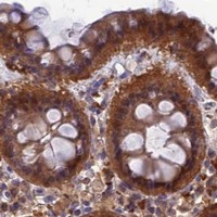

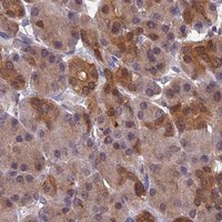


Comprehensive Human Protein Atlas IHC scoring per tissue (reliability: Approved — Medium consistency between antibody staining and RNA expression data.). Rows are taken directly from the HPA tissue chart — click any row's HPA link to view the source.
| Tissue | Cell type | Level | Evidence | Source |
|---|---|---|---|---|
| Epididymis | Glandular cells | High | Protein (IHC) | HPA → |
| Fallopian tube | Ciliated cells (cell body) | High | Protein (IHC) | HPA → |
| Gallbladder | Glandular cells | High | Protein (IHC) | HPA → |
| Kidney | Cells in tubules | High | Protein (IHC) | HPA → |
| Ovary | Follicle cells | High | Protein (IHC) | HPA → |
| Tissue | Cell type | Level | Evidence | Source |
|---|---|---|---|---|
| Heart muscle | Cardiomyocytes | Not detected | Protein (IHC) | HPA → |
| Lung | Alveolar cells | Not detected | Protein (IHC) | HPA → |
| Placenta | Trophoblastic cells | Not detected | Protein (IHC) | HPA → |
| Skeletal muscle | Myocytes | Not detected | Protein (IHC) | HPA → |
| Smooth muscle | Smooth muscle cells | Not detected | Protein (IHC) | HPA → |
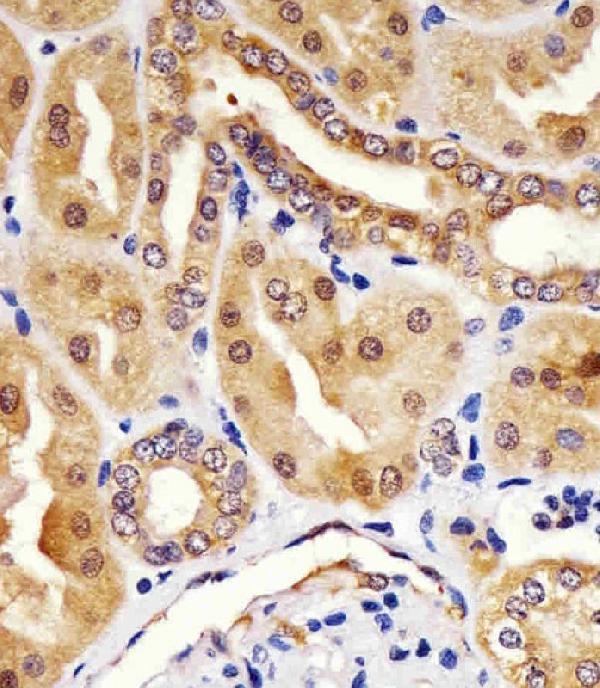
Use the selected paraffin-section image as the starting point for HINT1 IHC, then assess staining by cell type and compartment (datasheet PB9632; HPA tissue IHC).
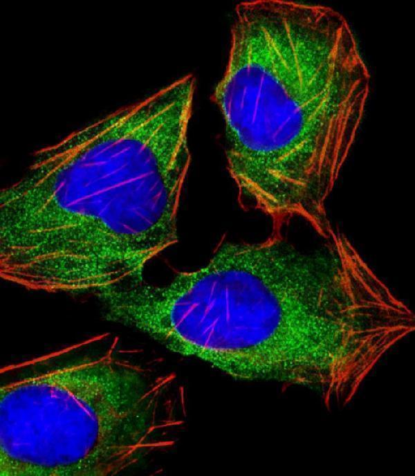
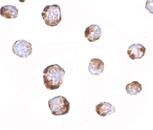
IHC images show HINT1 in rat and human kidney paraffin sections (PB9632 and M02557 captions); an ICC image shows Jurkat cells (A02557 caption). All three list human, mouse and rat reactivity (catalog reactivity).
PB9632 has IHC images of rat kidney, human intestinal cancer and mouse intestine paraffin sections, plus an IF image of A431 cells (PB9632 captions); M02557 has human kidney IHC-P and U-2 OS cell IF images (M02557 captions). A02557 has an ICC image of Jurkat cells and lists ICC among its applications (A02557 caption; catalog applications).
Which to pick: For tissue IHC across the three pictured species, choose PB9632: its IHC captions show rat kidney, human intestinal cancer and mouse intestine paraffin sections, and list 1 μg/ml primary antibody; the fixative is unreported (PB9632 IHC captions). For paraffin-section human kidney IHC-P, M02557 is a mouse monoclonal shown with citrate pH 6 retrieval at 1:25 (M02557 catalog clone and IHC caption). For cell IF, M02557 has a U-2 OS IF image at 1:25, while A02557 has a Jurkat ICC image at 2.5 μg/mL; choose according to the pictured assay (M02557 IF caption; A02557 ICC caption). The selected PB9632 tissue-IHC caption documents paraffin sections, but does not specify the fixative (selected-SKU IHC image PB9632).