This website uses cookies to ensure you get the best experience on our website.
- Table of Contents
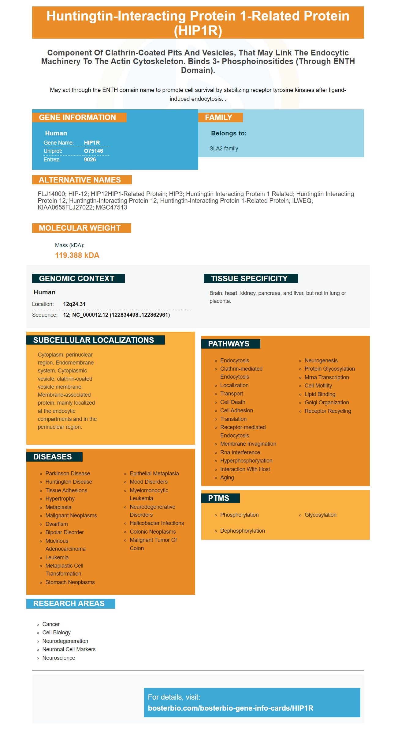
Facts about Huntingtin-interacting protein 1-related protein.
May act through the ENTH domain name to promote cell survival by stabilizing receptor tyrosine kinases after ligand-induced endocytosis. .
Human | |
---|---|
Gene Name: | HIP1R |
Uniprot: | O75146 |
Entrez: | 9026 |
Belongs to: |
---|
SLA2 family |
FLJ14000; HIP-12; HIP12HIP1-related protein; HIP3; huntingtin interacting protein 1 related; huntingtin interacting protein 12; Huntingtin-interacting protein 12; huntingtin-interacting protein 1-related protein; ILWEQ; KIAA0655FLJ27022; MGC47513
Mass (kDA):
119.388 kDA
Human | |
---|---|
Location: | 12q24.31 |
Sequence: | 12; NC_000012.12 (122834498..122862961) |
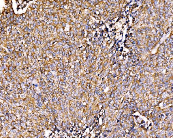
Brain, heart, kidney, pancreas, and liver, but not in lung or placenta.
Cytoplasm, perinuclear region. Endomembrane system. Cytoplasmic vesicle, clathrin-coated vesicle membrane. Membrane-associated protein, mainly localized at the endocytic compartments and in the perinuclear region.
Recombinant protein was captured by membrane staining using an anti-DDK affinity column and chromatography steps, including an ECL chemiluminescent detection system. The boster capture method has been validated and is applicable to scientists worldwide. The boster bio: HIP1R mAb is readily available in the market and is used for the detection of this protein.
Recombinant proteins are produced in large quantities at Boster Bio, a leading provider of recombinant protein production services. The company's expert scientists are trained to design efficient expression vectors and perform over 3000 protein expressions annually for their customers. The Boster protein production service also offers extensive consulting and consultation to help its clients choose the right recombinant protein production option for their needs.
Recombinant protein was purified using an anti-DDK affinity column. The resulting protein was used to determine the structure of Entamoeba histolytica fusion proteins. Afterwards, the protein was purified and identified as a native N-terminal fusion product. The boster protein capture method is a valuable tool for researchers worldwide, and is available at a price that suits the budget of every research institute.
Recombinant protein purification at low temperatures minimizes the effect of temperature on protein stability and structural integrity. High affinity of antibody-based purification resins often leads to incomplete elution, but at low temperatures, this problem is reduced even further. The elution of Spot-tagged proteins was optimized at +4 degC for optimal results.
Another popular method for protein production is the use of Escherichia coli. However, Escherichia coli has many disadvantages, such as low yield, inclusion bodies, and difficult purification procedures. The use of fusion proteins may solve many of these problems. The CusF3H+ metal-binding protein is one example. The CusF3H+ system produces high-quality soluble proteins with minimal inclusion bodies.
The Boster Bio recombinant protein is captured through membrane staining and was analyzed using conventional chromatography steps. The process can be used by scientists from all over the world as it uses a high-quality membrane. This technique allows scientists to capture peak antigen specificity and is applicable to all protein types. To maximize the quality of boster protein, the transfer buffer should be freshly prepared and the marker should be pre-stained.
A sandwich ELISA is another method used to capture Boster Bio recombinant protein through membrane staining. This technique is popular because of its high sensitivity and specificity. This method is also known as competitive ELISA. The primary antibody is labeled with the reagent to capture the recombinant protein, while the secondary antibody is unlabeled. This results in a decrease in the signal from the purified antigen, which indicates that the sample contains the antigen.
A boster biotechnology company has produced an ELISA kit based on the HIP1R marker for the study of human leukemia cells. The boster biotechnology company's proprietary ELISA platform is based on proprietary trade secrets and delivers high-sensitivity ELISA kits. The boster biotechnology company is an example of a global leader in antibody development and manufacturing.
Enhanced chemiluminescent detection (ECL) is a popular method to detect proteins in cell culture. This method uses a secondary antibody labeled with an enzyme (typically a horseradish peroxidase or an alkaline phosphatase). The light emitted from the protein is detected by exposing the Western blot to a CCD camera or X-ray film. The resulting band will be viewed as a signal that indicates that the antibody is binding to the target protein.
This method is superior to traditional colorimetric assays because it provides superior sensitivity for low and medium levels of protein expression. The signal can be detected even in low femtogram amounts of a protein, and it can be reimagined several times without losing signal intensity. The reagents for this technique are highly sensitive and can detect a wide range of proteins.
The C-DiGit(r) Blot Scanner is another important feature of the ECL chemiluminescent detection systems. This scanner eliminates the need for darkroom costs and developing reagents. Additionally, it has a wide dynamic range, which minimizes the need for multiple exposures. For more information, check out the Lambda U(r) education portal.
Enhanced chemiluminescent substrates have improved the sensitivity of the reaction. The Pierce ECL Western Blotting Substrate is a value-priced HRP substrate that can replace pricier products. The Pierce ECL Western Blotting Substrate offers reliable and high-quality results comparable to other standard ECL substrates. The ECL substrate is highly sensitive and produces low backgrounds, which makes it suitable for western blot applications. Its low background levels enable detection of HRP on x-ray film or CCD imaging systems.
A chromogenic detection system is critical for accurate detection of fluorescent proteins in biological samples. This process is based on a reaction between an enzyme known as alkaline phosphatase and the protein under investigation. Alkaline phosphatase is a key enzyme that induces a color shift from pink to blue. In this system, the protein of interest is stained with a chromogenic substrate. The reagents used for this technique are readily available through Boster Bio or other suppliers.
PMID: 11063258 by Chopra V.S., et al. HIP12 is a non-proapoptotic member of a gene family including HIP1, an interacting protein with huntingtin.
PMID: 9852681 by Seki N., et al. Cloning, expression analysis, and chromosomal localization of HIP1R, an isolog of huntingtin interacting protein (HIP1).