This website uses cookies to ensure you get the best experience on our website.


- Table of Contents
Plan HIP1R staining in paraffin sections using the observed cytoplasmic and occasional membranous tissue pattern (HPA tissue IHC). Compare cell types and include controls when interpreting staining intensity.

Expected localisation, validated protocols, controls and antibodies — the at-a-glance facts below, then the full design guide.
| Expected localisation | Cytoplasmic staining with some membranous staining (HPA tissue IHC) | |
| Staining pattern | Glandular cells: cytoplasmic, sometimes membranous staining (HPA tissue IHC) | |
| Antigen retrieval | EDTA pH 8.0 HIER, heat-mediated (datasheet A05059-2) | |
| Positive control | Appendix+4 more · see all | |
| Negative control | Adipose tissue+4 more · see all |
| Fixation | Keep fixation consistent across paraffin sections. (standard IHC practice; not target-specific) | |
| Caveat | Staining and RNA expression show medium consistency (HPA tissue IHC) | |
| Regulation | No expression regulator specified (UniProt) | |
| Isoform / epitope | 2 isoforms; epitope differences unspecified (UniProt) |
The catalog antibody protocol (datasheet A05059-2) is paired with published HIP1R IHC protocols for diffuse gliomas (PMC12063079) and lung cancer (PMC7291156).
| Sample | Paraffin-embedded human liver cancer tissue; fixative not specified (datasheet A05059-2) |
| Fixation | Image fixative and duration unreported (datasheet A05059-2); verify before use. |
| Sectioning | 4–5 µm sections on charged slides (standard) |
| Deparaffinisation | Xylene, graded ethanol series to water (standard) |
| Antigen retrieval | Heat retrieval: EDTA pH 8.0 (datasheet A05059-2); 20 min, 95–100 °C (standard) |
| Peroxidase block | 3% H2O2, 10 min, room temperature (standard) |
| Blocking | 10% goat serum (datasheet A05059-2) |
| Primary antibody | Rabbit anti-HIP1R, 2-5 μg/ml (datasheet A05059-2) |
| Primary incubation | Overnight at 4 °C (datasheet A05059-2) |
| Detection | Streptavidin-biotin complex (SABC), DAB chromogen (datasheet A05059-2) |
| Counterstain | Hematoxylin, blue, dehydrate and mount (standard) |
| Expected result | HIP1R-positive staining in glandular cells of appendix (HPA tissue IHC: High). HPA tissue profile: General cytoplasmic expression combined with membranous expression in several tissues. No signal in the no-primary control. |
HIP1R is membrane-associated at endocytic compartments and the perinuclear region, with no transmembrane segment (UniProt O75146 localization and topology). In paraffin-section IHC, expect cytoplasmic staining with membranous staining in some tissues (HPA: tissue IHC profile). High staining is reported in several glandular cell populations, bronchial goblet cells, cerebellar Purkinje cells, and fallopian-tube ciliated cell bodies (HPA: High). HPA rates tissue IHC reliability Enhanced, while reporting medium agreement with RNA expression (HPA: reliability).
| Cytoplasmic staining, sometimes with a membrane-associated component, is concentrated in the expected cells. | This fits the tissue IHC profile (HPA: general cytoplasmic expression with membranous expression in several tissues). Examples of strongly stained cells include colon and duodenal glandular cells and cerebellar Purkinje cells (HPA: High). Assess the named cell population, since neighboring cell types need not share its staining level (HPA: cell-specific tissue IHC observations). |
| The dominant signal is nuclear, with little cytoplasmic or membranous staining. | A dominant nuclear pattern conflicts with the reported compartments (UniProt O75146 localization; HPA: tissue IHC profile). Treat it as suspect, then compare morphology, the no-primary control, and an expected-positive area before assigning HIP1R positivity (general IHC practice). |
| Strong staining appears in adipocytes or skeletal-muscle myocytes. | Those specific cell populations were not detected in the HPA tissue observations (HPA: adipocytes and skeletal-muscle myocytes, Not detected). Check the no-primary control for endogenous detection activity and reassess antibody specificity if the signal persists (general IHC practice). A negative entry for one cell population does not classify every cell in that tissue (HPA: cell-specific tissue IHC observations). |
| Brown color spreads across unrelated structures and obscures cell boundaries. | This prevents a reliable compartment or cell-type call (general IHC practice). Possible contributors include nonspecific antibody binding, endogenous detection activity, or excessive chromogen development (general IHC practice). Compare controls and tissue morphology before calling the diffuse color HIP1R staining (general IHC practice). |
| No staining is visible in an expected-positive cell population. | First confirm that the selected population is actually present: appendix glandular cells and bronchial goblet cells are reported High, for example (HPA: tissue IHC). An absent signal warrants checking the IHC-validated antibody, detection reagents, and run controls (general IHC practice). HPA's Enhanced rating supports the observed pattern but does not guarantee signal in every preparation (HPA: reliability). |
| Compartment and topology | HIP1R is associated with clathrin-coated vesicle membranes and the perinuclear endomembrane system, without a transmembrane segment (UniProt O75146 localization and topology). Interpret a membranous component alongside cytoplasmic staining; do not require a sharp, continuous cell-surface rim to recognize the reported IHC pattern (HPA: tissue IHC profile). |
| Tissue and cell selection | HPA reports High staining in named glandular, goblet, Purkinje, and ciliated cell populations, but Not detected in specified adipocytes, myocytes, smooth-muscle cells, chondrocytes, and parathyroid glandular cells (HPA: tissue IHC). Select controls by cell population rather than tissue name alone (general IHC practice). |
| Evidence strength | HPA038135 and HPA038136 have Enhanced IHC validation, whereas CAB017187 is Approved (HPA: antibody validation). The tissue profile still has medium consistency with RNA expression (HPA: reliability). Use those ratings as context when weighing an unexpected result, rather than treating RNA or one stained section as decisive. |
| Isoforms and antibody coverage | UniProt lists 2 HIP1R isoforms (UniProt O75146 isoforms). Their existence alone does not establish which isoforms a given IHC antibody recognizes; resolve an isoform-specific question from that antibody's documented epitope or validation data before interpreting a discordant stain (general IHC practice). |
| IF/ICC Q: what localization is expected? | HPA reports mainly plasma membrane and cytosol, with additional vesicles, in its ICC-IF summary (HPA: subcellular ICC-IF). This supports a compartment comparison, while the paraffin-section chromogenic IHC call should follow the tissue IHC profile (HPA: tissue IHC). IF/ICC conditions belong to its separate guide. |
| Situation | Likely cause | Next action |
|---|---|---|
| Expected-positive cells are blank. | The population may be absent from the section, or an IHC staining step may have failed (general IHC practice). | Confirm the cells morphologically, then check a run control and the IHC-validated antibody's documented paraffin-section conditions (general IHC practice). Use a listed High population, such as colon glandular cells, when available (HPA: tissue IHC). |
| Only nuclei stain strongly. | The compartment conflicts with reported cytoplasmic and membrane-associated localization (UniProt O75146 localization; HPA: tissue IHC). | Inspect the counterstain and no-primary control; compare with the expected cytoplasmic pattern before scoring positive cells (general IHC practice; HPA: tissue IHC profile). |
| Listed Not detected cell populations stain strongly. | Nonspecific binding or endogenous detection activity is possible (general IHC practice); the result conflicts with the named HPA observations (HPA: tissue IHC). | Review cell identity and the no-primary control, then assess antibody specificity with the available validation information (general IHC practice; HPA: antibody validation). |
| Background masks a weak cellular pattern. | Nonspecific binding, endogenous detection activity, or overdeveloped chromogen can reduce interpretability (general IHC practice). | Use the relevant blocking and detection controls, review antibody dilution and development time, and score only clearly localized cellular signal (general IHC practice). |
| One tissue appears weaker than expected from a broad expression statement. | The records differ in granularity: UniProt lists tissue specificity, while HPA records staining levels for named cell types (UniProt O75146 tissue specificity; HPA: tissue IHC). | Compare the exact cell population with its HPA entry. For example, heart cardiomyocytes are Low and placental decidual cells are Low in the supplied HPA observations (HPA: tissue IHC). |
| Membranous staining seems inconsistent between IHC and IF images. | The summaries emphasize different observations: IHC is generally cytoplasmic with membranous staining in several tissues, while ICC-IF highlights plasma membrane, cytosol, and vesicles (HPA: tissue IHC; HPA: subcellular ICC-IF). | Interpret each image by its stated application and compare cell identity and compartment before calling disagreement (HPA: tissue IHC; HPA: subcellular ICC-IF; general IHC practice). |
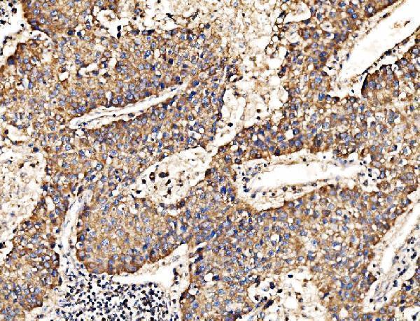
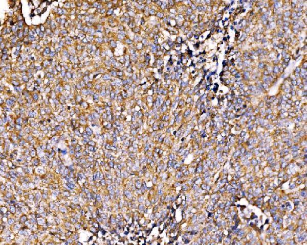
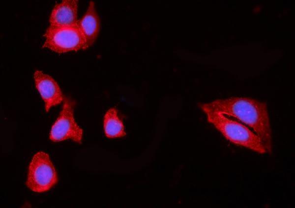


Comprehensive Human Protein Atlas IHC scoring per tissue (reliability: Enhanced — Medium consistency between antibody staining and RNA expression data.). Rows are taken directly from the HPA tissue chart — click any row's HPA link to view the source.
| Tissue | Cell type | Level | Evidence | Source |
|---|---|---|---|---|
| Appendix | Glandular cells | High | Protein (IHC) | HPA → |
| Bronchus | Goblet cells | High | Protein (IHC) | HPA → |
| Cerebellum | Purkinje cells | High | Protein (IHC) | HPA → |
| Colon | Glandular cells | High | Protein (IHC) | HPA → |
| Duodenum | Glandular cells | High | Protein (IHC) | HPA → |
| Tissue | Cell type | Level | Evidence | Source |
|---|---|---|---|---|
| Adipose tissue | Adipocytes | Not detected | Protein (IHC) | HPA → |
| Parathyroid gland | Glandular cells | Not detected | Protein (IHC) | HPA → |
| Skeletal muscle | Myocytes | Not detected | Protein (IHC) | HPA → |
| Smooth muscle | Smooth muscle cells | Not detected | Protein (IHC) | HPA → |
| Soft tissue | Chondrocytes | Not detected | Protein (IHC) | HPA → |
Troubleshoot HIP1R chromogenic IHC by checking retrieval, cytoplasmic and membranous staining, cell identity, and matched controls.
A05059-2 has IHC data from human paraffin sections (IHC image captions) and IF/ICC data from MCF-7 cells (IF image caption); catalog reactivity includes human, mouse, rat and monkey (catalog reactivity).
A05059-2 has IHC images from human paraffin sections of liver cancer, lung cancer, rectal cancer and renal clear cell carcinoma (IHC image captions). A05059-2 also has IF/ICC data in MCF-7 cells (IF image caption) and lists human, mouse, rat and monkey reactivity (catalog reactivity).
Which to pick: Choose A05059-2 for human paraffin-section IHC because its own IHC captions show that preparation; the fixative is unreported (IHC image captions). Choose A05059-2 for IF/ICC based on its MCF-7 image and listed IF/ICC applications (IF image caption; catalog applications). For other species, A05059-2 lists mouse, rat and monkey reactivity, but the supplied IHC and IF images show human samples only; clonality is unreported.