This website uses cookies to ensure you get the best experience on our website.


- Table of Contents
Plan chromogenic HLX IHC in paraffin sections with the catalog antibody's 1:100–1:300 dilution range (datasheet A30494). Compare the expected nuclear location (UniProt) with observed nuclear and cytoplasmic tissue staining, which has uncertain reliability (HPA tissue IHC).

Expected localisation, validated protocols, controls and antibodies — the at-a-glance facts below, then the full design guide.
| Expected localisation | Nuclear expected (UniProt); nuclear and cytoplasmic seen (HPA tissue IHC) | |
| Staining pattern | General nuclear and cytoplasmic staining (HPA tissue IHC) | |
| Antigen retrieval | Tris-EDTA pH 8.0 HIER, heat-mediated (datasheet A30494) | |
| Positive control | Adrenal gland+4 more · see all | |
| Negative control | Adipose tissue+4 more · see all |
| Fixation | Keep paraffin-section fixation consistent (standard IHC practice; not target-specific); Matched tissue-IHC evidence does not establish the fixation claim. Validate the specimen-specific method before use. (selected-SKU IHC image A30494) | |
| Caveat | Uncertain staining; low agreement with RNA (HPA tissue IHC) | |
| Regulation | High in activated lymphocytes (UniProt) | |
| Isoform / epitope | No isoforms or cleavage; one 1–488 chain (UniProt) |
Compare the catalog antibody’s IHC-P protocol with published HLX staining protocols for odontogenic cysts and mouse prostate cancer xenografts (PMC11857689; PMC13509987).
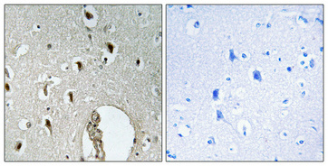
| Sample | Paraffin-embedded Human brain tissue; fixative not specified (datasheet A30494) |
| Fixation | Image fixative and duration unreported (datasheet A30494); verify before use. |
| Sectioning | 4–5 µm sections on charged slides (standard) |
| Deparaffinisation | Xylene, graded ethanol series to water (standard) |
| Antigen retrieval | Heat retrieval: Tris-EDTA pH 8.0 (datasheet A30494); 20 min, 95–100 °C (standard) |
| Peroxidase block | 3% H2O2, 10 min, room temperature (standard) |
| Blocking | 10% normal serum of the secondary host, 30 min, room temperature (standard) |
| Primary antibody | Rabbit anti-HLX, 1:100 - 1:300 (datasheet A30494) |
| Primary incubation | Overnight at 4 °C (standard) |
| Detection | HRP-polymer secondary, DAB chromogen 5–10 min (standard) |
| Counterstain | Hematoxylin, blue, dehydrate and mount (standard) |
| Expected result | HLX-positive staining in glandular cells of adrenal gland (HPA tissue IHC: High). HPA tissue profile: General cytoplasmic and nuclear expression. No signal in the no-primary control. |
HLX is a nuclear transcription factor with no transmembrane segment (UniProt Q14774). Expect nuclear staining in expressing cells, including bone marrow hematopoietic cells (HPA: High) and activated lymphocytes or monocytes (UniProt: tissue specificity). HPA also reports cytoplasmic and nuclear tissue staining, but rates its tissue IHC profile Uncertain because staining and RNA data have low consistency and external data contradict the reported location (HPA: tissue IHC reliability).
| Distinct nuclear staining in a subset of bone marrow hematopoietic cells, with restrained background. | This fits the reported high staining in bone marrow hematopoietic cells (HPA: High) and the nuclear location of HLX (UniProt Q14774). Record the fraction and intensity of positive cells separately; a whole-section average could hide a cell-restricted pattern (general IHC interpretation). HPA rates its tissue IHC profile Uncertain, so morphology alone does not establish antibody specificity (HPA: tissue IHC reliability). |
| Predominantly cytoplasmic staining, with little or no identifiable nuclear signal. | Treat this as ambiguous rather than an established HLX-positive result: UniProt places HLX in the nucleus (UniProt Q14774), while HPA reports general cytoplasmic and nuclear tissue staining with Uncertain reliability (HPA: tissue IHC). HPA separately approves nucleoplasm and cytosol localization in ICC-IF (HPA: subcellular), so cytoplasmic signal alone cannot prove an artefact. Check compartment boundaries and controls before scoring (general IHC practice). |
| Strong staining in a cell population selected as a low-expression or undetected comparator. | For example, adipocytes are reported as Not detected in adipose tissue (HPA: tissue IHC). Strong signal there warrants a cross-reactivity or endogenous detection-activity check (general IHC practice). It is a warning, not proof of either cause: HPA's tissue profile has Uncertain reliability (HPA: tissue IHC), and a comparator's reported level is specific to the listed cell population. |
| Diffuse color across cells and extracellular areas, obscuring nuclei. | A broad haze is difficult to interpret as cell-specific HLX staining (general IHC interpretation). Assess background with a no-primary control and inspect blocking, washes and chromogen development (general chromogenic IHC practice). Do not infer HLX expression from overall section color when the compartment and positive cells cannot be resolved; nuclear location is the UniProt expectation (UniProt Q14774). |
| No signal in bone marrow hematopoietic cells included as a positive comparator. | HPA reports High staining in these cells (HPA: tissue IHC), so first check that the comparator contains recognizable hematopoietic cells and that the staining run worked (general IHC practice). A blank result may reflect assay failure or sample variation; it cannot establish that all HLX expression is absent. Interpret cautiously because HPA rates the tissue profile Uncertain (HPA: tissue IHC reliability). |
| Cell population chosen for interpretation | Bone marrow hematopoietic cells are reported High, whereas adipocytes in adipose tissue are Not detected (HPA: tissue IHC). Compare the named cells, not entire tissues; activated lymphocytes and monocytes are described as high-level contexts by UniProt (UniProt Q14774: tissue specificity). These sources do not establish an intensity threshold for every cell in a section. |
| Compartment and evidence strength | UniProt places HLX in the nucleus (UniProt Q14774); HPA tissue IHC reports both cytoplasmic and nuclear expression but is Uncertain (HPA: tissue IHC). HPA005968 is Uncertain for IHC and Approved for ICC (HPA: antibody validation). Require compartment-resolved, controlled interpretation before treating chromogenic cytoplasmic signal as HLX. |
| Topology and processing | HLX has no transmembrane segment or signal peptide, and its annotated chain spans residues 1–488 (UniProt Q14774). These annotations support interpreting it as a cellular transcription factor rather than expecting a membrane outline or secreted deposit (UniProt Q14774: function and topology). They do not identify an antibody epitope or predict antigen retrieval performance. |
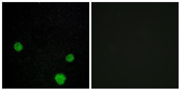
| IF/ICC: what location is reported? | HPA approves nucleoplasm and cytosol localization in ICC-IF and lists images from HEK293, U-251MG and U2OS (HPA: subcellular). That cell-imaging observation can inform compartment assessment, but it does not upgrade the Uncertain tissue IHC profile or supply an IF/ICC protocol (HPA: subcellular; HPA: tissue IHC reliability). |
| Situation | Likely cause | Next action |
|---|---|---|
| Bone marrow comparator is blank. | The run may have failed, or the examined area may lack the relevant hematopoietic cells (general IHC practice); HPA reports these cells High (HPA: tissue IHC). | Confirm the cell population on the section, review run controls, then optimize the routine IHC workflow if needed (general IHC practice). Do not assign an HLX-negative score from a failed comparator. |
| Signal is mainly cytoplasmic. | Tissue IHC localization is disputed: UniProt says nucleus, while HPA reports cytoplasmic and nuclear staining with Uncertain reliability (UniProt Q14774; HPA: tissue IHC). | Re-examine nuclear boundaries and controls; report the observed compartment explicitly (general IHC interpretation). Keep a cytoplasm-only result provisional, while acknowledging HPA's approved cytosol ICC-IF location (HPA: subcellular). |
| Adipocytes stain strongly in the negative comparator. | HPA reports adipocytes as Not detected (HPA: tissue IHC); cross-reactivity or endogenous detection activity is possible (general IHC practice). | Run a no-primary detection control and inspect whether signal follows cells or the detection chemistry (general IHC practice). Reassess specificity before counting these cells as HLX positive. |
| All compartments show diffuse chromogen. | Nonspecific background or excessive development may obscure cell boundaries (general chromogenic IHC practice). | Check no-primary background, blocking, washes and development time (general chromogenic IHC practice). Score only cells with resolvable staining; UniProt's nuclear location provides the primary compartment reference (UniProt Q14774). |
| Few cells stain despite a clearly stained bone marrow comparator. | HLX expression can vary by cell population: UniProt describes low levels in normal B and T cells and high levels in activated lymphocytes and monocytes (UniProt Q14774: tissue specificity). | Identify and score the relevant cell populations separately, retaining intensity and positive-cell fraction (general IHC interpretation). Do not apply the bone marrow High designation to every cell in the test section (HPA: tissue IHC). |
| A membrane rim or extracellular deposit dominates the slide. | That distribution conflicts with nuclear localization and the absence of a transmembrane segment or signal peptide (UniProt Q14774). | Check morphology and detection controls, then withhold a positive HLX call unless compartment-resolved cellular signal is present (general IHC practice; UniProt Q14774: nucleus). |
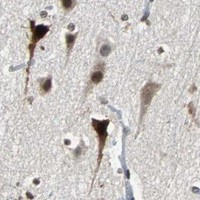


Comprehensive Human Protein Atlas IHC scoring per tissue (reliability: Uncertain — Low consistency between antibody staining and RNA expression data. Subcellular location is contradicted by external data. Pending external verification.). Rows are taken directly from the HPA tissue chart — click any row's HPA link to view the source.
| Tissue | Cell type | Level | Evidence | Source |
|---|---|---|---|---|
| Adrenal gland | Glandular cells | High | Protein (IHC) | HPA → |
| Bone marrow | Hematopoietic cells | High | Protein (IHC) | HPA → |
| Caudate | Neuronal cells | High | Protein (IHC) | HPA → |
| Cerebellum | Cells in molecular layer | High | Protein (IHC) | HPA → |
| Cerebral cortex | Neuronal cells | High | Protein (IHC) | HPA → |
| Tissue | Cell type | Level | Evidence | Source |
|---|---|---|---|---|
| Adipose tissue | Adipocytes | Not detected | Protein (IHC) | HPA → |
| Colon | Endothelial cells | Not detected | Protein (IHC) | HPA → |
| Duodenum | Glandular cells | Not detected | Protein (IHC) | HPA → |
| Esophagus | Squamous epithelial cells | Not detected | Protein (IHC) | HPA → |
| Small intestine | Glandular cells | Not detected | Protein (IHC) | HPA → |
Troubleshoot HLX staining in paraffin sections by checking retrieval, nuclear localisation, tissue context and controls before interpreting chromogenic signal.
The catalog lists anti-HLX antibodies for IHC and IF with human, mouse, and rat reactivity (catalog applications/reactivity); A30494 has pictured paraffin-section IHC and cell IF results (A30494 image captions).
A06177-1 lists IHC and IF applications and human, mouse, and rat reactivity, but provides no IHC or IF image caption (A06177-1 applications/reactivity/image alts). A30494 lists IHC, IF, and ICC and the same reactivity; its captions show paraffin-embedded human brain IHC and MCF7 cell IF (A30494 applications/reactivity/image captions).
Which to pick: For tissue IHC, pick A30494: its own caption documents paraffin-embedded human brain, 1:100 overnight at 4°C, and Tris-EDTA pH 8 retrieval; the fixative is unreported (A30494 IHC image caption). For IF/ICC, pick A30494 because its IF caption shows MCF7 cells and its application list includes both IF and ICC; it is described as polyclonal (A30494 IF image caption; A30494 applications; A30494 dilution_raw). For human, mouse, or rat samples, both SKUs list reactivity, while pictured results are available only for A30494 and clonality is unreported for A06177-1 (catalog reactivity/image alts; A30494 dilution_raw; A06177-1 clone).