HMGA2 / High mobility group protein HMGI-C · IHC design guide

Design Immunohistochemistry for HMGA2

Plan HMGA2 paraffin IHC around nuclear staining, using testis preleptotene spermatocytes as a strong positive reference (HPA tissue IHC). The guide covers the catalog antibody’s chromogenic workflow and cell-specific scoring, with adipocytes as a negative reference (datasheet A00436-3; HPA tissue IHC).

Evidence assembled Oct 2026 · For research use; verify linked source records and product datasheet before use
Immunohistochemistry protocol sheet for HMGA2 (IHC for HMGA2): expected localisation Nuclear staining in most tissues (HPA tissue IHC), antibody A00436-3, validated IHC image, and IHC protocol steps
Printable HMGA2 IHC protocol sheet — expected localisation Nuclear staining in most tissues (HPA tissue IHC), antibody A00436-3, controls and protocol steps. Open the full HMGA2 IHC guide →

HMGA2 Immunohistochemistry Experimental Design Guide

Expected localisation, validated protocols, controls and antibodies — the at-a-glance facts below, then the full design guide.

Must know before staining
Expected localisation Nuclear staining in most tissues (HPA tissue IHC)
Staining pattern Nuclear staining in most tissues; high in testis preleptotene spermatocytes (HPA tissue IHC)
Antigen retrieval EDTA pH 8.0 HIER, heat-mediated (datasheet A00436-3)
Positive control ⓘ Testis+4 more · see all
Negative control ⓘ Adipose tissue+1 more · see all
Important caveats
Reasons your staining may differ from the expected pattern.
Fixation Keep fixation consistent across paraffin sections (standard IHC practice; not target-specific)
Caveat Adipocytes have no detectable staining (HPA tissue IHC)
Regulation No expression regulator annotated (UniProt)
Isoform / epitope 6 isoforms; epitope coverage needs verification (UniProt)
Section 1

Recommended HMGA2 IHC & IF Protocols

Start with the catalog antibody’s IHC protocol, then compare four published HMGA2 staining protocols (PMC2376847; PMC5891726; PMC12086272; PMC12984743).

Recommended immunohistochemistry (IHC-P) protocol parameters
SampleParaffin-embedded human lung cancer tissue; fixative not specified (datasheet A00436-3)
FixationImage fixative and duration unreported (datasheet A00436-3); verify before use.
Sectioning4–5 µm sections on charged slides (standard)
DeparaffinisationXylene, graded ethanol series to water (standard)
Antigen retrievalHeat retrieval: EDTA pH 8.0 (datasheet A00436-3); 20 min, 95–100 °C (standard)
Peroxidase block3% H2O2, 10 min, room temperature (standard)
Blocking10% goat serum (datasheet A00436-3)
Primary antibodyRabbit anti-HMGA2, 1:50 recommended; image 1:100 (datasheet A00436-3)
Primary incubationOvernight at 4 °C (datasheet A00436-3)
DetectionHRP-conjugated secondary, DAB chromogen (datasheet A00436-3)
CounterstainHematoxylin, blue, dehydrate and mount (standard)
Expected resultHMGA2-positive staining in preleptotene spermatocytes of testis (HPA tissue IHC: High). HPA tissue profile: Nuclear expression in most tissues. No signal in the no-primary control.
💡Decision noteFor paraffin sections, try heat-mediated EDTA retrieval at pH 8.0 first (datasheet: A00436-3); a published alternative uses citrate at pH 6.0 (PMC12984743).
Section 2

What Is the Expected HMGA2 Staining Pattern?

HMGA2 should appear predominantly in nuclei across most tissues (HPA tissue IHC: Approved; nuclear expression in most tissues; UniProt P52926: nucleus). The clearest supplied positive is preleptotene spermatocytes in testis, with High staining (HPA tissue IHC: testis). HMGA2 has no transmembrane segment, so a membrane-only pattern is unexpected (UniProt P52926: topology). HPA reports no tissue-specificity annotation from UniProt (UniProt P52926: tissue specificity not annotated).

What am I looking at on my slide?
Distinct nuclear staining in preleptotene spermatocytes, with surrounding morphology preserved.This is the strongest listed positive pattern: HPA scores these testis cells High and describes nuclear expression in most tissues (HPA tissue IHC: testis; profile). Score the identified cell population, rather than treating every cell in the section as equally positive.
Nuclear staining in adrenal glandular or bronchial respiratory epithelial cells.These are plausible positive results at Medium intensity (HPA tissue IHC: adrenal gland; bronchus). Medium staining is also reported in selected glial, molecular-layer and glandular populations; intensity and cell identity should be read together (HPA tissue IHC: listed positive cells).
Predominantly membrane or cytoplasmic staining without a convincing nuclear component.Treat this as a localization mismatch requiring investigation: HMGA2 is nuclear and has no transmembrane segment (UniProt P52926: subcellular location; topology; HPA tissue IHC: nuclear profile). Uneven chromogen, section debris or nonspecific antibody binding are possible general IHC explanations, not established HMGA2-specific mechanisms.
Strong staining in adipocytes or seminal-vesicle glandular cells while nuclei in the expected positive population are unclear.HPA lists these cell types as Not detected (HPA tissue IHC: adipose tissue; seminal vesicle). Check for cross-reactivity or endogenous detection activity using appropriate controls (general IHC practice). An isolated positive cell is not, by itself, proof of a false positive.
A diffuse wash of color across cells and stroma, or no signal in testis preleptotene spermatocytes.Diffuse staining obscures the nuclear pattern; absent staining in the listed High population makes a negative result hard to interpret (HPA tissue IHC: profile; testis). Review controls and the IHC run before assigning either observation to HMGA2 biology (general IHC practice).
💡Expected HMGA2 appearanceCall a convincing positive when preleptotene spermatocyte nuclei show clear High staining (HPA tissue IHC: testis; nuclear profile); membrane-only color or dominant staining in HPA Not detected cell types warrants a control check (UniProt P52926: topology; HPA tissue IHC: adipocytes; seminal-vesicle glandular cells).
How each factor affects the staining
Tissue and cell selectionTestis preleptotene spermatocytes provide the listed High reference; adrenal glandular and bronchial respiratory epithelial cells are Medium (HPA tissue IHC). Low staining is reported in several other cell populations, so a weak field is less informative than a verified positive population (HPA tissue IHC: low list).
Localization and topologyNuclear localization is supported by both sources, while UniProt reports no transmembrane segment (HPA tissue IHC: profile; UniProt P52926: subcellular location; topology). Interpret compartment before intensity: strong color outside nuclei does not reproduce the stated tissue pattern.
Antibody evidenceThe HPA tissue pattern is rated Approved; its description cites external characterization and similar staining from paired antibodies (HPA tissue IHC: reliability). The listed antibodies HPA039076 and CAB017809 are each Approved for IHC (HPA antibodies). This supports the reference pattern without establishing performance for a different catalog antibody.
Isoforms and processingUniProt lists 6 isoforms, no signal peptide or propeptide, and a chain spanning residues 2–109 (UniProt P52926: isoforms; processing). The payload supplies no antibody epitope, so it cannot establish which isoforms a particular antibody detects or predict staining differences among them.
IF/ICC Q&A: where should fluorescence appear?HPA supports nucleoplasm, nucleoli and nucleoli-rim localization in ICC-IF, with images from A-431, U-251MG and U2OS (HPA ICC-IF: main location; image list). Use that as the IF/ICC compartment reference; the supplied tissue IHC profile establishes nuclear expression but does not resolve those subnuclear structures (HPA tissue IHC: profile).
Why is my staining missing, weak or wrong?
SituationLikely causeNext action
No nuclear signal in testis preleptotene spermatocytes.A failed or weak IHC run is possible; this population is listed High (HPA tissue IHC: testis). The supplied sources do not establish HMGA2-specific fixation sensitivity.Confirm the population and inspect positive-control staining, antibody dilution, retrieval conditions and detection reagents using the chosen assay's instructions (general IHC practice).
Only membrane or diffuse cytoplasmic color appears.The compartment conflicts with nuclear HMGA2 and its lack of a transmembrane segment (UniProt P52926: location; topology). Background or off-target binding remains possible (general IHC practice).Compare with a known-positive nuclear pattern and a no-primary control; reassess localization after background is addressed (HPA tissue IHC: nuclear profile; general IHC practice).
Strong staining appears in adipocytes or seminal-vesicle glandular cells.Both are listed Not detected in the supplied tissue observations (HPA tissue IHC: adipose tissue; seminal vesicle). Cross-reactivity or endogenous detection activity are possible (general IHC practice).Check a no-primary control, endogenous-enzyme blocking where applicable, and staining with an independently validated antibody before interpreting the signal (general IHC practice; HPA tissue IHC: paired-antibody support).
Color is diffuse across the entire section.Nonspecific background can obscure nuclear assessment (general IHC practice). The sources do not identify a target-specific cause for diffuse HMGA2 staining.Inspect negative controls and review blocking, antibody concentration, washes and chromogen development for the chosen assay (general IHC practice).
Staining is weak in a field selected as a positive control.The sampled population may have only Low HPA staining, unlike testis preleptotene spermatocytes at High (HPA tissue IHC: low list; testis).Verify the cell type and compare the run with the listed High testis population; interpret intensity within each identified population (HPA tissue IHC: testis; low list).
IF/ICC shows fluorescence outside the expected nuclear structures.The supported ICC-IF locations are nucleoplasm, nucleoli and nucleoli rim (HPA ICC-IF: main location); the supplied evidence does not explain an extranuclear pattern.Check channel controls and image alignment, then compare nuclear counterstain and fluorescence in the IF/ICC assay (general IF practice; HPA ICC-IF: main location).

Sample controls for HMGA2 IHC & IF

🧪Run testis first and look for nuclear staining in preleptotene spermatocytes (HPA: High in preleptotene spermatocytes; UniProt P52926: nucleus). Use adipose tissue as the negative comparator, expecting adipocytes to remain at background (HPA: Not detected in adipocytes); on the testis slide, treat cells outside the identified preleptotene population as internal background comparators only where they lack nuclear signal, since their negative status is unreported (HPA: testis row).
Positive control tissue: Testis (Preleptotene spermatocytes, HPA High)
Negative control tissue: Adipose tissue (HPA Not detected)
ICC-IF cell lines (HPA subcellular resource): HPA ICC-IF images show HMGA2 in A-431, U-251MG, U2OS, with annotated localisation: Nucleoplasm (supported), Nucleoli (supported), Nucleoli rim (supported) (HPA subcellular).
Technical controls: Include a no-primary, secondary-only control, a rabbit IgG isotype control matched to the primary antibody’s clonality where known, and HMGA2-knockout material as a biological negative (selected tissue-IHC caption: rabbit primary antibody). For testis sections, quench endogenous peroxidase and check background in the DAB controls; assess autofluorescence if using IF (selected tissue-IHC caption: peroxidase and DAB detection; HPA: testis positive tissue).
⚠️Feasibility: A target-specific fixation window and fixation effect are unreported in the supplied evidence, and the exact selected-SKU tissue-IHC caption does not state the fixative (selected tissue-IHC caption: fixative not stated). Heat-mediated retrieval in EDTA at pH 8.0 was used for the paraffin-section example, but retrieval dependency is unreported (selected tissue-IHC caption: EDTA pH 8.0). The supplied evidence does not establish whether frozen sections or IF/ICC are easier than paraffin IHC, and it documents no testis-specific artefact; check nuclear background in the testis controls (HPA: testis positive tissue; selected tissue-IHC caption: paraffin-section IHC).

HPA tissue IHC evidence for HMGA2

Comprehensive Human Protein Atlas IHC scoring per tissue (reliability: Approved — External characterization data and paired antibodies with high similarity supports the antibody staining pattern.). Rows are taken directly from the HPA tissue chart — click any row's HPA link to view the source.

Positive expression · recommended positive controls

TissueCell typeLevelEvidenceSource
Testis Preleptotene spermatocytes High Protein (IHC) HPA →
Adrenal gland Glandular cells Medium Protein (IHC) HPA →
Bronchus Respiratory epithelial cells Medium Protein (IHC) HPA →
Caudate Glial cells Medium Protein (IHC) HPA →
Cerebellum Cells in molecular layer Medium Protein (IHC) HPA →

Undetected expression · recommended negative controls

TissueCell typeLevelEvidenceSource
Adipose tissue Adipocytes Not detected Protein (IHC) HPA →
Seminal vesicle Glandular cells Not detected Protein (IHC) HPA →
Section 3

Advanced HMGA2 IHC Tips

Troubleshoot HMGA2 staining by checking nuclear localisation, documented processing conditions, cell identity, and controls before interpreting signal intensity.

Which retrieval conditions should I start with for paraffin section HMGA2 IHC?
Start with heat mediated antigen retrieval in EDTA at pH 8.0 (datasheet A00436-3). The catalog antibody detected HMGA2 in a paraffin section of human lung cancer tissue after that retrieval, followed by a 1:100 primary dilution overnight at 4°C (datasheet A00436-3). If staining is weak, adjust heating duration and cooling consistently across test sections before considering another buffer as a fallback (standard IHC practice). Compare nuclear signal with a no primary control and a reference section processed in the same run; HMGA2 is a nuclear protein (UniProt P52926 localisation; standard IHC practice).
How should I troubleshoot variable HMGA2 staining after fixation?
HMGA2 specific sensitivity to fixation is unknown from the supplied evidence (datasheet A00436-3: fixative not stated). The catalog image documents a paraffin section, but its caption does not identify the fixative or fixation duration (datasheet A00436-3). Record those variables for each specimen and compare sections with matched processing and retrieval when investigating weak or uneven nuclear staining (standard IHC practice; UniProt P52926 localisation). Examine morphology and include a no primary control before attributing a difference to HMGA2 abundance (standard IHC practice). The documented starting retrieval remains heat mediated EDTA at pH 8.0 (datasheet A00436-3).
Where should convincing HMGA2 staining appear in tissue sections?
Prioritise staining within nuclei when assessing chromogenic HMGA2 IHC (UniProt P52926 localisation; HPA tissue IHC: nuclear expression in most tissues). Cellular imaging also places HMGA2 in the nucleoplasm, nucleoli, and nucleoli rim, so nuclear subpatterns can vary (HPA subcellular: supported locations). Testis preleptotene spermatocytes show high staining, while adrenal glandular cells and bronchial respiratory epithelial cells show medium staining in the supplied tissue profile (HPA tissue IHC). If strong diffuse cytoplasmic or extracellular DAB dominates, inspect the no primary control, counterstain, and section integrity before scoring it as HMGA2 (UniProt P52926 localisation; standard IHC practice).
Can this IHC antibody distinguish HMGA2 isoforms or modified epitopes?
Do not assign an isoform from a positive chromogenic section: HMGA2 has 6 listed isoforms, and the supplied catalog caption does not identify the antibody epitope or isoform coverage (UniProt P52926 isoforms; datasheet A00436-3). HMGA2 also has annotated phosphorylation at residues 40, 44, 101, and 105, but the staining example does not establish modification selectivity (UniProt P52926 modified residues; datasheet A00436-3). Obtain the antibody epitope specification and check which isoforms contain it before making isoform claims (standard IHC practice). Report the observed result as HMGA2 immunoreactivity until epitope and specificity evidence supports a narrower interpretation (standard IHC practice).
How can I adapt the HMGA2 localisation check for multiplex IF?
For multiplex IF, pair HMGA2 with a marker that identifies the expected cell population and score overlap at the single cell level; preleptotene spermatocytes are one high staining reference population in the supplied tissue profile (HPA tissue IHC; standard IF practice). Select fluorophores after inspecting unstained tissue autofluorescence, and assign the weaker channel to a spectrally cleaner region where feasible (standard IF practice). HMGA2 is nuclear and has no transmembrane segment, so use permeabilisation that permits antibody access to intracellular nuclei and check that nuclear morphology remains intact (UniProt P52926 localisation and topology; standard IF practice). Include single colour and no primary controls to assess bleed through and background (standard IF practice).
What should I check when DAB background obscures HMGA2 nuclei?
First compare the no primary section with the stained section to identify secondary reagent or detection background (standard IHC practice). For the documented workflow, separate effects of serum blocking, peroxidase detection, and DAB development: the caption specifies 10% goat serum, a peroxidase conjugated secondary, and DAB (datasheet A00436-3). Add an endogenous peroxidase block where appropriate and keep development times matched across sections (standard chromogenic IHC practice). Review antibody dilution and washing if nuclear and extranuclear haze rises together; the documented primary starting point is 1:100 overnight at 4°C (datasheet A00436-3; standard IHC practice).
How should I quantify HMGA2 staining across tissue regions? ⚠ ANSWER MARKED FOR VERIFICATION
Define the cell population and tissue region before scoring, then count evaluable nuclei rather than total stained area because HMGA2 is nuclear (UniProt P52926 localisation; standard IHC practice). Report the percentage of positive nuclei and an intensity based H-score, calculated as the sum of each intensity category multiplied by its percentage of cells (standard IHC practice). For spatial comparisons, positive nuclei per mm² can supplement the score when region area is recorded (standard IHC practice). Normalise each result to the number of intact, evaluable nuclei in the same region and document the positivity threshold, counterstain, and batch controls (standard IHC practice).
How do I separate true HMGA2 positivity from staining artefacts?
A convincing positive signal is nuclear and occurs in identifiable cells; the supplied profile reports high staining in testis preleptotene spermatocytes and no detected staining in adipocytes (UniProt P52926 localisation; HPA tissue IHC). Compare that pattern with a no primary control and inspect whether DAB also appears in irrelevant compartments or cells (standard IHC practice). Exclude tissue edges, folds, and necrotic areas from scoring when their staining or morphology is unreliable (standard IHC practice). If diffuse signal persists in controls, assess endogenous peroxidase activity and detection background before interpreting the section biologically (standard chromogenic IHC practice).
Boster reagents

Best HMGA2 / High mobility group protein HMGI-C IHC Antibodies

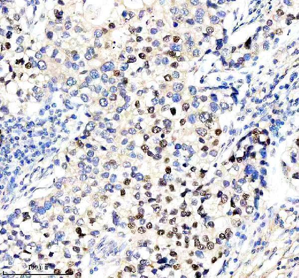
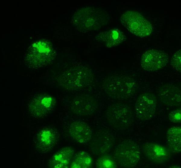
A00436-3 has real IHC data from paraffin-embedded human lung cancer tissue (IHC image caption) and IF data from A431 cells (IF image caption); listed reactivity covers human, mouse, and rat (datasheet).

Real IHC data IHC analysis of HMGA2 using anti-HMGA2 antibody (A00436-3). HMGA2 was detected in a paraffin-embedded section of human lung cancer tissue. Heat mediated antigen retrieval was performed in EDTA buffer (pH 8.0, epitope retrieval solution). The tissue section was blocked with 10% goat serum. The tissue section was then incubated with 1:100 rabbit anti-HMGA2 Antibody (A00436-3) overnight at 4°C. Peroxidase Conjugated Goat Anti-rabbit IgG was used as secondary antibody and incubated for 30 minutes at 37°C. The tissue section was developed using HRP Conjugated Rabbit IgG Super Vision Assay Kit (Catalog # SV0002) with DAB as the chromogen.
Anti-HMGA2 Antibody
Cat # A00436-3

A00436-3 will render with an IHC figure from paraffin-embedded human lung cancer tissue (IHC image caption). It is also listed for IF/ICC (datasheet), with an IF example in A431 cells (IF image caption).

Which to pick: For tissue IHC, choose A00436-3: its own figure shows EDTA pH 8.0 retrieval and 1:100 primary antibody on a paraffin-embedded section; the fixative is unreported (IHC image caption). For IF/ICC, choose A00436-3 based on its listed applications (datasheet) and IF example in A431 cells (IF image caption). For human, mouse, or rat work, A00436-3 is listed as reactive with all three species and is rabbit polyclonal (datasheet); the IHC figure demonstrates human tissue only (IHC image caption).

Each figure is that product's own IHC / IF validation image from its datasheet.

References

  1. UniProt Consortium. UniProt entry P52926 (HMGA2_HUMAN, High mobility group protein HMGI-C).
  2. Human Protein Atlas. HMGA2 tissue IHC expression (reliability: Approved).
  3. Human Protein Atlas. HMGA2 subcellular location (ICC-IF): Localized to the nucleoplasm, nucleoli and nucleoli rim..
  4. Human Protein Atlas. HMGA2 antibody validation summary (2 antibodies).
  5. An increased high-mobility group A2 expression level is associated with malignant phenotype in pancreatic exocrine tissue. British journal of cancer 2003 — PMC2376847.
  6. Intravenous leiomyomatosis: an unusual intermediate between benign and malignant uterine smooth muscle tumors. Modern pathology : an official journal of the United States and Canadian Academy of Pathology, Inc 2016 — PMC5891726.
  7. Assessing the potential of high-mobility group AT-hook 2 immunohistochemical staining as a prognostic marker of metastatic recurrence in follicular thyroid cancer: a retrospective cohort study. Endocrine journal 2025 — PMC12086272.
  8. Comparative Evaluation of HMG Family Proteins and miR-106a-5p in Low-Grade Non-Invasive and High-Grade Muscle-Invasive Papillary Urothelial Carcinoma. International journal of molecular sciences 2026 — PMC12984743.
  9. PubMed PMID:8198613 — UniProt-cited evidence.
  10. PubMed PMID:7670494 — UniProt-cited evidence.
  11. PubMed PMID:7501444 — UniProt-cited evidence.