Click image to see more details
Product Info Summary
| SKU: | A00436-3 |
|---|---|
| Size: | 100 μl/vial |
| Reactive Species: | Human, Mouse, Rat |
| Host: | Rabbit |
| Application: | ELISA, IP, IF, IHC, ICC, WB |
Customers Who Bought This Also Bought

Product info
Product Name
Anti-HMGA2 Antibody
SKU/Catalog Number
A00436-3
Size
100 μl/vial
Form
Liquid
Description
Boster Bio Anti-HMGA2 Antibody catalog # A00436-3. Tested in WB, IHC, ICC, IF, IP, ELISA applications. This antibody reacts with Human, Mouse, Rat.
Storage & Handling
12 months from date of receipt,-20℃ as supplied. 6 months 2 to 8℃ after reconstitution. Avoid repeated freezing and thawing.
Cite This Product
Anti-HMGA2 Antibody (Boster Biological Technology, Pleasanton CA, USA, Catalog # A00436-3)
Host
Rabbit
Contents
500 μg/ml antibody with PBS, 0.02% NaN3, 1 mg stabilizing protein and 50% glycerol
*This antibody is supplied in a stabilized formulation.
Compatibility with conjugation reactions depends on the chemistry of the conjugation method used.
For conjugation methods that are not compatible with the stabilizing components present in this formulation, a carrier-free antibody format is required.
Clonality
Polyclonal
Immunogen
E.coli-derived human HMGA2 recombinant protein (Position: E6-D109).
Reactive Species
A00436-3 is reactive to HMGA2 in Human, Mouse, Rat
Calculated molecular weight
11.8 kDa
Background of HMGA2
This gene encodes a protein that belongs to the non-histone chromosomal high mobility group (HMG) protein family. HMG proteins function as architectural factors and are essential components of the enhancesome. This protein contains structural DNA-binding domains and may act as a transcriptional regulating factor. Identification of the deletion, amplification, and rearrangement of this gene that are associated with myxoid liposarcoma suggests a role in adipogenesis and mesenchymal differentiation. A gene knock out study of the mouse counterpart demonstrated that this gene is involved in diet-induced obesity. Alternate transcriptional splice variants, encoding different isoforms, have been characterized.
Antibody Validation
Boster validates all antibodies on WB, IHC, ICC, Immunofluorescence, and ELISA with known positive control and negative samples to ensure specificity and high affinity, including thorough antibody incubations.
Application & Images
Applications
A00436-3 is guaranteed for ELISA, IP, IF, IHC, ICC, WB Boster Guarantee
Recommend Dilution
| Application | Dilution | Species |
|---|---|---|
| Western blot | 1:500-2000 | |
| Immunohistochemistry | 1:50-400 | |
| Immunocytochemistry/Immunofluorescence | 1:50-400 | |
| Immunoprecipitation | 1:50 | |
| ELISA | 1:100-1000 |
Validation Images & Assay Conditions
Click image to see more details
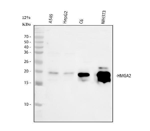
Western blot analysis of HMGA2 using anti-HMGA2 antibody (A00436-3).
Electrophoresis was performed on a 10% SDS-PAGE gel at 80V (Stacking gel) / 120V (Resolving gel) for 2 hours. The sample well of each lane was loaded with 30 ug of sample under reducing conditions.
Lane 1: human A549 whole cell lysates,
Lane 2: human HepG2 whole cell lysates,
Lane 3: rat C6 whole cell lysates,
Lane 4: mouse NIH/3T3 whole cell lysates.
After electrophoresis, proteins were transferred to a nitrocellulose membrane at 150 mA for 50-90 minutes. Blocked the membrane with 5% non-fat milk/TBS for 1.5 hour at RT. The membrane was incubated with rabbit anti-HMGA2 antigen affinity purified polyclonal antibody (A00436-3) at 1:1000 overnight at 4°C, then washed with TBS-0.1%Tween 3 times with 5 minutes each and probed with a goat anti-rabbit IgG-HRP secondary antibody at a dilution of 1:5000 for 1.5 hour at RT. The signal is developed using an ECL Plus Western Blotting Substrate (Catalog # AR1196-200) with Tanon 5200 system. A specific band was detected for HMGA2 at approximately 18-20 kDa. The expected band size for HMGA2 is at 12 kDa.

Click image to see more details
IHC analysis of HMGA2 using anti-HMGA2 antibody (A00436-3).
HMGA2 was detected in a paraffin-embedded section of human lung cancer tissue. Heat mediated antigen retrieval was performed in EDTA buffer (pH 8.0, epitope retrieval solution). The tissue section was blocked with 10% goat serum. The tissue section was then incubated with 1:100 rabbit anti-HMGA2 Antibody (A00436-3) overnight at 4°C. Peroxidase Conjugated Goat Anti-rabbit IgG was used as secondary antibody and incubated for 30 minutes at 37°C. The tissue section was developed using HRP Conjugated Rabbit IgG Super Vision Assay Kit (Catalog # SV0002) with DAB as the chromogen.

Click image to see more details
IF analysis of HMGA2 using anti-HMGA2 antibody (A00436-3).
HMGA2 was detected in an immunocytochemical section of A431 cells. Enzyme antigen retrieval was performed using IHC enzyme antigen retrieval reagent (AR0022) for 15 mins. The cells were blocked with 10% goat serum. And then incubated with 1:100 rabbit anti-HMGA2 Antibody (A00436-3) overnight at 4°C. DyLight®488 Conjugated Goat Anti-Rabbit IgG (BA1127) was used as secondary antibody at 1:500 dilution and incubated for 30 minutes at 37°C. Visualize using a fluorescence microscope and filter sets appropriate for the label used.
Specific Publications For Anti-HMGA2 Antibody (A00436-3)
Loading publications
Recommended Resources
Here are featured tools and databases that you might find useful.
- Boster's Pathways Library
- Protein Databases
- Bioscience Research Protocol Resources
- Data Processing & Analysis Software
- Photo Editing Software
- Scientific Literature Resources
- Research Paper Management Tools
- Molecular Biology Software
- Primer Design Tools
- Bioinformatics Tools
- Phylogenetic Tree Analysis
Customer Reviews
Have you used Anti-HMGA2 Antibody?
Share your experimental results or join a short interview to earn up to $1,000 in product credits or other rewards.
0 Reviews For Anti-HMGA2 Antibody
Customer Q&As
Have a question?
Find answers in Q&As, reviews.
Can't find your answer?
Submit your question
