This website uses cookies to ensure you get the best experience on our website.


- Table of Contents
Plan chromogenic HMGCS1 IHC on paraffin sections with the catalog antibody at 2–5 μg/mL (datasheet A05313-2). Assess cytoplasmic and nuclear staining while accounting for low consistency between staining and RNA expression (HPA tissue IHC).

Expected localisation, validated protocols, controls and antibodies — the at-a-glance facts below, then the full design guide.
| Expected localisation | Cytoplasmic expected; cytoplasmic/nuclear in tissue (UniProt; HPA tissue IHC) | |
| Staining pattern | Selected cells show variable cytoplasmic/nuclear staining (HPA tissue IHC) | |
| Antigen retrieval | EDTA pH 8.0 HIER, heat-mediated (datasheet A05313-2) | |
| Positive control | Appendix+4 more · see all | |
| Negative control | Adipose tissue+4 more · see all |
| Fixation | Keep fixation consistent across paraffin sections. (standard IHC practice; not target-specific) | |
| Caveat | Staining and RNA show low consistency; verify independently (HPA tissue IHC) | |
| Regulation | Liver-enhanced RNA expression (HPA tissue RNA) | |
| Isoform / epitope | No annotated isoforms or processing to alter the epitope map (UniProt) |
The catalog antibody protocol uses EDTA pH 8.0 retrieval (datasheet: A05313-2). The published option covers prostate tissue microarrays (PMC5686443).
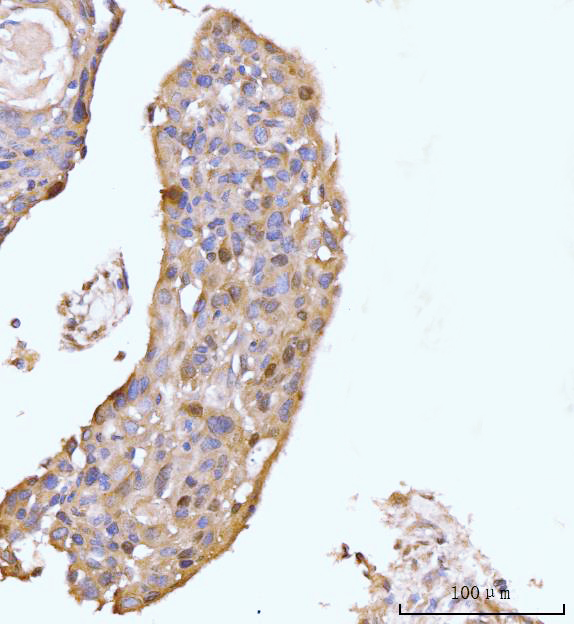
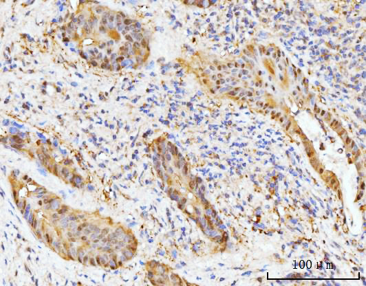
| Sample | Paraffin-embedded human esophageal squamous carcinoma tissue; fixative not specified (datasheet A05313-2) |
| Fixation | Image fixative and duration unreported (datasheet A05313-2); verify before use. |
| Sectioning | 4–5 µm sections on charged slides (standard) |
| Deparaffinisation | Xylene, graded ethanol series to water (standard) |
| Antigen retrieval | Heat retrieval: EDTA pH 8.0 (datasheet A05313-2); 20 min, 95–100 °C (standard) |
| Peroxidase block | 3% H2O2, 10 min, room temperature (standard) |
| Blocking | 10% goat serum (datasheet A05313-2) |
| Primary antibody | Rabbit anti-HMGCS1, 2-5 μg/ml (datasheet A05313-2) |
| Primary incubation | Overnight at 4 °C (datasheet A05313-2) |
| Detection | Streptavidin-biotin complex (SABC), DAB chromogen (datasheet A05313-2) |
| Counterstain | Hematoxylin, blue, dehydrate and mount (standard) |
| Expected result | HMGCS1-positive staining in glandular cells of appendix (HPA tissue IHC: High). HPA tissue profile: Cytoplasmic and nuclear expression of varying intensity in selected tissues. No signal in the no-primary control. |
HMGCS1 is a cytoplasmic enzyme with no transmembrane segment or signal peptide (UniProt Q01581). In paraffin-section IHC, expect staining in hepatocytes and gastrointestinal glandular cells, with high staining also reported in esophageal squamous epithelial cells (HPA tissue IHC: High). Nuclear staining can accompany cytoplasmic staining (HPA tissue IHC: cytoplasmic and nuclear expression). HPA rates its tissue IHC profile Approved, while reporting low agreement with RNA data and pending external verification (HPA tissue IHC).
| Clear cytoplasmic staining in hepatocytes or colon, duodenal, or stomach glandular cells; some nuclei also stain. | This fits HPA's High staining in those cell types and its description of variable cytoplasmic and nuclear expression (HPA tissue IHC). Compare the named cells within the section; a positive tissue label does not imply that every cell should stain equally (HPA tissue IHC: selected tissues and varying intensity). |
| Signal is confined to extracellular spaces, with little or none in the expected cells. | An extracellular-only pattern conflicts with the cytoplasmic localisation and absence of a signal peptide or transmembrane segment (UniProt Q01581). Check morphology and detection controls before calling it HMGCS1; misplaced chromogen or nonspecific staining is possible (general IHC practice). Nuclear staining alone needs more cautious review because HPA does report a nuclear component (HPA tissue IHC). |
| Adipocytes or bone-marrow hematopoietic cells stain strongly while the expected positive cells do not. | HPA reports HMGCS1 as Not detected in those cell types (HPA tissue IHC). The mismatch raises concern for antibody cross-reactivity or endogenous detection activity (general IHC practice). It does not prove those mechanisms or establish that every adipocyte or hematopoietic cell is biologically negative (HPA tissue IHC: observational profile). |
| A weak, even haze covers cells and empty areas, obscuring cell borders. | That distribution cannot be scored confidently as the selected-cell cytoplasmic and nuclear pattern reported by HPA (HPA tissue IHC). In chromogenic IHC, incomplete blocking, residual detection reagent, or inadequate washing can produce diffuse background (general IHC practice); assess a no-primary control before interpreting faint staining. |
| Hepatocytes and gastrointestinal glandular cells show no convincing signal. | These are useful positive comparison cells because HPA reports High staining in them (HPA tissue IHC). First consider section quality and assay performance (general IHC practice). A negative result in one run does not overturn the tissue profile, particularly given HPA's reported low staining–RNA agreement and pending external verification (HPA tissue IHC). |
| Cell compartment and topology | UniProt places HMGCS1 in the cytoplasm and lists no signal peptide or transmembrane segment (UniProt Q01581). HPA tissue IHC also reports variable nuclear expression (HPA tissue IHC). Evaluate cytoplasmic signal first while recording any nuclear component; the sources do not require one fixed cytoplasmic-to-nuclear ratio. |
| Choice of comparison cells | Hepatocytes, gastrointestinal glandular cells, and esophageal squamous epithelial cells are reported High, whereas adipocytes and bone-marrow hematopoietic cells are Not detected (HPA tissue IHC). Use these named cell populations when judging selectivity; do not treat the entire organ as a uniform positive or negative control. |
| Strength of the tissue evidence | The tissue IHC reliability is Approved, but HPA reports low consistency between antibody staining and RNA expression and says external verification is pending (HPA tissue IHC). Interpret an unexpected distribution as a reason to check controls and independent evidence, rather than as proof of a new HMGCS1 expression pattern. |
| Processing and epitope assumptions | UniProt lists a single 1–520 chain, no propeptide, and no annotated isoforms for this record (UniProt Q01581). It also lists modified residues (UniProt Q01581). These annotations do not identify the antibody epitope or establish a retrieval condition or target-specific fixation sensitivity; optimize those variables using assay controls (general IHC practice). |
| IF/ICC Q: What compartment should appear in the separate IF/ICC guide? | A: HPA reports supported nucleoplasm and cytosol localisation, with additional plasma-membrane localisation, in ICC-IF images (HPA subcellular). The two listed antibodies with ICC ratings are Uncertain, while their IHC ratings are Approved (HPA antibodies: HPA036913, HPA036914). These ICC observations should not be substituted for this paraffin-section IHC pattern. |
| Situation | Likely cause | Next action |
|---|---|---|
| No staining in a hepatocyte or gastrointestinal glandular positive comparison. | Assay failure, weak detection, or unsuitable retrieval is possible (general IHC practice); these cells are reported High (HPA tissue IHC). | Check section integrity, staining controls, and the IHC-validated antibody's established paraffin-section procedure; repeat with a documented positive section (general IHC practice). Do not infer HMGCS1 absence from a failed control. |
| Nuclear staining appears alongside a clear cytoplasmic signal. | HPA describes both nuclear and cytoplasmic expression in selected tissues (HPA tissue IHC). | Record each compartment and compare the named cell types with the HPA pattern (HPA tissue IHC). Investigate only if the distribution, controls, or tissue morphology is inconsistent (general IHC practice). |
| Extracellular deposits dominate the slide. | That pattern conflicts with UniProt's cytoplasmic localisation and lack of a signal peptide or transmembrane segment (UniProt Q01581); detection artifact is possible (general IHC practice). | Inspect a no-primary control, reagent carryover, and washing; then reassess whether signal is intracellular and follows intact cell morphology (general IHC practice). |
| Adipocytes or bone-marrow hematopoietic cells stain strongly. | HPA reports Not detected in those cells (HPA tissue IHC). Cross-reactivity or endogenous chromogenic activity could account for the discrepancy (general IHC practice). | Compare with a no-primary control and appropriate detection controls, and check whether an HPA High cell population stains as expected (general IHC practice; HPA tissue IHC). |
| The whole section has a diffuse brown haze. | Nonselective background can arise from blocking, washing, or detection steps in chromogenic IHC (general IHC practice). | Review no-primary and detection controls; adjust blocking, antibody concentration, washing, and development time within the validated assay workflow (general IHC practice). Score only cell-associated staining that remains distinguishable from background. |
| A low-staining tissue disagrees with RNA expectations or another assay. | HPA reports low consistency between its antibody staining and RNA expression data, with external verification pending (HPA tissue IHC). | Document the tissue, cell type, compartment, and controls; compare independent evidence before assigning a new positive or negative pattern (general IHC practice). Keep the HPA observation separate from any claim of confirmed absence. |
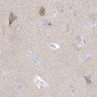
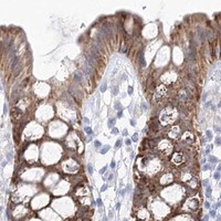
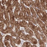
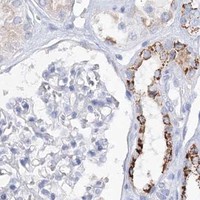


Comprehensive Human Protein Atlas IHC scoring per tissue (reliability: Approved — Low consistency between antibody staining and RNA expression data. Pending external verification.). Rows are taken directly from the HPA tissue chart — click any row's HPA link to view the source.
| Tissue | Cell type | Level | Evidence | Source |
|---|---|---|---|---|
| Appendix | Glandular cells | High | Protein (IHC) | HPA → |
| Colon | Glandular cells | High | Protein (IHC) | HPA → |
| Duodenum | Glandular cells | High | Protein (IHC) | HPA → |
| Esophagus | Squamous epithelial cells | High | Protein (IHC) | HPA → |
| Liver | Hepatocytes | High | Protein (IHC) | HPA → |
| Tissue | Cell type | Level | Evidence | Source |
|---|---|---|---|---|
| Adipose tissue | Adipocytes | Not detected | Protein (IHC) | HPA → |
| Bone marrow | Hematopoietic cells | Not detected | Protein (IHC) | HPA → |
| Breast | Adipocytes | Not detected | Protein (IHC) | HPA → |
| Cerebellum | Cells in granular layer | Not detected | Protein (IHC) | HPA → |
| Heart muscle | Cardiomyocytes | Not detected | Protein (IHC) | HPA → |
Use the catalog antibody’s tissue result as the starting point for HMGCS1 IHC, and interpret compartment and cell type with appropriate controls.
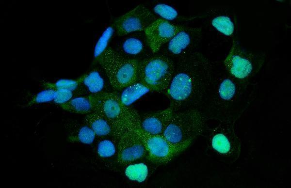
Anti-HMGCS1 antibodies have human tissue IHC images and A431 cell IF data (catalog image captions); listed reactivity spans human, mouse and rat (catalog reactivity).
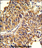
A05313-2 will render with an IHC image from a human esophageal squamous carcinoma paraffin section; its separate IF image uses A431 cells (A05313-2 image captions). A05313-1 will render with an IHC image from formalin-fixed, paraffin-embedded human lung carcinoma (A05313-1 IHC image caption).
Which to pick: For human tissue IHC, choose A05313-2 for its documented paraffin-section procedure; its image caption does not report the fixative (A05313-2 IHC image caption). For IF/ICC, choose A05313-2, which lists both applications and has an A431 cell IF image (A05313-2 applications and IF image caption). For cross-species IHC, A05313-1 lists human and mouse reactivity, while monoclonal M05313 lists human, mouse and rat; the supplied IHC image documents human tissue for A05313-1, and none is supplied for M05313 (catalog reactivity, clone and IHC image captions).