This website uses cookies to ensure you get the best experience on our website.


- Table of Contents
Plan HNF4G IHC-P around nuclear staining in small-intestine endocrine cells (HPA tissue IHC). Use duodenum as a positive reference (HPA tissue IHC) and optimize the catalog antibody within 1:10–1:50 (datasheet: A09682-1).

Expected localisation, validated protocols, controls and antibodies — the at-a-glance facts below, then the full design guide.
| Expected localisation | Nuclear staining in intestinal endocrine cells (HPA tissue IHC) | |
| Staining pattern | Strong nuclear staining in duodenal and small-intestine endocrine cells (HPA tissue IHC) | |
| Antigen retrieval | Tris-EDTA pH 9.0 HIER, 95–98 °C, 20 min (rule: nuclear antigen) | |
| Positive control | Duodenum+2 more · see all | |
| Negative control | Adipose tissue+4 more · see all |
| Fixation | Keep formalin fixation consistent across sections. (standard IHC practice; not target-specific) | |
| Caveat | Rectal endocrine staining is weaker than small-intestine staining (HPA tissue IHC) | |
| Regulation | Expression regulation not reported (UniProt) | |
| Isoform / epitope | 2 isoforms; N-terminal epitope coverage is unspecified (UniProt; datasheet: A09682-1) |
The catalog antibody protocol is provided separately. These four published HNF4G IHC protocols provide additional conditions for lung, prostate and pancreatic cancer samples (PMC5915054; PMC5728174; PMC7862466; PMC12695649).
| Sample | FFPE human testis tissue (datasheet A09682-1) |
| Fixation | Image formalin-fixed; duration unreported (datasheet A09682-1); verify before use. |
| Sectioning | 4–5 µm sections on charged slides (standard) |
| Deparaffinisation | Xylene, graded ethanol series to water (standard) |
| Antigen retrieval | Heat-induced epitope retrieval in Tris-EDTA buffer, pH 9.0, 20 min at 95–98 °C (standard rule: nuclear antigen) |
| Peroxidase block | 3% H2O2, 10 min, room temperature (standard) |
| Blocking | 10% normal serum of the secondary host, 30 min, room temperature (standard) |
| Primary antibody | Rabbit anti-HNF4G, 1:10-1:50 (datasheet A09682-1) |
| Primary incubation | Overnight at 4 °C (standard) |
| Detection | HRP-polymer secondary, DAB chromogen 5–10 min (standard) |
| Counterstain | Hematoxylin, blue, dehydrate and mount (standard) |
| Expected result | HNF4G-positive staining in endocrine cells of duodenum (HPA tissue IHC: High). HPA tissue profile: Small intestine-specific nuclear expression. No signal in the no-primary control. |
HNF4G is a nuclear transcription factor with no transmembrane segment (UniProt Q14541). In tissue IHC, expect nuclear staining in small intestinal endocrine cells, including those in duodenum (HPA: High; tissue IHC reliability Enhanced). HPA reports consistency between staining and RNA expression, with external verification pending (HPA: reliability description).
| Distinct nuclear staining in duodenal or small intestinal endocrine cells, with little surrounding stain. | This matches the reported High endocrine cell signal and small intestine-specific nuclear profile (HPA: tissue IHC). Score the fraction and intensity of stained nuclei in the relevant cells; a clear counterstain helps distinguish nuclei from adjacent cytoplasm (general IHC practice). |
| Predominantly cytoplasmic staining, a membrane rim, or stain confined to extracellular material. | That distribution does not establish the expected IHC pattern: UniProt places HNF4G in the nucleus and records no transmembrane segment (UniProt Q14541). Check morphology and detection controls before assigning such staining to HNF4G (general IHC practice). |
| Strong staining in cells outside the expected endocrine population, especially in a negative reference section. | Consider cross-reactivity or endogenous detection activity rather than calling every stained cell positive (general IHC practice). Interpret against the specific reference cell type: appendix endocrine cells were not detected, while duodenal endocrine cells were High (HPA: tissue IHC). |
| Diffuse brown haze across cells, stroma, or the entire section, without resolvable stained nuclei. | This cannot be scored as the reported nuclear pattern (HPA: tissue IHC). Background can arise from blocking, wash, reagent, or chromogen conditions; compare a no-primary control and optimize the general IHC workflow (general IHC practice). |
| No nuclear signal in a duodenal or small intestinal section expected to contain endocrine cells. | First confirm that the sampled section contains identifiable endocrine cells; HPA's High designation applies to those cells, not every cell in the tissue (HPA: tissue IHC). If present, review antibody dilution, retrieval, detection, and control performance as general IHC variables (general IHC practice). |
| Reference tissue and cell type (HPA: tissue IHC) | Duodenal and small intestinal endocrine cells are reported High; rectal endocrine cells are Medium, and appendix endocrine cells are not detected (HPA: tissue IHC). Compare like cells across sections: a whole-tissue label does not mean every nucleus should stain (HPA: cell-level entries). |
| Evidence strength (HPA: tissue IHC; antibody validation) | The tissue profile is Enhanced because antibody staining is consistent with RNA expression, with external verification pending (HPA: reliability description). HPA005438 has Enhanced IHC validation; HPA010610 has no listed IHC validation status (HPA: antibody records). |
| Tissue expression versus observed staining (UniProt Q14541; HPA: tissue IHC) | UniProt lists expression in pancreas, kidney, small intestine, and testis, with weak colon expression (UniProt Q14541). The supplied HPA IHC observations specify endocrine cell staining in duodenum, small intestine, and rectum; they do not establish an IHC pattern for every UniProt-listed tissue (HPA: tissue IHC). |
| Isoforms and antibody epitope (UniProt Q14541) | Two isoforms are recorded (UniProt Q14541). No epitope location or isoform coverage is supplied, so do not infer that a staining difference identifies an isoform or that either antibody detects both (UniProt Q14541; HPA: antibody records). |
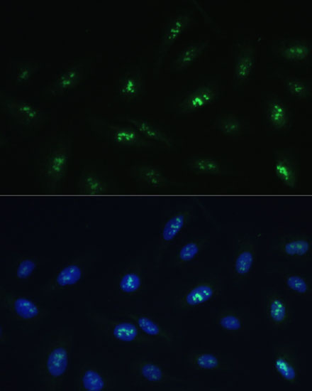
| IF/ICC Q: Should cytosolic signal be interpreted like IHC nuclear staining? | A: HPA reports nucleoplasm as the main ICC-IF location, with additional cytosol, cytokinetic bridge, and mitotic spindle locations (HPA: subcellular ICC-IF). Use that observation on the separate IF/ICC guide; the supplied tissue IHC profile remains nuclear in small intestinal endocrine cells (HPA: tissue IHC). |
| Situation | Likely cause | Next action |
|---|---|---|
| Positive tissue is uniformly blank. | The sampled level may lack recognizable endocrine cells, or an IHC workflow step may have failed (HPA: endocrine cell-specific pattern; general IHC practice). | Confirm the target cell population in the section; then check the positive control, primary antibody dilution, retrieval, and detection reagents under the chosen IHC procedure (general IHC practice). No HNF4G-specific retrieval or fixation effect is supplied. |
| Nuclei stain weakly in an expected positive section. | Rectal endocrine cells are reported Medium, whereas duodenal and small intestinal endocrine cells are High (HPA: tissue IHC); weak staining in the latter also warrants a workflow check (general IHC practice). | Record the tissue and cell type before comparing intensity. Review counterstain balance and the positive control, then optimize standard IHC conditions if the expected High cells are faint (general IHC practice). |
| Only cytoplasm or cell borders stain. | That appearance conflicts with the nuclear tissue profile and nuclear UniProt annotation (HPA: tissue IHC; UniProt Q14541); a cell-border pattern also lacks support from the recorded topology (UniProt Q14541). | Do not score it as confirmed HNF4G-positive IHC. Inspect a no-primary control, tissue morphology, and detection background; seek a reproducible nuclear pattern in the positive reference cells (general IHC practice; HPA: tissue IHC). |
| A negative reference shows convincing brown signal. | Cross-reactivity or endogenous detection activity is possible (general IHC practice); HPA reports no detectable staining in appendix endocrine cells and adipose adipocytes, among other listed cells (HPA: tissue IHC). | Match the observed cell type to the HPA negative entry, inspect no-primary and detection controls, and assess whether the signal is nuclear before changing the interpretation (HPA: tissue IHC; general IHC practice). |
| Broad diffuse stain obscures nuclear boundaries. | Nonspecific reagent binding, incomplete washing, or detection background can obscure localisation (general IHC practice); diffuse staining is not the reported small intestine-specific nuclear pattern (HPA: tissue IHC). | Compare the no-primary section, review blocking and washes, and adjust general chromogenic detection conditions until nuclei and counterstained tissue remain distinguishable (general IHC practice). |
| Two antibodies give different IHC distributions. | The supplied validation records differ: HPA005438 is Enhanced for IHC, while HPA010610 has no listed IHC status (HPA: antibody records); epitope and isoform coverage are unspecified (UniProt Q14541; HPA: antibody records). | Compare each result with the HPA nuclear endocrine cell pattern and suitable controls. Treat an unvalidated distribution as unresolved rather than attributing the discrepancy to a particular isoform (HPA: tissue IHC; general IHC practice). |



Comprehensive Human Protein Atlas IHC scoring per tissue (reliability: Enhanced — High consistency between antibody staining and RNA expression data. Pending external verification.). Rows are taken directly from the HPA tissue chart — click any row's HPA link to view the source.
| Tissue | Cell type | Level | Evidence | Source |
|---|---|---|---|---|
| Duodenum | Endocrine cells | High | Protein (IHC) | HPA → |
| Small intestine | Endocrine cells | High | Protein (IHC) | HPA → |
| Rectum | Endocrine cells | Medium | Protein (IHC) | HPA → |
| Tissue | Cell type | Level | Evidence | Source |
|---|---|---|---|---|
| Adipose tissue | Adipocytes | Not detected | Protein (IHC) | HPA → |
| Adrenal gland | Glandular cells | Not detected | Protein (IHC) | HPA → |
| Appendix | Endocrine cells | Not detected | Protein (IHC) | HPA → |
| Bone marrow | Hematopoietic cells | Not detected | Protein (IHC) | HPA → |
| Breast | Adipocytes | Not detected | Protein (IHC) | HPA → |
Troubleshoot HNF4G staining by checking nuclear signal in expected cells, then evaluating retrieval, detection background and scoring consistency (UniProt Q14541; HPA tissue IHC).
The catalog pairs an IHC-P antibody with a chromogenic image of formalin-fixed, paraffin-embedded human testis and an IF/ICC antibody with an image of U-2 OS cells (catalog: applications; image captions).
A09682-1 is listed for human IHC-P, with DAB staining shown in formalin-fixed, paraffin-embedded human testis (catalog: applications and reactivity; IHC image caption). A09682 is listed for IF/ICC in Human, Mouse and Rat, with IF shown in U-2 OS cells (catalog: applications and reactivity; IF image caption).
Which to pick: Choose A09682-1 for human tissue IHC-P: its own image documents formalin-fixed, paraffin-embedded testis stained with DAB, and its listed dilution is 1:10–1:50 (IHC image caption; datasheet: IHC-P dilution). Choose A09682 for IF/ICC: its own image shows U-2 OS cells with DAPI nuclear staining, and its listed dilution is 1:50–1:100 (IF image caption; datasheet: ICC/IF dilution). For Mouse or Rat samples, A09682 has listed reactivity and a polyclonal format, but its supplied IF image documents U-2 OS cells only (catalog: reactivity and clonality; IF image caption).