HNRNPA2B1 / Heterogeneous nuclear ribonucleoproteins A2/B1 · IHC design guide

Design Immunohistochemistry for HNRNPA2B1

Plan paraffin IHC for HNRNPA2B1 around widespread nuclear staining (HPA tissue IHC). Start with 0.5–1 μg/mL of the catalog antibody (datasheet A00396-1), and compare nuclear staining across sections prepared under consistent conditions.

Evidence assembled Oct 2026 · For research use; verify linked source records and product datasheet before use
Immunohistochemistry protocol sheet for HNRNPA2B1 (IHC for HNRNPA2B1): expected localisation Ubiquitous nuclear staining (HPA tissue IHC), antibody A00396-1, validated IHC image, and IHC protocol steps
Printable HNRNPA2B1 IHC protocol sheet — expected localisation Ubiquitous nuclear staining (HPA tissue IHC), antibody A00396-1, controls and protocol steps. Open the full HNRNPA2B1 IHC guide →

HNRNPA2B1 Immunohistochemistry Experimental Design Guide

Expected localisation, validated protocols, controls and antibodies — the at-a-glance facts below, then the full design guide.

Must know before staining
Expected localisation Ubiquitous nuclear staining (HPA tissue IHC)
Staining pattern Nuclear signal across many cell types (HPA tissue IHC)
Antigen retrieval EDTA pH 8.0 HIER, heat-mediated (datasheet A00396-1)
Positive control ⓘ Adipose tissue+4 more · see all
Negative control ⓘ None in HPA (detected in all 45 tissues); use no-primary + isotype controls
Important caveats
Reasons your staining may differ from the expected pattern.
Fixation Keep fixation conditions consistent across sections. (standard IHC practice; not target-specific)
Caveat Staining may include proteins from more than one gene (HPA tissue IHC)
Regulation Viral DNA triggers cytoplasmic translocation (UniProt)
Isoform / epitope 2 isoforms, A2 and B1; epitope effects unknown (UniProt)
Section 1

Recommended HNRNPA2B1 IHC & IF Protocols

The catalog antibody uses EDTA pH 8.0 retrieval (datasheet A00396-1). The published IHC protocols below provide mouse liver cancer and paraffin-section examples (PMC10683354; PMC11430354).

Recommended immunohistochemistry (IHC-P) protocol parameters
SampleParaffin-embedded human intestinal cancer tissue; fixative not specified (datasheet A00396-1)
FixationImage fixative and duration unreported (datasheet A00396-1); verify before use.
Sectioning4–5 µm sections on charged slides (standard)
DeparaffinisationXylene, graded ethanol series to water (standard)
Antigen retrievalHeat retrieval: EDTA pH 8.0 (datasheet A00396-1); 20 min, 95–100 °C (standard)
Peroxidase block3% H2O2, 10 min, room temperature (standard)
Blocking10% goat serum (datasheet A00396-1)
Primary antibodyRabbit anti-HNRNPA2B1, 0.5-1μg/ml (datasheet A00396-1)
Primary incubationOvernight at 4 °C (datasheet A00396-1)
DetectionStreptavidin-biotin complex (SABC), DAB chromogen (datasheet A00396-1)
CounterstainHematoxylin, blue, dehydrate and mount (standard)
Expected resultHNRNPA2B1-positive staining in adipocytes of adipose tissue (HPA tissue IHC: High). HPA tissue profile: Ubiquitous nuclear expression. No signal in the no-primary control.
💡Decision noteStart with EDTA pH 8.0 for the catalog antibody (datasheet A00396-1); citrate retrieval is a published alternative with a different antibody (PMC11430354).
Section 2

What Is the Expected HNRNPA2B1 Staining Pattern?

HNRNPA2B1 should appear predominantly in nuclei, especially the nucleoplasm (UniProt P22626: subcellular location; HPA: ubiquitous nuclear expression; HPA ICC-IF: nucleoplasm). HPA reports High staining in adipocytes, respiratory epithelial cells, glial cells and hematopoietic cells, among others (HPA: tissue IHC). Its tissue IHC profile is Supported, with a caution that the staining may include protein from more than one gene (HPA: reliability). HNRNPA2B1 has no transmembrane segment (UniProt P22626: topology).

What am I looking at on my slide?
Many intact nuclei stain, with signal concentrated away from nucleoli; the expected cell population is readily identifiable (HPA: ubiquitous nuclear expression; UniProt P22626: not found in nucleoli).This fits the reported pattern. Compare nuclei within the same section before judging intensity: HPA calls staining High in several named cell populations, but its tissue profile does not set a numerical scoring threshold (HPA: tissue IHC).
Signal is predominantly cytoplasmic or concentrated in nucleoli, with little nucleoplasmic staining.Check the staining before calling it HNRNPA2B1: nucleoplasm is the principal reported location (HPA: ICC-IF), and UniProt reports absence from nucleoli. Some cytoplasmic localization is biologically reported, so cytoplasmic signal alone cannot establish artefact (UniProt P22626: subcellular location).
Staining appears confined to an unexpected cell population while the expected cells remain unstained.Consider antibody cross-reactivity or endogenous detection activity (general IHC practice). HPA describes ubiquitous nuclear expression and cautions that its tissue staining may include protein from more than one gene; an unexpected stained cell type is therefore a reason to investigate, not proof of nonspecificity (HPA: tissue IHC).
Color spreads across extracellular space or tissue without resolving individual nuclei.Treat this as background until cellular localization is clear (general IHC practice). HNRNPA2B1 can occur in exosomes after sumoylation, but that annotation does not establish a diffuse extracellular pattern in paraffin section IHC (UniProt P22626: subcellular location).
No nuclear signal appears in a section containing a cell population reported as High, such as bone marrow hematopoietic cells (HPA: tissue IHC).The result is inconclusive. Check tissue preservation, retrieval, antibody performance and detection controls as general IHC troubleshooting steps; HPA staining levels do not identify a target-specific fixation failure (general IHC practice; HPA: tissue IHC).
💡Expected HNRNPA2B1 appearanceCall a positive result when distinct nucleoplasmic staining is visible in expected cells, including populations reported as High (HPA: tissue IHC; HPA: ICC-IF); diffuse color lacking nuclear outlines is suspect background (general IHC practice).
How each factor affects the staining
Tissue and cell contextHPA reports ubiquitous nuclear expression with low tissue RNA specificity, plus High staining in the listed cell populations (HPA: tissue IHC). Its supplied profile contains no negative or low-staining tissue examples; use named positive cells to assess a run.
Compartment and redistributionNucleoplasm is the enhanced ICC-IF location (HPA: ICC-IF). UniProt also reports cytoplasm and cytoplasmic mRNP granules, so a cytoplasmic component requires context and confirmation rather than an automatic negative call (UniProt P22626: subcellular location).
Antibody specificityThe tissue profile is Supported but cautions that staining may include protein from more than one gene (HPA: tissue IHC). HPA lists IHC Supported status for HPA001666 and CAB012403; validation status does not resolve every ambiguous cell or compartment in a new specimen (HPA: antibodies).
Isoform coverageUniProt lists 2 isoforms, A2 and B1 (UniProt P22626: isoforms). The supplied evidence does not map the IHC antibodies' epitopes to either isoform, so staining cannot be assigned to one isoform from this record alone.
IF/ICC Q&A: where should fluorescence appear?Expect nucleoplasmic fluorescence in HPA's imaged cell lines, including A-431, U2OS and A-549 (HPA: ICC-IF). That localization supports interpretation of nuclear staining in IHC; the supplied ICC-IF record gives no basis for an IF protocol here.
Why is my staining missing, weak or wrong?
SituationLikely causeNext action
A known positive cell population has no visible nuclear staining.A failed IHC workflow or detection step is possible (general IHC practice); the supplied sources do not identify HNRNPA2B1-specific fixation sensitivity.Verify the positive tissue, controls, retrieval conditions and detection reagents under the laboratory's IHC workflow (general IHC practice). Bone marrow hematopoietic cells are one reported High population (HPA: tissue IHC).
Cytoplasmic staining dominates and nuclei are faint.The result differs from HPA's ubiquitous nuclear tissue pattern, although UniProt also records cytoplasmic localization (HPA: tissue IHC; UniProt P22626: subcellular location).Check nuclear morphology and compare with a positive control; seek independent antibody or orthogonal support before interpreting redistribution (general IHC practice; HPA: antibody validation caveat).
Nucleoli appear darker than the surrounding nucleoplasm.A nucleolar dominant pattern conflicts with UniProt's report that HNRNPA2B1 is not found in the nucleolus (UniProt P22626: subcellular location).Confirm that the dark structures are nucleoli using morphology and counterstain, then review background and antibody controls (general IHC practice).
An unexpected cell population stains while reported High cells do not.Cross-reactivity or endogenous detection activity is possible (general IHC practice). HPA's caution about staining protein from more than one gene limits assignment to HNRNPA2B1 alone (HPA: tissue IHC).Inspect cell identity and nuclear localization, review detection controls, and compare an independently validated antibody where available (general IHC practice; HPA: antibody validation caveat).
Diffuse chromogen obscures nuclear boundaries.Background from the staining workflow can prevent localization calls (general IHC practice); the supplied target sources do not attribute this appearance to HNRNPA2B1.Review blocking, washes, chromogen development and a detection control, then score only interpretable cells (general IHC practice).
Two IHC antibodies give different apparent distributions.HPA reports IHC Supported status for HPA001666 and CAB012403, while its tissue profile cautions about protein from more than one gene (HPA: antibodies; HPA: tissue IHC).Compare the same tissue and cell compartments with matched controls; treat a disputed pattern as unresolved until corroborated (general IHC practice).

Sample controls for HNRNPA2B1 IHC & IF

🧪Run bone marrow first; hematopoietic cells should stain (HPA: High in hematopoietic cells). HPA detects HNRNPA2B1 in all 45 scored tissues (HPA), so there is no supported negative tissue or identified negative cell population on this slide; use no-primary and isotype controls to define background, and expect cells without specific signal to show counterstain only.
Positive control tissue: Adipose tissue (Adipocytes, HPA High)
Negative control tissue: None in HPA: HNRNPA2B1 is detected in all 45 scored tissues. Use a no-primary (secondary-only) and an isotype control instead.
ICC-IF cell lines (HPA subcellular resource): HPA ICC-IF images show HNRNPA2B1 in A-431, U-251MG, U2OS, A-549, PC-3, SH-SY5Y, hTERT-RPE1 (serum starved), with annotated localisation: Nucleoplasm (enhanced) (HPA subcellular).
Technical controls: Include a no-primary, secondary-only section and a section incubated with concentration-matched nonimmune rabbit IgG; the catalog primary is rabbit (selected-SKU IHC caption). Use HNRNPA2B1 knockout material as a biological specificity control, and block endogenous peroxidase in bone marrow before chromogenic detection (standard IHC practice).
⚠️Feasibility: The selected-SKU paraffin-section caption does not report a fixative, and the supplied evidence reports no target-specific fixation window or fixation effect (selected-SKU IHC caption). EDTA retrieval at pH 8.0 was used in that caption, but retrieval dependence is unreported (selected-SKU IHC caption). Paraffin IHC has a documented example (selected-SKU IHC caption), while ICC-IF images exist (HPA: subcellular); the evidence does not establish that frozen sections or IF are easier, and endogenous peroxidase in marrow cells can cause chromogenic background (standard IHC practice).

HPA tissue IHC evidence for HNRNPA2B1

Comprehensive Human Protein Atlas IHC scoring per tissue (reliability: Supported — High consistency between antibody staining and RNA expression data. Caution, targets protein from more than one gene.). Rows are taken directly from the HPA tissue chart — click any row's HPA link to view the source.

Positive expression · recommended positive controls

TissueCell typeLevelEvidenceSource
Adipose tissue Adipocytes High Protein (IHC) HPA →
Adrenal gland Glandular cells High Protein (IHC) HPA →
Appendix Glandular cells High Protein (IHC) HPA →
Bone marrow Hematopoietic cells High Protein (IHC) HPA →
Breast Adipocytes High Protein (IHC) HPA →

Undetected expression · recommended negative controls

TissueCell typeLevelEvidenceSource
None in HPA: HNRNPA2B1 is detected in all 45 scored tissues. Use a no-primary (secondary-only) and an isotype control instead.
Section 3

Advanced HNRNPA2B1 IHC Tips

Use nuclear staining as the main reference when evaluating HNRNPA2B1 in chromogenic paraffin section IHC (HPA tissue IHC; HPA subcellular).

What retrieval should I start with if HNRNPA2B1 staining is weak?
Start with heat mediated retrieval in EDTA at pH 8.0 for paraffin sections (datasheet A00396-1). The selected tissue image used that retrieval before overnight incubation at 4°C with 1 μg/ml primary antibody, so keep those conditions together for the first comparison (datasheet A00396-1). If staining remains weak, compare a second retrieval condition on adjacent sections while holding detection and exposure to chromogen constant (standard IHC practice). Review nuclear signal alongside tissue structure, because excessive retrieval can impair morphology and complicate compartment scoring (standard IHC practice; HPA tissue IHC). Include a known staining control in each retrieval comparison (standard IHC practice).
Can I infer a suitable fixative from the published paraffin section image?
No fixative is stated for the selected paraffin section image, so target specific fixation sensitivity is unknown (datasheet A00396-1). Record the actual fixative, fixation duration, and section processing for each specimen before comparing HNRNPA2B1 intensity (standard IHC practice). Test processing conditions on matched sections using the documented EDTA pH 8.0 retrieval and 1 μg/ml primary concentration as reference conditions (datasheet A00396-1; standard IHC practice). Compare nuclear staining with morphology and a consistently processed control tissue, since processing differences can alter accessible epitopes in IHC (standard IHC practice). Do not assign a specific fixation effect to this target without a direct comparison (datasheet A00396-1).
Should cytoplasmic staining be scored as positive in paraffin sections?
Use nucleoplasmic staining as the primary localisation reference: tissue IHC shows ubiquitous nuclear expression, and subcellular imaging reports enhanced nucleoplasmic localisation (HPA tissue IHC; HPA subcellular). HNRNPA2B1 can also occur in cytoplasmic mRNP granules, while the nucleolus is excluded in the cited localisation record (UniProt P22626). Score nuclear and cytoplasmic compartments separately rather than combining their chromogenic intensity into one value (standard IHC practice). For unexpected cytoplasmic signal, compare adjacent well preserved cells, control sections, and staining outside tissue edges before assigning biological significance (standard IHC practice). A compartment shift requires supporting evidence beyond a single brown deposit pattern (standard IHC practice).
Can this stain distinguish HNRNPA2B1 isoforms or modified epitopes?
The record lists 2 isoforms, B1 and A2, but the selected IHC caption does not specify an isoform selective epitope (UniProt P22626; datasheet A00396-1). HNRNPA2B1 contains 2 RNA recognition motifs and multiple modified residues, so an epitope claim needs direct antibody mapping rather than an inference from staining pattern (UniProt P22626; standard IHC practice). Treat nuclear DAB staining as antibody reactivity under the documented conditions, without assigning it to B1, A2, or a phosphorylation state (datasheet A00396-1; standard IHC practice). If isoform resolution is essential, obtain documented epitope specificity and validate it with an appropriate isoform specific control (standard IHC practice). Report the antibody identity and retrieval conditions with the score (datasheet A00396-1; standard IHC practice).
How should I assess an IF result alongside this IHC stain?
Use the paraffin section chromogenic result as an IHC reference; its caption does not establish an IF fixation or permeabilisation method for this antibody (datasheet A00396-1). In a separate IF assessment, multiplex HNRNPA2B1 with a marker identifying the expected cell type, such as adipocytes when examining adipose tissue, and check whether signal lies in nuclei (HPA tissue IHC; HPA subcellular; standard IF practice). Choose a fluorophore channel separated from the specimen's autofluorescence and examine an unstained control in that channel (standard IF practice). Because the expected signal is nucleoplasmic and the protein has no transmembrane segment, assess permeabilisation that permits antibody access to the nucleus (HPA subcellular; UniProt P22626; standard IF practice). Interpret IF compartment patterns separately from chromogenic intensity scores (standard IHC/IF practice).
How can I reduce diffuse brown staining without losing nuclear signal?
Compare the stained section with a no primary control to identify signal from the secondary reagent, endogenous enzyme activity, or chromogen deposition (standard IHC practice). The selected image used 10% goat serum blocking, a biotinylated goat anti rabbit secondary, a streptavidin biotin complex, and DAB (datasheet A00396-1). Check peroxidase blocking and consider endogenous biotin when troubleshooting this detection chemistry, particularly if background persists in the no primary control (standard IHC practice; datasheet A00396-1). Titrate primary exposure around the documented 1 μg/ml condition while keeping retrieval and DAB development consistent (datasheet A00396-1; standard IHC practice). Preserve the expected nuclear pattern when judging whether background has improved (HPA tissue IHC; standard IHC practice).
How should I quantify HNRNPA2B1 across paraffin sections? ⚠ ANSWER MARKED FOR VERIFICATION
Define the cell population and region before scoring, because tissue IHC describes broad nuclear expression across cell types (HPA tissue IHC; standard IHC practice). For nuclear DAB, record the percentage of positive nuclei and intensity categories, then calculate an H score if intensity comparisons are needed (standard IHC practice). Alternatively, report positive nuclei per mm² when cell density is the question, with the counted area and exclusion rules stated (standard IHC practice). Normalise to evaluable nuclei within the same annotated cell population and compare sections processed with the same retrieval and detection conditions (standard IHC practice; datasheet A00396-1). Score cytoplasmic staining separately and document the threshold using control sections (UniProt P22626; standard IHC practice).
What distinguishes convincing HNRNPA2B1 positivity from section artefact?
Convincing chromogenic staining follows a reproducible nuclear pattern in intact cells, consistent with ubiquitous nuclear tissue staining and enhanced nucleoplasmic localisation (HPA tissue IHC; HPA subcellular; standard IHC practice). HNRNPA2B1 may enter cytoplasmic granules, so isolated cytoplasmic deposits need corroboration before being interpreted as redistribution (UniProt P22626; standard IHC practice). Check whether apparent positivity tracks section edges, necrotic areas, or the no primary control, which can reveal processing or detection artefacts (standard IHC practice). Check endogenous peroxidase background when interpreting DAB, and compare nuclear signal in the intended cell population with matched control sections (standard IHC practice). The HPA tissue assessment cautions that its antibody staining may reflect proteins from more than 1 gene, so it alone cannot establish this antibody's specificity (HPA tissue IHC).
Boster reagents

Best HNRNPA2B1 / Heterogeneous nuclear ribonucleoproteins A2/B1 IHC Antibodies

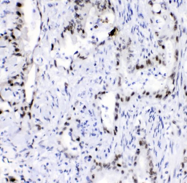
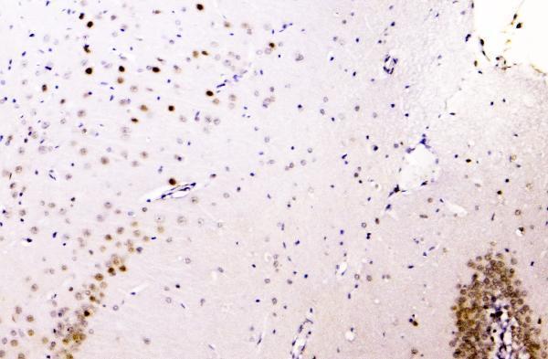
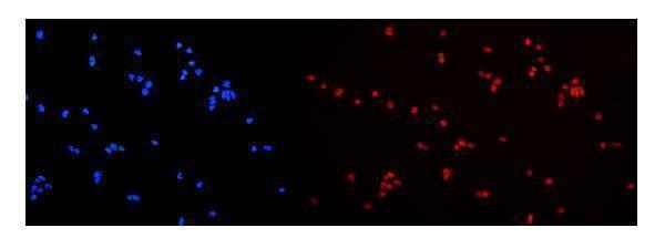
A00396-1 has IHC images from paraffin sections of human intestinal cancer tissue and mouse and rat brain, plus IF data from U20S cells (catalog image captions).

Real IHC data IHC analysis of hnRNP A2B1 using anti-hnRNP A2B1 antibody (A00396-1). hnRNP A2B1 was detected in paraffin-embedded section of human intestinal cancer tissue. Heat mediated antigen retrieval was performed in EDTA buffer (pH8.0, epitope retrieval solution). The tissue section was blocked with 10% goat serum. The tissue section was then incubated with 1μg/ml rabbit anti-hnRNP A2B1 Antibody (A00396-1) overnight at 4°C. Biotinylated goat anti-rabbit IgG was used as secondary antibody and incubated for 30 minutes at 37°C. The tissue section was developed using Strepavidin-Biotin-Complex (SABC) (Catalog # SA1022) with DAB as the chromogen.
Anti-hnRNP A2B1/HNRNPA2B1 Antibody ®
Cat # A00396-1

A00396-1 is listed for IHC, IF and ICC in human, mouse and rat samples (catalog applications and reactivity). Its IHC captions show paraffin sections of human intestinal cancer tissue and mouse and rat brain; its IF caption shows U20S cells (catalog image captions).

Which to pick: Choose A00396-1 for paraffin-section IHC: its own caption documents EDTA pH 8 retrieval, 1 μg/ml primary antibody and DAB detection (A00396-1 IHC caption); the fixative is unreported (A00396-1 IHC caption). For IF/ICC, A00396-1 is listed for both applications and its cell image uses 2 μg/ml primary antibody (catalog applications; A00396-1 IF caption). For cross-species IHC, its reactivity list includes human, mouse and rat, with paraffin-section images for each (catalog reactivity; A00396-1 IHC captions); clonality is unreported (catalog clone field).

Each figure is that product's own IHC / IF validation image from its datasheet.

References

  1. UniProt Consortium. UniProt entry P22626 (ROA2_HUMAN, Heterogeneous nuclear ribonucleoproteins A2/B1).
  2. Human Protein Atlas. HNRNPA2B1 tissue IHC expression (reliability: Supported).
  3. Human Protein Atlas. HNRNPA2B1 subcellular location (ICC-IF): Localized to the nucleoplasm..
  4. Human Protein Atlas. HNRNPA2B1 antibody validation summary (4 antibodies).
  5. HNRNPA2B1 is a potential biomarker of breast cancer related to prognosis and immune infiltration. Aging 2023 — PMC10522385.
  6. The Potential Impact of HNRNPA2B1 on Human Cancers Prognosis and Immune Microenvironment. Journal of immunology research 2024 — PMC11392580.
  7. hnRNPA2B1 promotes the occurrence and progression of hepatocellular carcinoma by downregulating PCK1 mRNA via a m6A RNA methylation manner. Journal of translational medicine 2023 — PMC10683354.
  8. Melatonin Regulates Osteoblast Differentiation through the m6A Reader hnRNPA2B1 under Simulated Microgravity. Current issues in molecular biology 2024 — PMC11430354.
  9. PubMed PMID:2557628 — UniProt-cited evidence.
  10. PubMed PMID:8029005 — UniProt-cited evidence.
  11. PubMed PMID:7789969 — UniProt-cited evidence.