HNRNPD / Heterogeneous nuclear ribonucleoprotein D0 · IHC design guide

Design Immunohistochemistry for HNRNPD

Plan chromogenic HNRNPD IHC in paraffin sections using its generally nuclear tissue pattern (HPA tissue IHC). Use high-staining colon glandular cells and undetected parathyroid glandular cells as reference tissues (HPA tissue IHC), and start the catalog antibody at 0.5–1 μg/mL (datasheet A09982-1).

Evidence assembled Oct 2026 · For research use; verify linked source records and product datasheet before use
Immunohistochemistry protocol sheet for HNRNPD (IHC for HNRNPD): expected localisation Generally nuclear tissue staining (HPA tissue IHC); cytoplasmic localisation is also annotated (UniProt), antibody A09982-1, validated IHC image, and IHC protocol steps
Printable HNRNPD IHC protocol sheet — expected localisation Generally nuclear tissue staining (HPA tissue IHC); cytoplasmic localisation is also annotated (UniProt), antibody A09982-1, controls and protocol steps. Open the full HNRNPD IHC guide →

HNRNPD Immunohistochemistry Experimental Design Guide

Expected localisation, validated protocols, controls and antibodies — the at-a-glance facts below, then the full design guide.

Must know before staining
Expected localisation Generally nuclear tissue staining (HPA tissue IHC); cytoplasmic localisation is also annotated (UniProt)
Staining pattern Generally nuclear staining across tissue cell types (HPA tissue IHC)
Antigen retrieval EDTA pH 8.0 HIER, heat-mediated (datasheet A09982-1)
Positive control ⓘ Adrenal gland+4 more · see all
Negative control ⓘ Parathyroid gland
Important caveats
Reasons your staining may differ from the expected pattern.
Fixation Keep fixation consistent across sections. (standard IHC practice; not target-specific)
Caveat Parathyroid glandular cells have no detected staining (HPA tissue IHC)
Regulation Cytoplasmic localisation varies diurnally (UniProt)
Isoform / epitope Four isoforms; epitope coverage needs checking (UniProt)
Section 1

Recommended HNRNPD IHC & IF Protocols

The catalog antibody protocol is followed by two published HNRNPD IHC protocols with reported staining parameters (PMC9221583; PMC4553214).

Recommended immunohistochemistry (IHC-P) protocol parameters
SampleParaffin-embedded mouse intestine tissue; fixative not specified (datasheet A09982-1)
FixationImage fixative and duration unreported (datasheet A09982-1); verify before use.
Sectioning4–5 µm sections on charged slides (standard)
DeparaffinisationXylene, graded ethanol series to water (standard)
Antigen retrievalHeat retrieval: EDTA pH 8.0 (datasheet A09982-1); 20 min, 95–100 °C (standard)
Peroxidase block3% H2O2, 10 min, room temperature (standard)
Blocking10% goat serum (datasheet A09982-1)
Primary antibodyRabbit anti-HNRNPD, 0.5-1μg/ml (datasheet A09982-1)
Primary incubationOvernight at 4 °C (datasheet A09982-1)
DetectionStreptavidin-biotin complex (SABC), DAB chromogen (datasheet A09982-1)
CounterstainHematoxylin, blue, dehydrate and mount (standard)
Expected resultHNRNPD-positive staining in glandular cells of adrenal gland (HPA tissue IHC: High). HPA tissue profile: General nuclear expression. No signal in the no-primary control.
💡Decision noteStart with heat-mediated EDTA retrieval at pH 8.0 (datasheet A09982-1); both published protocols report retrieval without specifying its conditions (PMC9221583; PMC4553214).
Section 2

What Is the Expected HNRNPD Staining Pattern?

HNRNPD should stain mainly nuclei in paraffin sections, consistent with HPA’s general nuclear IHC pattern and enhanced nucleoplasmic ICC-IF localization (HPA: tissue IHC Supported; HPA: ICC Enhanced). Cytoplasmic staining can also be biologically plausible because HNRNPD occurs in cytoplasmic mRNP granules (UniProt Q14103: subcellular location). It has no transmembrane segment, so an exclusively membranous pattern is unexpected (UniProt Q14103: topology).

What am I looking at on my slide?
Distinct nuclear stain in glandular cells of adrenal gland, appendix, cervix, or colon.This fits HPA’s High staining in those cell populations and its general nuclear IHC profile (HPA: tissue IHC). Judge the named cells, rather than the whole section, as the positive population (HPA: tissue IHC).
Strong, exclusively membranous staining with little nuclear signal.This conflicts with the reported nuclear pattern and the absence of a transmembrane segment (HPA: tissue IHC; UniProt Q14103: topology). Review morphology and controls for nonspecific staining (general IHC practice).
Prominent stain in a cell population that the chosen reference tissue lists as Low or Not detected.Compare the same cell type: HPA lists parathyroid glandular cells as Not detected and thyroid glandular cells as Low (HPA: tissue IHC). Unexpected stain warrants control review; it alone does not prove cross-reactivity because tissue IHC reliability is Supported, with medium RNA–staining consistency (HPA: tissue IHC).
Diffuse colour covers nuclei, cytoplasm, and areas between cells.Loss of compartment and cell boundaries makes localization hard to score (general IHC practice). Compare a section processed without primary antibody to check detection background; do not classify all cytoplasmic signal as artefact, since cytoplasmic mRNP localization is reported (UniProt Q14103: subcellular location).
No detectable stain in adrenal glandular cells or bone marrow hematopoietic cells.Both are listed as High by HPA (HPA: tissue IHC). An absent result calls for checking tissue identity, detection controls, antibody dilution, and the validated IHC-P workflow before concluding that the sample lacks HNRNPD (general IHC practice).
💡Expected HNRNPD appearanceA positive IHC section shows clear, predominantly nuclear staining in HPA High cell populations such as colon glandular cells; an exclusively membranous signal or diffuse colour without cell boundaries is suspect (HPA: tissue IHC; UniProt Q14103: topology; general IHC practice).
How each factor affects the staining
Compartment and topologyHPA reports general nuclear tissue staining, while UniProt also places HNRNPD in the cytoplasm and cytoplasmic mRNP granules (HPA: tissue IHC; UniProt Q14103: subcellular location). Score nuclear and cytoplasmic compartments separately; no transmembrane segment supports questioning an exclusively membranous result (UniProt Q14103: topology).
Choice of reference cellsUse a named High population, such as appendix glandular cells or bone marrow hematopoietic cells, to assess whether staining is detectable (HPA: tissue IHC). Parathyroid glandular cells are listed as Not detected, but that designation does not describe every cell in the tissue (HPA: tissue IHC).
Antibody evidenceHPA lists HPA004911 IHC as Supported and ICC as Enhanced (HPA: antibody validation). The tissue IHC profile has medium consistency between staining and RNA expression (HPA: tissue IHC); interpret an unexpected result alongside cell morphology and controls (general IHC practice).
Isoforms and epitope coverageUniProt lists four HNRNPD isoforms and two RNA recognition motifs (UniProt Q14103: isoforms and domains). Antibody epitope coverage is not supplied here, so isoform-specific staining cannot be inferred from these data (UniProt Q14103: isoforms).
Processing and fixation evidenceUniProt lists no signal peptide or propeptide and no transmembrane segment (UniProt Q14103: processing and topology). The supplied sources report no target-specific fixation sensitivity; assess retrieval with the validated IHC-P instructions and run controls without assuming a particular fixation effect (general IHC practice).
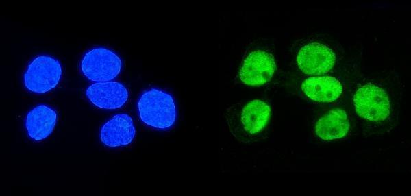
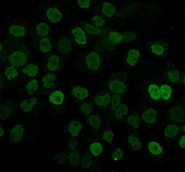
IF/ICC Q: What localization should I expect?A: HPA reports enhanced nucleoplasmic localization in ICC-IF images and lists HPA004911 ICC validation as Enhanced (HPA: subcellular; HPA: antibody validation). UniProt also reports cytoplasmic localization, so a cytoplasmic component is plausible, but HPA’s ICC-IF summary identifies the nucleoplasm as the main location (UniProt Q14103: subcellular location; HPA: subcellular).
Why is my staining missing, weak or wrong?
SituationLikely causeNext action
No nuclear stain in a chosen High reference population.The reference cells may be absent from the section, or an IHC workflow step may have failed (HPA: tissue IHC; general IHC practice).Verify the cell type on the slide, then check the catalog antibody’s IHC-P instructions, retrieval, dilution, and detection controls (general IHC practice). Do not infer target absence from a single failed run (general IHC practice).
Stain appears only at cell membranes.An exclusively membranous pattern conflicts with HPA’s general nuclear pattern and UniProt’s topology (HPA: tissue IHC; UniProt Q14103: topology).Compare with a High reference population and a primary-omission control; review whether the colour follows cell boundaries or deposits outside cells (HPA: tissue IHC; general IHC practice).
Colour develops in the primary-omission control.The signal can arise from the detection system or endogenous activity rather than primary-antibody binding (general IHC practice).Review the detection chemistry and its applicable blocking controls, then repeat the comparison on matched sections (general IHC practice). Interpret tissue-cell staining only after control background is resolved (general IHC practice).
Low or Not detected reference cells stain strongly.The result differs from the reported level for that cell population; identity, nonspecific binding, or detection background may need review (HPA: tissue IHC; general IHC practice).Confirm the exact cell population and compare a High reference and primary-omission control (HPA: tissue IHC; general IHC practice). Treat the discrepancy as unresolved until controls support its interpretation (general IHC practice).
Nuclear staining is visible, but diffuse colour obscures cell boundaries.Background or excess detection product may prevent compartment-level assessment (general IHC practice). A cytoplasmic component is possible for HNRNPD, but diffuse colour alone cannot establish it (UniProt Q14103: subcellular location; general IHC practice).Inspect counterstained morphology and the primary-omission control; review the catalog antibody’s IHC-P dilution and detection instructions before rescoring localization (general IHC practice).

Sample controls for HNRNPD IHC & IF

🧪Run bone marrow first: its hematopoietic cells should stain (HPA: High in bone-marrow hematopoietic cells). Use parathyroid gland as the negative tissue (HPA: Not detected in parathyroid glandular cells); on the positive slide, cells without target staining should show only background chromogen and no discrete nuclear signal, though the supplied HPA row does not identify an internal negative cell type.
Positive control tissue: Adrenal gland (Glandular cells, HPA High)
Negative control tissue: Parathyroid gland (HPA Not detected)
ICC-IF cell lines (HPA subcellular resource): HPA ICC-IF images show HNRNPD in A-431, U-251MG, U2OS, U2OS, siRNA 1 (10x), U2OS, siRNA 1 (40x), U2OS, scrambled (10x), U2OS, scrambled (40x), with annotated localisation: Nucleoplasm (enhanced) (HPA subcellular).
Technical controls: Include a no-primary, secondary-only control, a concentration-matched rabbit IgG isotype control, and knockout tissue if available as a biological negative (selected SKU caption: rabbit primary; standard IHC practice). In bone marrow, block endogenous peroxidase before DAB detection and assess endogenous biotin if using the caption’s biotin-based detection system (selected SKU caption: biotinylated secondary, SABC and DAB; standard IHC practice).
⚠️Feasibility: No target-specific fixation window or fixation effect is reported, and the A09982-1 paraffin-section caption does not state the fixative (selected SKU caption). That mouse-intestine example uses heat retrieval in EDTA at pH 8.0; whether retrieval is required in the selected control tissues is unreported (selected SKU caption). For IF/ICC, is it easier than frozen-section IHC? The supplied evidence does not establish which is easier; HPA reports enhanced nucleoplasmic ICC-IF localization, while endogenous peroxidase in bone marrow can produce misleading DAB signal (HPA subcellular; standard IHC practice).

HPA tissue IHC evidence for HNRNPD

Comprehensive Human Protein Atlas IHC scoring per tissue (reliability: Supported — Medium consistency between antibody staining and RNA expression data.). Rows are taken directly from the HPA tissue chart — click any row's HPA link to view the source.

Positive expression · recommended positive controls

TissueCell typeLevelEvidenceSource
Adrenal gland Glandular cells High Protein (IHC) HPA →
Appendix Glandular cells High Protein (IHC) HPA →
Bone marrow Hematopoietic cells High Protein (IHC) HPA →
Caudate Glial cells High Protein (IHC) HPA →
Cerebellum Cells in granular layer High Protein (IHC) HPA →

Undetected expression · recommended negative controls

TissueCell typeLevelEvidenceSource
Parathyroid gland Glandular cells Not detected Protein (IHC) HPA →
Section 3

Advanced HNRNPD IHC Tips

Troubleshoot HNRNPD staining in paraffin sections using the catalog antibody’s tissue IHC conditions and the expected nuclear pattern (caption A09982-1; HPA: general nuclear expression).

What retrieval should I use when nuclear HNRNPD staining is weak?
Start with heat-mediated EDTA pH 8.0 antigen retrieval for paraffin sections (datasheet A09982-1). The selected mouse intestine image used that retrieval, followed by 10% goat serum and 1 μg/ml primary antibody overnight at 4°C (caption A09982-1). If nuclear DAB remains faint, compare retrieval durations on adjacent sections while keeping section thickness, detection chemistry, and development time matched (standard IHC practice). Only if staining remains weak, trial another validated buffer or pH on paired sections, recording tissue preservation and nonspecific signal (standard IHC practice). Judge the result against predominantly nuclear staining, while allowing for documented cytoplasmic localization (HPA: general nuclear expression; UniProt Q14103).
How can I assess whether fixation is reducing HNRNPD staining?
The selected paraffin section caption does not report its fixative, so target-specific fixation sensitivity is unknown (caption A09982-1). Compare specimens with documented fixation intervals and process them together before attributing a staining difference to fixation (standard IHC practice). Hold section thickness, EDTA pH 8.0 retrieval, primary concentration, and DAB development constant during that comparison (datasheet A09982-1; standard IHC practice). Assess nuclear detail and tissue morphology alongside signal intensity, since damaged sections make nuclear staining difficult to score (standard IHC practice). Include a matched positive section and a negative reagent control in each run to distinguish processing variation from nonspecific detection (standard IHC practice).
Should I count cytoplasmic staining as HNRNPD positive?
Score nuclear staining separately because tissue IHC shows general nuclear expression and cell imaging places HNRNPD in the nucleoplasm (HPA: tissue IHC; HPA: subcellular). Cytoplasmic staining is biologically plausible: HNRNPD also localizes to cytoplasmic mRNP granules containing untranslated mRNAs (UniProt Q14103). Examine whether cytoplasmic signal occurs within intact cells and follows their boundaries, rather than appearing as diffuse extracellular DAB (standard IHC practice). Record nuclear and cytoplasmic percentages independently, using the same threshold across matched sections (standard IHC practice). Avoid treating every cytoplasmic deposit as a localization shift; compare controls and tissue morphology first (standard IHC practice).
Can this stain distinguish HNRNPD isoforms or reveal epitope masking?
HNRNPD has 4 annotated isoforms and two RNA recognition motifs at residues 97–179 and 182–261 (UniProt Q14103). The supplied tissue image does not map the antibody epitope or establish which isoforms it detects (caption A09982-1). Therefore, report chromogenic staining as HNRNPD immunoreactivity rather than assigning a specific isoform (standard IHC interpretation). If signal varies after retrieval, compare adjacent sections under matched conditions before attributing the change to epitope masking (standard IHC practice). Its annotated phosphorylation and other modifications provide biological context, but this IHC evidence cannot establish modification-specific recognition (UniProt Q14103; caption A09982-1).
How should I check HNRNPD localization in a multiplex IF experiment?
Treat IF as a separate assay and validate its staining and controls independently of the paraffin IHC image (caption A09982-1; standard IF practice). Pair HNRNPD with a validated marker of the expected cell type, such as glandular cells in colon, and inspect nuclear overlap (HPA: high in colon glandular cells; HPA: general nuclear expression). Choose a fluorophore channel with low measured tissue autofluorescence and include single-channel controls to assess bleed-through (standard IF practice). Permeabilize sufficiently for antibody access to nuclear and cytoplasmic epitopes, then check that cell boundaries remain interpretable (UniProt Q14103 localization; standard IF practice). Score nuclear and cytoplasmic fluorescence separately (standard IF practice).
What should I change when DAB background obscures nuclear staining?
First inspect a no-primary control to locate signal from secondary detection, endogenous enzyme activity, or tissue deposits (standard IHC practice). The selected image used a biotinylated secondary, streptavidin–biotin complex, and DAB, so assess background in that complete detection system (caption A09982-1). Use a peroxidase block, appropriate serum blocking, and thorough washes as general chromogenic IHC steps (standard IHC practice). Compare primary antibody concentration and DAB development on adjacent sections, keeping EDTA pH 8.0 retrieval constant (datasheet A09982-1; standard IHC practice). Preserve a clean nuclear signal while reducing diffuse or edge-associated staining, rather than judging improvement by total darkness alone (HPA: general nuclear expression; standard IHC practice).
How should I quantify HNRNPD across paraffin tissue sections? ⚠ ANSWER MARKED FOR VERIFICATION
Define the cell population and compartment before scoring; tissue IHC describes general nuclear expression, with high staining in colon glandular cells (HPA: tissue IHC). Record the percentage of positive nuclei and, if intensity is reproducible, calculate an H-score from the proportions at each intensity level (standard IHC practice). For spatial comparisons, report positive nuclei per mm² of viable tissue and normalize to counted nuclei or analyzable tissue area (standard IHC practice). Keep thresholds, image settings, and DAB development consistent across slides (standard IHC practice). Score cytoplasmic signal separately because HNRNPD can also occupy cytoplasmic mRNP granules (UniProt Q14103).
How can I distinguish genuine HNRNPD staining from an artefact?
A credible result should include staining in intact nuclei, consistent with general nuclear tissue expression and nucleoplasmic localization (HPA: tissue IHC; HPA: subcellular). Cytoplasmic staining may be real when it is confined to intact cells, since HNRNPD occurs in cytoplasmic mRNP granules (UniProt Q14103). Check the stained cell type against tissue context; colon glandular cells show high staining in the supplied tissue profile (HPA: high in colon glandular cells). Distrust signal restricted to section edges, necrotic areas, or extracellular deposits, and compare those regions with morphology (standard IHC practice). Use a no-primary control to assess endogenous enzyme or detection-system staining before interpreting DAB as HNRNPD (standard IHC practice).
Boster reagents

Best HNRNPD / Heterogeneous nuclear ribonucleoprotein D0 IHC Antibodies

Three anti-HNRNPD antibodies have IHC images from paraffin sections and IF images from cells or paraffin sections (catalog image captions). The catalog lists human, mouse and rat reactivity for each (catalog: reactivity).

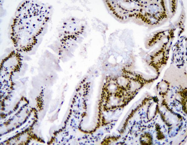
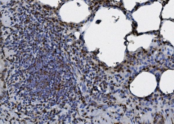
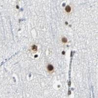
Real IHC data IHC analysis of HNRNPD using anti-HNRNPD antibody (A09982-1). HNRNPD was detected in paraffin-embedded section of mouse intestine tissue tissue. Heat mediated antigen retrieval was performed in EDTA buffer (pH8.0, epitope retrieval solution). The tissue section was blocked with 10% goat serum. The tissue section was then incubated with 1μg/ml rabbit anti-HNRNPD Antibody (A09982-1) overnight at 4°C. Biotinylated goat anti-rabbit IgG was used as secondary antibody and incubated for 30 minutes at 37°C. The tissue section was developed using Strepavidin-Biotin-Complex (SABC) (Catalog # SA1022) with DAB as the chromogen.
Anti-hnRNP D/AUF1/HNRNPD Antibody ®
Cat # A09982-1
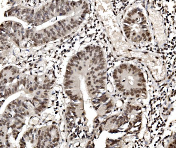
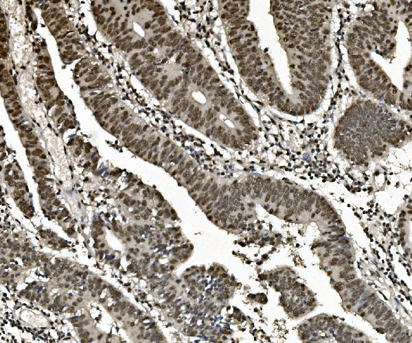
Real IHC data IHC analysis of hnRNP D/AUF1/HNRNPD using anti-hnRNP D/AUF1/HNRNPD antibody (M09982). hnRNP D/AUF1/HNRNPD was detected in paraffin-embedded section of human rectal cancer tissue. Heat mediated antigen retrieval was performed in EDTA buffer (pH8.0, epitope retrieval solution). The tissue section was blocked with 10% goat serum. The tissue section was then incubated with 1μg/ml mouse anti-hnRNP D/AUF1/HNRNPD Antibody (M09982) overnight at 4°C. Biotinylated goat anti-mouse IgG was used as secondary antibody and incubated for 30 minutes at 37°C. The tissue section was developed using Strepavidin-Biotin-Complex (SABC) (Catalog # SA1021) with DAB as the chromogen.
Anti-hnRNP D/AUF1/HNRNPD Antibody ® (monoclonal, 2B12)
Cat # M09982
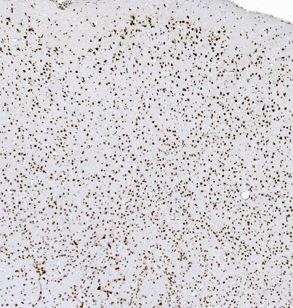
Real IHC data IHC analysis of hnRNP D/AUF1/HNRNPD using anti-hnRNP D/AUF1/HNRNPD antibody (M09982-1). hnRNP D/AUF1/HNRNPD was detected in paraffin-embedded section of human rectal cancer tissue. Heat mediated antigen retrieval was performed in EDTA buffer (pH8.0, epitope retrieval solution). The tissue section was blocked with 10% goat serum. The tissue section was then incubated with 1μg/ml mouse anti-hnRNP D/AUF1/HNRNPD Antibody (M09982-1) overnight at 4°C. Biotinylated goat anti-mouse IgG was used as secondary antibody and incubated for 30 minutes at 37°C. The tissue section was developed using Strepavidin-Biotin-Complex (SABC) (Catalog # SA1021) with DAB as the chromogen.
Anti-hnRNP D/AUF1/HNRNPD Antibody ® (monoclonal, 4F3)
Cat # M09982-1

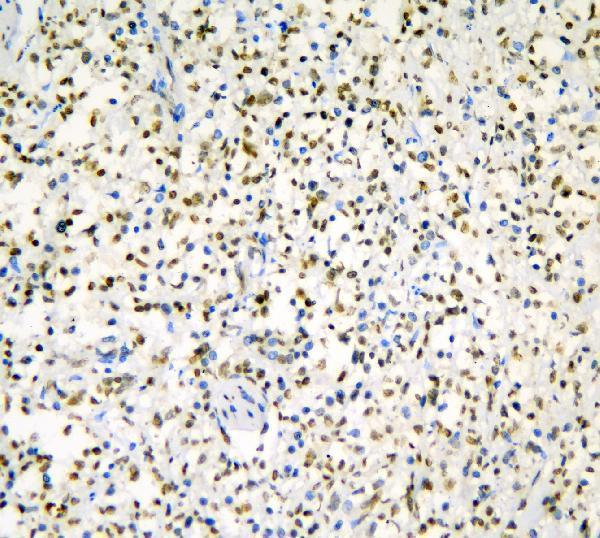
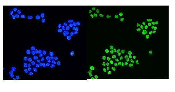
A09982-1 has IHC images from mouse intestine and human lung and renal cancer sections, plus IF images from A431 cells and human intestine cancer sections (A09982-1 image captions). M09982 has IHC images from human rectal cancer and rat and mouse lung sections; M09982-1 has IHC images from human rectal cancer and rat brain sections, and both have IF images from A431 cells and human rectal cancer sections (M09982 and M09982-1 image captions).

Which to pick: For tissue IHC across human, mouse and rat samples, choose M09982: its own paraffin-section images cover all three species (M09982 IHC captions); the fixative is unreported (M09982 IHC captions). For IF/ICC, choose A09982-1 if a rabbit primary and a listed 2 μg/ml starting concentration suit your detection setup (catalog: A09982-1 host, applications and IF dilution). For a mouse monoclonal, M09982 is clone 2B12 and has both IF/ICC applications and IHC images across all three species (catalog: M09982 clone and applications; M09982 IHC captions); M09982-1 is clone 4F3, with its own IHC images from human and rat sections (catalog: M09982-1 clone; M09982-1 IHC captions).

Each figure is that product's own IHC / IF validation image from its datasheet.

References

  1. UniProt Consortium. UniProt entry Q14103 (HNRPD_HUMAN, Heterogeneous nuclear ribonucleoprotein D0).
  2. Human Protein Atlas. HNRNPD tissue IHC expression (reliability: Supported).
  3. Human Protein Atlas. HNRNPD subcellular location (ICC-IF): Localized to the nucleoplasm..
  4. Human Protein Atlas. HNRNPD antibody validation summary (1 antibodies).
  5. Evaluation of Heterogeneous Nuclear Ribonucleoprotein D Expression as a Diagnostic Marker for Oral Squamous Cell Carcinoma. Diagnostics (Basel, Switzerland) 2022 — PMC9221583.
  6. Nuclear heterogeneous nuclear ribonucleoprotein D is associated with poor prognosis and interactome analysis reveals its novel binding partners in oral cancer. Journal of translational medicine 2015 — PMC4553214.
  7. Silencing hnRNPD inhibits gastric cancer growth by increasing TXNIP-mediated oxidative stress. Discover oncology 2025 — PMC12085443.
  8. The expression and biological effect of NR2F6 in non-small cell lung cancer. Frontiers in oncology 2022 — PMC9478584.
  9. PubMed PMID:7673195 — UniProt-cited evidence.
  10. PubMed PMID:9615222 — UniProt-cited evidence.
  11. PubMed PMID:14702039 — UniProt-cited evidence.