This website uses cookies to ensure you get the best experience on our website.


- Table of Contents
Plan paraffin-section HOOK2 IHC using the reported cytoplasmic tissue pattern (HPA tissue IHC). Compare strong staining in glandular cells with the absence of detectable staining in adipocytes, and assess localization alongside intensity (HPA tissue IHC).

Expected localisation, validated protocols, controls and antibodies — the at-a-glance facts below, then the full design guide.
| Expected localisation | Cytoplasmic staining in tissue sections (HPA tissue IHC) | |
| Staining pattern | Widespread cytoplasmic staining across cell types (HPA tissue IHC) | |
| Antigen retrieval | EDTA pH 8.0 HIER, heat-mediated (datasheet A08854-1) | |
| Positive control | Appendix+4 more · see all | |
| Negative control | Adipose tissue |
| Fixation | Matched tissue-IHC evidence does not establish the fixation claim. Validate the specimen-specific method before use. (selected-SKU IHC image A08854-1) | |
| Caveat | Adipocytes may show no detectable staining (HPA tissue IHC) | |
| Regulation | Low tissue specificity; no regulator reported (HPA tissue IHC) | |
| Isoform / epitope | 2 isoforms; confirm epitope coverage (UniProt) |
The catalog antibody uses EDTA pH 8.0 retrieval (datasheet A08854-1). The published HOOK2 IHC methods below describe free-floating brain sections (PMC4370497) and antibody dilutions for endogenous and overexpressed Hook proteins (PMC1896156).
| Sample | Paraffin-embedded human ovarian cancer tissue; fixative not specified (datasheet A08854-1) |
| Fixation | Image fixative and duration unreported (datasheet A08854-1); verify before use. |
| Sectioning | 4–5 µm sections on charged slides (standard) |
| Deparaffinisation | Xylene, graded ethanol series to water (standard) |
| Antigen retrieval | Heat retrieval: EDTA pH 8.0 (datasheet A08854-1); 20 min, 95–100 °C (standard) |
| Peroxidase block | 3% H2O2, 10 min, room temperature (standard) |
| Blocking | 10% goat serum (datasheet A08854-1) |
| Primary antibody | Rabbit anti-HOOK2, 2-5μg/ml (datasheet A08854-1) |
| Primary incubation | Overnight at 4 °C (datasheet A08854-1) |
| Detection | HRP-conjugated secondary, DAB chromogen (datasheet A08854-1) |
| Counterstain | Hematoxylin, blue, dehydrate and mount (standard) |
| Expected result | HOOK2-positive staining in glandular cells of appendix (HPA tissue IHC: High). HPA tissue profile: Ubiquitous cytoplasmic expression. No signal in the no-primary control. |
HOOK2 should appear mainly in the cytoplasm of many cell types, including strongly stained glandular cells and kidney tubule cells (HPA tissue IHC: ubiquitous cytoplasmic expression; High in listed cells). Perinuclear or punctate signal is plausible (UniProt Q96ED9: centrosome, trans-Golgi network and cytoplasmic foci). HOOK2 has no transmembrane segment, so a crisp plasma-membrane outline is unexpected (UniProt Q96ED9 topology). HPA rates the tissue IHC profile Approved, with medium consistency against RNA data (HPA tissue IHC).
| Chromogenic cytoplasmic staining is High in colon glandular cells or kidney tubule cells; nearby cell-free areas remain clear. | This matches two HPA positive examples (HPA tissue IHC: High in colon glandular cells and kidney tubule cells). Compare cells within the section rather than requiring every cell to stain equally: the profile is ubiquitous, while the reported intensity varies by cell type (HPA tissue IHC: profile and staining levels). |
| Fine perinuclear or punctate cytoplasmic staining accompanies broader cytoplasmic signal; ciliary rootlets stain in fallopian tube or nasopharynx. | These locations can fit HOOK2 biology and observed tissue staining (UniProt Q96ED9: centrosome, trans-Golgi network and cytoplasmic foci; HPA tissue IHC: High in ciliated-cell rootlets). A punctum alone is less convincing than a pattern with the expected cells and a clean control (general IHC interpretation practice). |
| Signal is chiefly nuclear or forms a sharp plasma-membrane rim, with little cytoplasmic staining. | Treat this as a discordant compartment pattern and investigate artefact or nonspecific binding (HPA tissue IHC: ubiquitous cytoplasmic expression; UniProt Q96ED9: cytoplasmic locations and no transmembrane segment). Chromogenic morphology alone cannot identify which staining step caused the discrepancy (general IHC interpretation practice). |
| Adipocytes stain strongly while expected positive cells do not, or colour appears in a section lacking primary antibody. | Strong adipocyte staining conflicts with the reported HPA example, where adipocytes are Not detected (HPA tissue IHC: adipose tissue). Colour without primary antibody points to endogenous detection activity or reagent background; a tissue pattern alone cannot distinguish cross-reactivity from detection artefact (general IHC control interpretation). |
| Uniform haze covers tissue and cell-free spaces, or an expected positive section has no detectable signal. | Haze extending beyond cells is background rather than interpretable HOOK2 localisation (general IHC interpretation practice). No signal in colon glandular cells or kidney tubules conflicts with HPA positive examples, but does not by itself prove HOOK2 absence; assay performance needs checking first (HPA tissue IHC: High in both). |
| Tissue and cell selection | Use a named positive cell population for interpretation: appendix, colon, duodenum, gallbladder and rectum glandular cells, kidney tubules, and ciliated-cell rootlets in fallopian tube or nasopharynx are High (HPA tissue IHC). Adipocytes are a useful contrasting population because HPA reports them as Not detected (HPA tissue IHC). |
| Compartment and topology | HOOK2 is associated with cytoplasm, centrosome, microtubules and trans-Golgi network; cytoplasmic foci are also reported (UniProt Q96ED9). It has no transmembrane segment (UniProt Q96ED9 topology). Judge a focal pattern in its cell context; the protein record does not make every isolated punctum specific. |
| IHC antibody evidence | HPA043519 and HPA050351 each have IHC status Approved (HPA antibodies). The supplied record does not label either antibody IHC Enhanced (HPA antibodies). HPA describes medium consistency between tissue staining and RNA data, so use the listed cell patterns as reference observations, not an absolute rule for every section (HPA tissue IHC). |
| IF/ICC Q: What localisation is reported? | HPA reports vesicles and cytosol as supported ICC-IF locations (HPA subcellular). UniProt also reports centrosome, trans-Golgi network and cytoplasmic foci (UniProt Q96ED9). Those sources describe localisation; they do not provide an IF/ICC protocol for this IHC section. |
| Isoforms and modifications | UniProt lists 2 isoforms and phosphorylation sites at residues 163, 230 and 710 (UniProt Q96ED9). The supplied sources do not establish which isoforms or modified states the IHC antibodies detect. Avoid assigning intensity differences to isoforms or phosphorylation without separate validation. |
| Situation | Likely cause | Next action |
|---|---|---|
| No stain in colon glandular cells or kidney tubules. | The result misses HPA High reference populations (HPA tissue IHC); a failed staining run remains possible (general IHC practice). | Confirm the positive cells are present, then check primary-antibody use, chromogen development and run controls before interpreting the section as HOOK2 negative (general IHC practice). |
| Strong stain in adipocytes, especially when expected positive cells are weak. | HPA reports adipocytes as Not detected (HPA tissue IHC); nonspecific binding or detection background may explain discordant colour (general IHC practice). | Compare a no-primary control and an HPA positive cell population in the same run; reassess antibody dilution and blocking if the discrepancy persists (general IHC practice). |
| Dominant nuclear staining or a continuous membrane rim. | The pattern conflicts with cytoplasmic HOOK2 localisation and absent transmembrane topology (HPA tissue IHC; UniProt Q96ED9 topology). | Check the counterstain and no-primary control; repeat with the IHC-validated antibody and compare cellular compartments in a known positive tissue (general IHC practice; HPA antibodies: IHC Approved). |
| Brown haze extends through tissue and cell-free regions. | A spatially uniform deposit is compatible with background from blocking, washing or detection chemistry (general IHC practice). | Review the no-primary control; improve washing or blocking and inspect chromogen development before scoring cytoplasmic signal (general IHC practice). |
| Only tiny perinuclear dots appear in an otherwise negative section. | Centrosomal or punctate localisation is plausible (UniProt Q96ED9), but dots alone do not establish antibody specificity (general IHC interpretation practice). | Check whether dots occur in HPA positive cell populations, then compare with the no-primary control and broader cytoplasmic staining (HPA tissue IHC: positive cells; general IHC practice). |
| Ciliary rootlet signal is absent in fallopian tube or nasopharynx. | HPA reports High staining in ciliated-cell rootlets at both sites (HPA tissue IHC), so missing signal may reflect sampling or assay performance (general IHC practice). | Verify that ciliated cells and their rootlets are represented in the section; assess the run against another HPA positive population before calling the tissue negative (HPA tissue IHC; general IHC practice). |
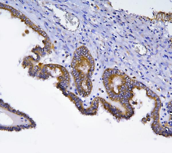
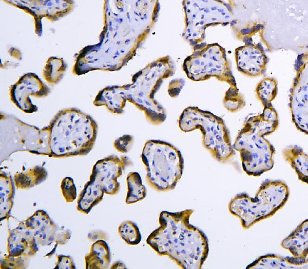
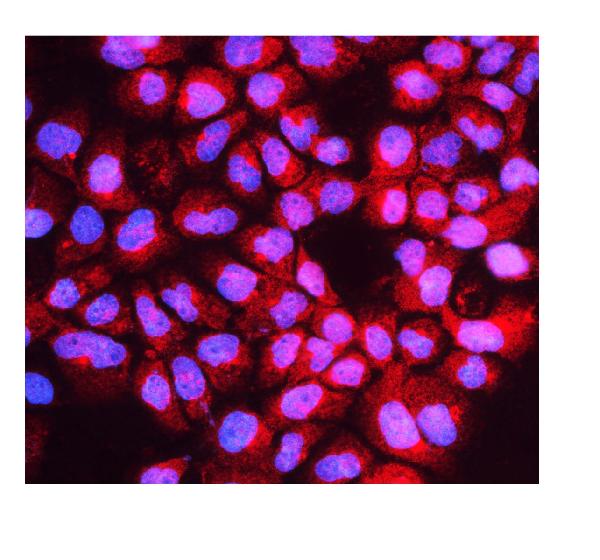


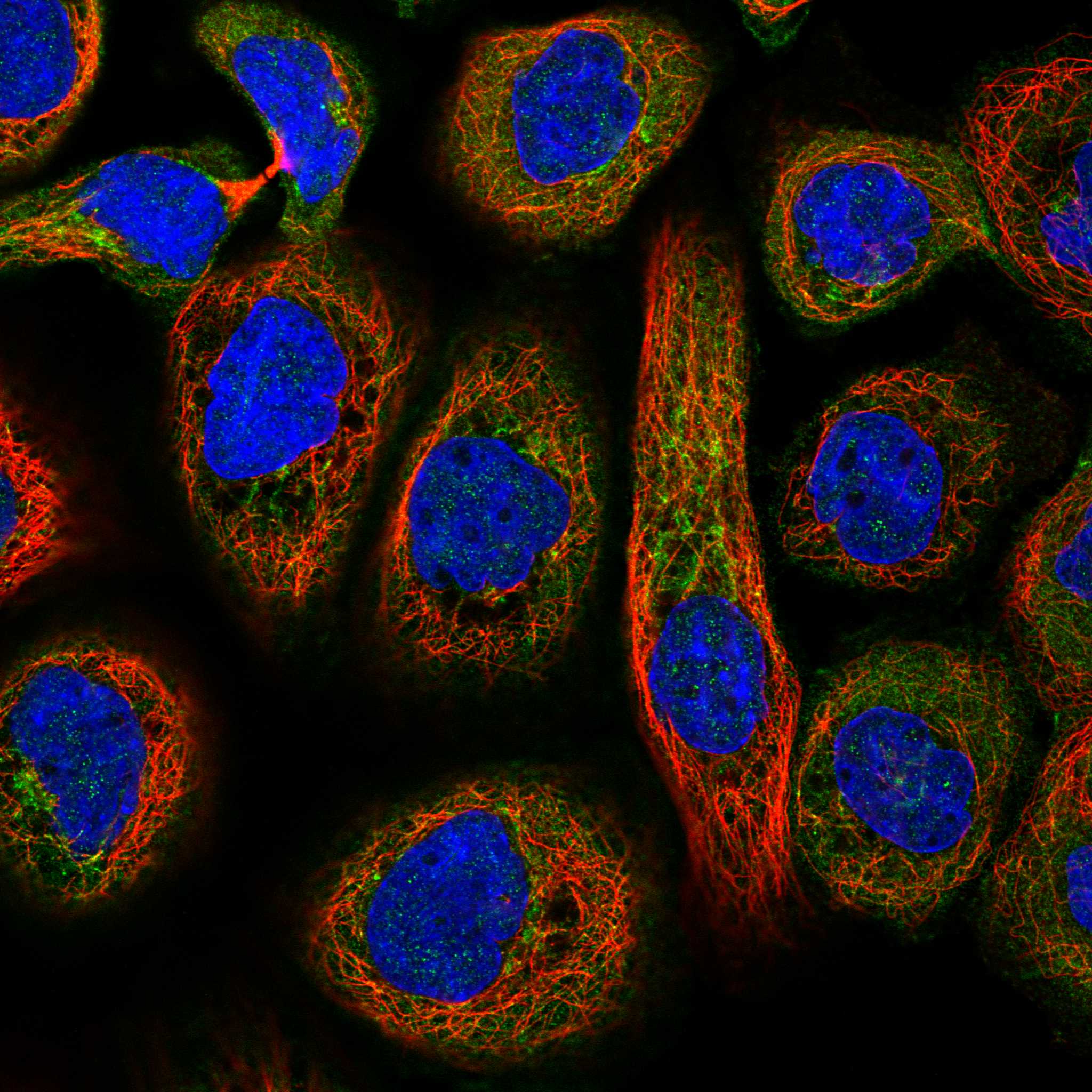
Comprehensive Human Protein Atlas IHC scoring per tissue (reliability: Approved — Medium consistency between antibody staining and RNA expression data.). Rows are taken directly from the HPA tissue chart — click any row's HPA link to view the source.
| Tissue | Cell type | Level | Evidence | Source |
|---|---|---|---|---|
| Appendix | Glandular cells | High | Protein (IHC) | HPA → |
| Colon | Glandular cells | High | Protein (IHC) | HPA → |
| Duodenum | Glandular cells | High | Protein (IHC) | HPA → |
| Fallopian tube | Ciliated cells (ciliary rootlets) | High | Protein (IHC) | HPA → |
| Gallbladder | Glandular cells | High | Protein (IHC) | HPA → |
| Tissue | Cell type | Level | Evidence | Source |
|---|---|---|---|---|
| Adipose tissue | Adipocytes | Not detected | Protein (IHC) | HPA → |
Troubleshoot HOOK2 staining in paraffin sections by checking retrieval, cytoplasmic localisation, cell type, and chromogenic controls before scoring.
A08854-1 has IHC images from human ovarian cancer and placenta paraffin sections and an IF/ICC image from A431 cells (catalog image captions).
A08854-1 is the SKU with a rendered card: its IHC figure shows a human ovarian cancer paraffin section (A08854-1 IHC caption). Its catalog also documents IHC in human placenta and IF/ICC in A431 cells (A08854-1 image captions).
Which to pick: For tissue IHC, choose A08854-1: its rabbit antibody has a documented paraffin-section result using EDTA retrieval at pH 8.0 and 2 μg/ml primary antibody; the fixative is unreported (A08854-1 IHC caption). For IF/ICC, choose A08854-1 because its A431 image documents staining at 5 μg/ml; M08854 lists IHC but has no supplied IHC figure or IF/ICC application (A08854-1 IF caption; M08854 catalog entry). Neither SKU has documented cross-species reactivity; both list Human only (catalog reactivity).