This website uses cookies to ensure you get the best experience on our website.


- Table of Contents
Plan HOPX IHC in paraffin sections using the human-reactive A05019 antibody, starting at 5 μg/mL (datasheet: A05019). Compare nuclear and cytoplasmic staining with tissue patterns, including high staining in skin granular-layer cells (HPA tissue IHC).

Expected localisation, validated protocols, controls and antibodies — the at-a-glance facts below, then the full design guide.
| Expected localisation | Nuclear and cytoplasmic staining (HPA tissue IHC) | |
| Staining pattern | Nuclear/cytoplasmic in squamous cells, glands and pneumocytes (HPA tissue IHC) | |
| Antigen retrieval | Tris-EDTA pH 9.0 HIER, 95–98 °C, 20 min (rule: nuclear antigen) | |
| Positive control | Duodenum+4 more · see all | |
| Negative control | Adipose tissue+4 more · see all |
| Fixation | Matched tissue-IHC evidence does not establish the fixation claim. Validate the specimen-specific method before use. (selected-SKU IHC image A05019) | |
| Caveat | Staining varies by cell population within a tissue (HPA tissue IHC) | |
| Regulation | Down-regulated in some cancers (UniProt) | |
| Isoform / epitope | 4 isoforms; epitope coverage is unspecified (UniProt) |
Compare the catalog antibody's IHC-P protocol with published chromogenic HOPX IHC protocols for thyroid, pancreatic, and head and neck tissues (PMC6800262; PMC3488580; PMC5146930).
| Sample | Tissue sections; selected-image fixative not specified (standard IHC workflow) |
| Fixation | Image fixative and duration unreported (datasheet A05019); verify before use. |
| Sectioning | 4–5 µm sections on charged slides (standard) |
| Deparaffinisation | Xylene, graded ethanol series to water (standard) |
| Antigen retrieval | Heat-induced epitope retrieval in Tris-EDTA buffer, pH 9.0, 20 min at 95–98 °C (standard rule: nuclear antigen) |
| Peroxidase block | 3% H2O2, 10 min, room temperature (standard) |
| Blocking | 10% normal serum of the secondary host, 30 min, room temperature (standard) |
| Primary antibody | Rabbit anti-HOPX, 5 μg/mL (datasheet A05019) |
| Primary incubation | Overnight at 4 °C (standard) |
| Detection | HRP-polymer secondary, DAB chromogen 5–10 min (standard) |
| Counterstain | Hematoxylin, blue, dehydrate and mount (standard) |
| Expected result | HOPX-positive staining in endocrine cells of duodenum (HPA tissue IHC: High). HPA tissue profile: Nuclear and cytoplasmic expression in squamous epithelia, glandular cells of thyroid and epididymis and in pneumocytes. No signal in the no-primary control. |
HOPX should appear in nuclei and cytoplasm, with staining interpreted by cell type rather than as a uniform tissue-wide signal (UniProt Q9BPY8; HPA tissue IHC). HPA reports nuclear and cytoplasmic staining in squamous epithelia, thyroid and epididymal glandular cells, and pneumocytes; its tissue IHC reliability is Enhanced, with medium consistency between staining and RNA expression (HPA tissue IHC). HOPX has no transmembrane segment (UniProt Q9BPY8 topology).
| Distinct nuclear staining, with possible cytoplasmic staining, appears in skin granular-layer cells. | This fits a high-staining cell population (HPA: High in skin granular-layer cells) and the documented nuclear and cytoplasmic locations (HPA tissue IHC; UniProt Q9BPY8). Judge the relevant cells rather than the darkness of the whole section. |
| A sharply outlined, membrane-only signal dominates otherwise unstained skin epithelial cells. | That is atypical for the expected nuclear and cytoplasmic pattern (HPA tissue IHC; UniProt Q9BPY8). Check staining specificity and detection artifacts before scoring it as HOPX. Interpret other tissues separately: HPA labels cerebellar granular-cell staining as cytoplasm/membrane (HPA: Medium in cerebellum). |
| Strong staining appears mainly in bronchial respiratory epithelial cells or adipocytes. | These cell populations are listed as not detected (HPA: bronchus respiratory epithelial cells; adipose tissue adipocytes). Consider cross-reactivity or endogenous detection activity, and compare the same run with a known-positive section. A single unexpected cell population does not establish a new HOPX pattern. |
| Color spreads across stroma, empty spaces, and many unrelated cells without clear cellular boundaries. | A diffuse field cannot be assigned confidently to the documented nuclear and cytoplasmic locations (HPA tissue IHC; UniProt Q9BPY8). In general chromogenic IHC practice, incomplete blocking, residual detection reagent, and inadequate washing can create background; inspect controls before interpreting weak cells. |
| A skin section lacks staining in its granular-layer cells. | HPA reports high staining in those cells (HPA: High in skin granular layer). First assess section quality, retrieval and detection controls under the assay's validated conditions. Absence in one run may reflect technical failure; it cannot alone establish absent HOPX expression. |
| Cell population and tissue | Expected intensity changes by cell population: duodenal and small-intestinal endocrine cells are High, epididymal glandular cells are Medium, and bronchial respiratory epithelial cells are Not detected (HPA tissue IHC). Select and score controls at the cell level. |
| Compartment and topology | UniProt lists nucleus and cytoplasm and no transmembrane segment (UniProt Q9BPY8). HPA tissue IHC reports both compartments; compartment alone is insufficient without the expected cell type (HPA tissue IHC). |
| Antibody evidence | HPA030180 and CAB018632 each have Enhanced IHC status (HPA antibody validation). HPA describes tissue IHC reliability as Enhanced but reports only medium consistency with RNA expression (HPA tissue IHC); retain that qualification when judging an unexpected pattern. |
| Isoforms and processing | Four isoforms are listed, while the supplied record gives no isoform-specific IHC pattern (UniProt Q9BPY8). It lists a 1–73 chain, no signal peptide or propeptide, and no annotated glycosylation sites (UniProt Q9BPY8); these facts do not predict retrieval behavior. |
| IF/ICC: where should signal appear? | Mainly in nuclear bodies, with additional cytosolic signal (HPA ICC-IF: approved locations). That observation answers the localisation question for IF/ICC; use its separate guide for assay setup. It does not override the tissue-specific chromogenic IHC pattern (HPA tissue IHC). |
| Situation | Likely cause | Next action |
|---|---|---|
| No color in granular-layer cells of a skin positive control. | The expected population is High (HPA: skin granular layer); a failed control leaves staining sensitivity unresolved. | Check the run's reagent and detection controls, then review section integrity, retrieval conditions and primary-antibody dilution against the validated IHC procedure (general IHC practice). Do not assign a HOPX-negative result from this run. |
| Signal is nuclear but appears chiefly in cells listed as not detected. | Correct-looking localisation does not establish cell-type specificity (UniProt Q9BPY8; HPA tissue IHC). Off-target binding or endogenous detection activity is possible (general IHC practice). | Compare a known-positive tissue in the same run and inspect appropriate primary-omission and detection controls (general IHC practice). Report the unexpected cell population separately rather than treating all nuclear color as HOPX. |
| Broad brown haze obscures nuclei and cytoplasmic boundaries. | Diffuse color is difficult to reconcile with cellular HOPX localisation (HPA tissue IHC; UniProt Q9BPY8); nonspecific detection or inadequate washing can contribute (general IHC practice). | Review blocking, washes, detection-reagent exposure and negative controls under the laboratory's validated chromogenic IHC workflow (general IHC practice). Score only cells whose signal can be localised reliably. |
| A membrane-only outline is scored as positive in skin. | That appearance conflicts with the expected nuclear and cytoplasmic pattern in squamous epithelium and with the absence of a transmembrane segment (HPA tissue IHC; UniProt Q9BPY8). | Reassess morphology and background, then compare staining with the documented granular-layer pattern (HPA: High in skin granular layer). Do not apply this skin rule to cerebellar granular cells, for which HPA reports cytoplasm/membrane staining (HPA: Medium in cerebellum). |
| A low-staining or not-detected population is used as the only positive control. | Esophageal squamous epithelial cells are Low, while bronchial respiratory epithelial cells are Not detected (HPA tissue IHC). Either choice gives limited evidence that the run can reveal a High population. | Include a section containing skin granular-layer cells or duodenal endocrine cells, both reported High (HPA tissue IHC). Evaluate those specific cells before interpreting weak or absent staining in the test tissue. |
| IF/ICC nuclear-body signal is used to reject a chromogenic tissue result. | HPA's approved nuclear-body and cytosolic locations come from ICC-IF, while its tissue IHC profile describes nuclear and cytoplasmic expression (HPA ICC-IF; HPA tissue IHC). | Judge the paraffin-section result against the tissue IHC cell population and compartment evidence (HPA tissue IHC). Consult the separate IF/ICC guide when interpreting fluorescent cell images. |


Comprehensive Human Protein Atlas IHC scoring per tissue (reliability: Enhanced — Medium consistency between antibody staining and RNA expression data.). Rows are taken directly from the HPA tissue chart — click any row's HPA link to view the source.
| Tissue | Cell type | Level | Evidence | Source |
|---|---|---|---|---|
| Duodenum | Endocrine cells | High | Protein (IHC) | HPA → |
| Skin | Cells in granular layer | High | Protein (IHC) | HPA → |
| Small intestine | Endocrine cells | High | Protein (IHC) | HPA → |
| Vagina | Squamous epithelial cells | High | Protein (IHC) | HPA → |
| Cerebellum | Granular cells - cytoplasm/membrane | Medium | Protein (IHC) | HPA → |
| Tissue | Cell type | Level | Evidence | Source |
|---|---|---|---|---|
| Adipose tissue | Adipocytes | Not detected | Protein (IHC) | HPA → |
| Adrenal gland | Glandular cells | Not detected | Protein (IHC) | HPA → |
| Bone marrow | Hematopoietic cells | Not detected | Protein (IHC) | HPA → |
| Breast | Adipocytes | Not detected | Protein (IHC) | HPA → |
| Bronchus | Respiratory epithelial cells | Not detected | Protein (IHC) | HPA → |
Troubleshoot HOPX staining in paraffin sections by checking retrieval, cell identity, subcellular pattern and controls before interpreting chromogenic signal.
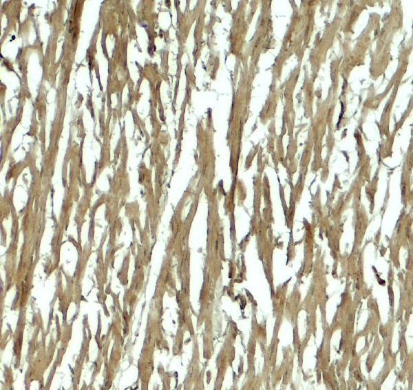
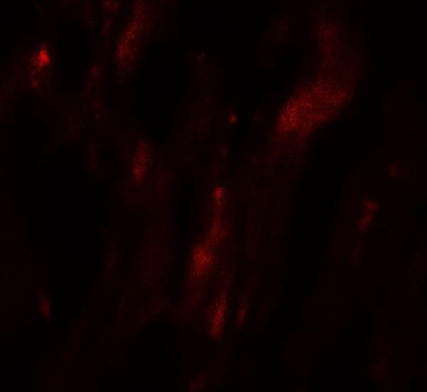
A05019 has IHC-P and IF listings for human samples, with IHC and IF images from human heart tissue (A05019 catalog; A05019 image captions).
A05019 is listed for human IHC-P and IF (A05019 catalog: applications, reactivity). Its own images show human heart tissue stained by IHC at 5 μg/mL and IF at 20 μg/mL (A05019 IHC and IF image captions).
Which to pick: Choose A05019 for paraffin-section tissue IHC based on its IHC-P listing; its own IHC image shows human heart tissue at 5 μg/mL, but the fixative is unreported (A05019 catalog: applications; A05019 IHC image caption). Choose A05019 for tissue IF at 20 μg/mL; ICC validation is unreported (A05019 IF image caption; A05019 catalog: applications). No cross-species option is supported: A05019 lists Human reactivity, has a Rabbit host, and has no reported clonality (A05019 catalog: reactivity, host, clone).