This website uses cookies to ensure you get the best experience on our website.


- Table of Contents
Plan HOXA4 paraffin-section IHC around general nuclear staining (HPA tissue IHC) and a catalog antibody dilution of 0.5–1 μg/mL (datasheet: PA1603 IHC-P). Compare high-staining glandular or neuronal cells with parathyroid glandular cells reported as undetected, while accounting for low staining–RNA concordance (HPA tissue IHC).

Expected localisation, validated protocols, controls and antibodies — the at-a-glance facts below, then the full design guide.
| Expected localisation | General nuclear staining (HPA tissue IHC) | |
| Staining pattern | Nuclear signal in glandular and neuronal cells (HPA tissue IHC) | |
| Antigen retrieval | Tris-EDTA pH 9.0 HIER, 95–98 °C, 20 min (rule: nuclear antigen) | |
| Positive control | Adrenal gland+4 more · see all | |
| Negative control | Parathyroid gland |
| Fixation | Matched tissue-IHC evidence does not establish the fixation claim. Validate the specimen-specific method before use. (selected-SKU IHC image PA1603) | |
| Caveat | Staining and RNA levels have low concordance (HPA tissue IHC) | |
| Regulation | Embryonic nervous system expression (UniProt) | |
| Isoform / epitope | No isoforms or processing variants annotated (UniProt) |
The catalog antibody’s IHC-P protocol is accompanied by three published HOXA4 IHC protocols (PMC3125234; PMC4750173; PMC9859321).
| Sample | Tissue sections; selected-image fixative not specified (standard IHC workflow) |
| Fixation | Image fixative and duration unreported (datasheet PA1603); verify before use. |
| Sectioning | 4–5 µm sections on charged slides (standard) |
| Deparaffinisation | Xylene, graded ethanol series to water (standard) |
| Antigen retrieval | Heat-induced epitope retrieval in Tris-EDTA buffer, pH 9.0, 20 min at 95–98 °C (standard rule: nuclear antigen) |
| Peroxidase block | 3% H2O2, 10 min, room temperature (standard) |
| Blocking | 10% normal serum of the secondary host, 30 min, room temperature (standard) |
| Primary antibody | Rabbit anti-HOXA4, 0.5-1μg/ml (datasheet PA1603) |
| Primary incubation | Overnight at 4 °C (standard) |
| Detection | HRP-polymer secondary, DAB chromogen 5–10 min (standard) |
| Counterstain | Hematoxylin, blue, dehydrate and mount (standard) |
| Expected result | HOXA4-positive staining in glandular cells of adrenal gland (HPA tissue IHC: High). HPA tissue profile: General nuclear expression. No signal in the no-primary control. |
HOXA4 is a nuclear transcription factor with no transmembrane segment (UniProt Q00056). In tissue IHC, HPA reports general nuclear staining, including high staining in selected glandular, neuronal, and squamous epithelial cells (HPA tissue IHC). HPA rates the tissue staining Approved but reports low consistency with RNA expression and pending external verification (HPA tissue IHC). Interpret a positive stain in its tissue and cell context.
| Distinct nuclear staining in the expected cells. | This matches the reported compartment (UniProt Q00056; HPA tissue IHC). High staining in cerebellar Purkinje cells or colon glandular cells is an observed reference pattern (HPA tissue IHC). Compare nuclear signal with neighboring cells on the same section; intensity alone does not establish specificity (general IHC practice). |
| Predominantly cytoplasmic or membranous staining. | That distribution conflicts with the reported nuclear localization and absence of a transmembrane segment (UniProt Q00056). Treat it as suspect; review morphology, detection background, and an appropriate control before scoring it as HOXA4 (general IHC practice). |
| Strong signal in a cell population outside the expected pattern. | HPA's listed high staining applies to particular cells within each tissue, not every cell in that tissue (HPA tissue IHC). Cross-reactivity or endogenous detection activity are possible explanations (general IHC practice); check the cell identity and controls before assigning the signal to HOXA4. |
| Diffuse color across nuclei, cytoplasm, and surrounding tissue. | This does not resolve the reported nuclear pattern (HPA tissue IHC). Background from detection reagents, inadequate blocking, or excessive antibody concentration can obscure localization (general IHC practice). Judge the stain against a reagent control processed with the section. |
| No nuclear signal in a listed high-staining tissue. | An absent signal in the relevant cell population warrants a technical check, although HPA's Approved rating has an RNA-consistency caveat (HPA tissue IHC). Confirm the cells are present, then review controls and the catalog antibody's IHC-P instructions (general IHC practice). |
| Tissue and cell selection | HPA reports high staining in adrenal and appendix glandular cells, cerebellar Purkinje cells, cerebral cortex neuronal cells, and esophageal squamous epithelial cells (HPA tissue IHC). Its parathyroid glandular cells are listed as not detected (HPA tissue IHC). Use the stated cell population when comparing sections. |
| Strength of the tissue evidence | The tissue profile is Approved, with low consistency between antibody staining and RNA expression and external verification pending (HPA tissue IHC). The listed staining is an observed reference, not proof that every positive cell contains HOXA4. HPA lists HPA060088 as IHC Approved; the payload does not report IHC Enhanced validation (HPA antibodies). |
| Compartment and topology | UniProt places HOXA4 in the nucleus and lists no transmembrane segment (UniProt Q00056). HPA describes general nuclear tissue staining (HPA tissue IHC). Score the nuclear compartment first; a strong signal elsewhere needs independent scrutiny before interpretation (general IHC practice). |
| Processing and annotated forms | UniProt lists one chain spanning residues 1–320, no signal peptide or propeptide, no annotated isoforms, and no glycosylation sites (UniProt Q00056). These annotations provide no basis for predicting a secreted or membrane staining pattern; they do not predict formalin sensitivity or retrieval requirements. |
| IF/ICC: what pattern is reported? | HPA reports HOXA4 in nuclear bodies in ICC-IF, with images from HEK293, Hep-G2, and U2OS (HPA subcellular). This supports a punctate nuclear pattern in that application. It does not establish an IHC-P staining intensity or an IF/ICC protocol. |
| Situation | Likely cause | Next action |
|---|---|---|
| Expected nuclei appear unstained in a listed high-staining tissue. | The relevant cells may be absent from the field, or the staining run may have failed (general IHC practice); HPA's tissue result also has an RNA-consistency caveat (HPA tissue IHC). | Identify the specified cells on the counterstain, check a run control, and compare the procedure with the catalog antibody's IHC-P instructions (general IHC practice). Do not infer a HOXA4-specific fixation effect from this result. |
| The section stains mainly at cell borders or in cytoplasm. | That compartment disagrees with nuclear HOXA4 localization and its lack of a transmembrane segment (UniProt Q00056); nonspecific staining is possible (general IHC practice). | Check a reagent control and the detection system, then rescore only morphologically identifiable nuclei (general IHC practice). Do not count border staining as a HOXA4-positive cell. |
| Many unrelated cell types show equally strong color. | The pattern lacks the cell specificity needed to compare with HPA's cell-level observations (HPA tissue IHC). Cross-reactivity or endogenous detection activity may contribute (general IHC practice). | Compare the named cell population with adjacent cells; check a control omitting primary antibody and assess endogenous detection activity as appropriate to the chromogen system (general IHC practice). |
| Hematoxylin obscures a weak nuclear signal. | A heavy counterstain can make low chromogenic signal difficult to judge (general IHC practice). HPA also lists low staining in hippocampal neuronal cells and pancreatic exocrine glandular cells (HPA tissue IHC). | Review counterstain balance and use a listed high-staining cell population to assess the run before calling a low-staining population negative (general IHC practice; HPA tissue IHC). |
| Diffuse background prevents compartment scoring. | Excess detection signal, insufficient blocking, or concentrated primary antibody can raise background (general IHC practice); the supplied sources give no HOXA4-specific background mechanism. | Check reagent controls, blocking, washes, and the dilution specified for the IHC-validated antibody (general IHC practice). Score only when nuclei can be distinguished from surrounding tissue. |
| Parathyroid glandular cells stain strongly. | HPA lists this population as not detected (HPA tissue IHC), but one discordant section does not establish whether the signal is biological or technical (general IHC practice). | Confirm cell identity, examine a reagent control, and compare a listed high-staining population in the same run (general IHC practice; HPA tissue IHC). Report the discrepancy rather than treating the HPA negative call as an absolute exclusion. |
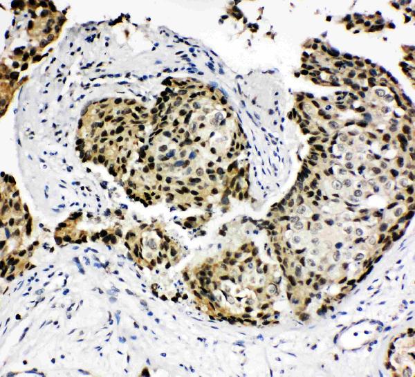
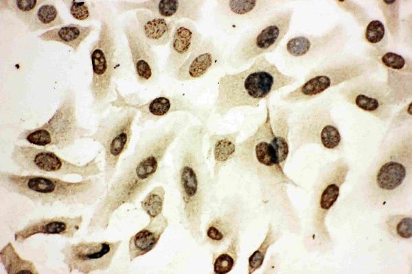


Comprehensive Human Protein Atlas IHC scoring per tissue (reliability: Approved — Low consistency between antibody staining and RNA expression data. Pending external verification.). Rows are taken directly from the HPA tissue chart — click any row's HPA link to view the source.
| Tissue | Cell type | Level | Evidence | Source |
|---|---|---|---|---|
| Adrenal gland | Glandular cells | High | Protein (IHC) | HPA → |
| Appendix | Glandular cells | High | Protein (IHC) | HPA → |
| Cerebellum | Purkinje cells | High | Protein (IHC) | HPA → |
| Cerebral cortex | Neuronal cells | High | Protein (IHC) | HPA → |
| Cervix | Glandular cells | High | Protein (IHC) | HPA → |
| Tissue | Cell type | Level | Evidence | Source |
|---|---|---|---|---|
| Parathyroid gland | Glandular cells | Not detected | Protein (IHC) | HPA → |
Troubleshoot HOXA4 staining by checking nuclear localisation, section handling and assay controls before interpreting staining intensity (UniProt Q00056; HPA tissue IHC).
PA1603 has IHC image data from human mammary cancer tissue and ICC image data from HeLa cells (catalog image captions); its listed reactivity is human, mouse and rat (catalog: reactivity).
PA1603 is listed for IHC and ICC, with an IHC(P) image of human mammary cancer tissue (catalog: applications; PA1603 IHC image caption). Its ICC image shows HeLa cells, and its listed reactivity includes human, mouse and rat (PA1603 ICC image caption; catalog: reactivity).
Which to pick: Choose PA1603 for paraffin-section IHC: its tissue image is labeled IHC(P), and its IHC dilution is 0.5–1 μg/ml (PA1603 IHC image caption; datasheet: IHC dilution). For ICC, PA1603 has a HeLa cell image and a listed dilution of 0.5–1 μg/ml; IF is not separately listed as an application (PA1603 ICC image caption; datasheet: ICC dilution; catalog: applications). PA1603 also lists mouse and rat reactivity, but its IHC image documents human tissue only; the caption does not report a fixative, and clonality is unreported (catalog: reactivity; PA1603 IHC image caption; catalog: clone).