This website uses cookies to ensure you get the best experience on our website.


- Table of Contents
Plan HSD3B1 paraffin IHC around strong cytoplasmic staining in placental syncytiotrophoblasts (HPA tissue IHC). Start with the IHC-validated antibody A02856-2 at 0.5–1 μg/mL and include a negative control (datasheet: A02856-2; standard IHC practice).

Expected localisation, validated protocols, controls and antibodies — the at-a-glance facts below, then the full design guide.
| Expected localisation | Cytoplasmic staining in placenta and adrenal gland (HPA tissue IHC) | |
| Staining pattern | Syncytiotrophoblast and adrenal cell cytoplasm (HPA tissue IHC) | |
| Antigen retrieval | EDTA pH 8.0 HIER, heat-mediated (datasheet A02856-2) | |
| Positive control | Placenta+1 more · see all | |
| Negative control | Adipose tissue+4 more · see all |
| Fixation | Keep paraffin tissue fixation consistent across samples. (standard IHC practice; not target-specific) | |
| Caveat | Presumed off-target binding may mimic positivity (HPA tissue IHC) | |
| Regulation | Regulatory drivers are not specified (UniProt) | |
| Isoform / epitope | No annotated isoforms; epitope sidedness unresolved (UniProt) |
The catalog antibody’s IHC-P protocol (datasheet: A02856-2) is accompanied by published HSD3B1 staining methods for human adenoma, rat testis, and cat testis (PMC4821076; PMC5643909; PMC13313884).
| Sample | Paraffin-embedded human placenta tissue; fixative not specified (datasheet A02856-2) |
| Fixation | Image fixative and duration unreported (datasheet A02856-2); verify before use. |
| Sectioning | 4–5 µm sections on charged slides (standard) |
| Deparaffinisation | Xylene, graded ethanol series to water (standard) |
| Antigen retrieval | Heat retrieval: EDTA pH 8.0 (datasheet A02856-2); 20 min, 95–100 °C (standard) |
| Peroxidase block | 3% H2O2, 10 min, room temperature (standard) |
| Blocking | 10% goat serum (datasheet A02856-2) |
| Primary antibody | Rabbit anti-HSD3B1, 0.5-1μg/ml (datasheet A02856-2) |
| Primary incubation | Overnight at 4 °C (datasheet A02856-2) |
| Detection | Streptavidin-biotin complex (SABC), DAB chromogen (datasheet A02856-2) |
| Counterstain | Hematoxylin, blue, dehydrate and mount (standard) |
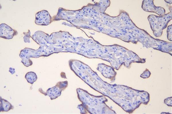
| Expected result | HSD3B1-positive staining in syncytiotrophoblasts of placenta (HPA tissue IHC: High). HPA tissue profile: Cytoplasmic expression in placenta and adrenal gland. No signal in the no-primary control. |
HSD3B1 is an endoplasmic reticulum and mitochondrial membrane protein with one transmembrane segment at residues 288–308 (UniProt P14060 topology). In paraffin-section IHC, expect cytoplasmic staining, strongest in placental syncytiotrophoblasts (HPA tissue IHC: High; reliability Supported). Interpret that pattern with caution: HPA reports antibodies targeting proteins from more than one gene and presumed off-target binding that was disregarded in its expression profile (HPA tissue IHC: reliability description).
| Strong cytoplasmic staining in placental syncytiotrophoblasts. | This is the clearest supplied positive-tissue pattern (HPA tissue IHC: Syncytiotrophoblasts High). Record the cell type and cytoplasmic distribution together; intensity alone is insufficient because HPA flags antibodies that target proteins from more than one gene (HPA tissue IHC: reliability description). |
| Predominantly nuclear or other noncytoplasmic staining in placental syncytiotrophoblasts. | A nuclear-only IHC pattern does not match the reported cytoplasmic tissue pattern or membrane localization (HPA tissue IHC: cytoplasmic expression; UniProt P14060 topology). Review staining controls and interpretation before calling it HSD3B1; the ICC-IF nucleolar annotation is uncertain (HPA subcellular: Nucleoli uncertain). |
| Strong staining in a cell population HPA reports as not detected, such as adipocytes in adipose tissue. | That result conflicts with the supplied cell-specific reference (HPA tissue IHC: Adipocytes Not detected). Consider antibody cross-reactivity, which is a particular concern given HPA's multi-gene targeting caution, or endogenous chromogenic detection activity as a general IHC cause (HPA tissue IHC: reliability description; general IHC practice). |
| Weak, widespread chromogen obscures cell boundaries across the section. | Treat this as background until controls establish a cell-specific signal (general IHC practice). It cannot be scored as the reported placental pattern merely because the section contains placenta (HPA tissue IHC: Syncytiotrophoblasts High); inspect a no-primary control and the distribution of staining (general IHC practice). |
| No cytoplasmic signal in placental syncytiotrophoblasts. | A blank known-positive cell population conflicts with the HPA reference pattern (HPA tissue IHC: Syncytiotrophoblasts High). Check section identity and IHC run controls, then review antigen retrieval, primary-antibody conditions and detection as general workflow variables; the supplied sources do not establish HSD3B1-specific fixation sensitivity (general IHC practice). |
| Membrane localization and topology | UniProt places HSD3B1 at endoplasmic reticulum and mitochondrial membranes and annotates one transmembrane segment, residues 288–308 (UniProt P14060 topology). These facts support a cytoplasmic IHC interpretation; they do not identify this antibody's epitope or predict an antigen-retrieval condition. |
| Cell-specific reference levels | HPA reports High staining in placental syncytiotrophoblasts, Medium in testicular spermatogonia and Low in adrenal zona fasciculata cells (HPA tissue IHC). Compare like cell populations when assessing intensity; do not expect uniform staining across a tissue from a cell-specific rating. |
| Strength and limit of IHC evidence | The tissue profile is rated Supported, and three listed antibodies have Supported IHC status (HPA tissue IHC: reliability Supported; HPA antibodies: HPA043261, HPA043264, HPA044028). HPA also warns of multi-gene targeting and presumed off-target binding, so agreement with the expected pattern supports interpretation without proving molecular specificity (HPA tissue IHC: reliability description). |
| IF/ICC Q&A: What localization should be expected? | The supported ICC-IF location is endoplasmic reticulum (HPA subcellular: Endoplasmic reticulum supported). Nucleoli, primary cilium and other listed locations are uncertain, with a multi-gene antibody caution (HPA subcellular). Use the separate IF/ICC guide for that application; these annotations are not an IHC protocol. |
| Situation | Likely cause | Next action |
|---|---|---|
| Placental syncytiotrophoblasts are unstained. | The expected High cell-specific signal is absent (HPA tissue IHC: Syncytiotrophoblasts High); a failed staining run or incorrect section identification are general IHC possibilities. | Confirm tissue and cell identity, examine run controls, then review retrieval, primary-antibody and detection steps using the validated IHC procedure (general IHC practice). Do not infer an HSD3B1-specific fixation problem from these sources. |
| Placenta shows staining, but it is chiefly nuclear. | This conflicts with the cytoplasmic tissue profile (HPA tissue IHC: cytoplasmic expression). The ICC-IF nucleolar report is uncertain and does not validate nuclear IHC staining (HPA subcellular: Nucleoli uncertain). | Check the counterstain and chromogen distribution, compare with the positive-tissue reference and repeat with appropriate controls if the compartment remains unclear (general IHC practice). |
| Adipocytes or other HPA-negative cell populations stain strongly. | Their staining conflicts with HPA's Not detected ratings; antibody cross-reactivity is plausible given the documented multi-gene targeting caution (HPA tissue IHC: negative cells; reliability description). | Compare the precise cell population with HPA's reference and examine a no-primary control for endogenous detection activity (general IHC practice). Avoid scoring unexpected staining as HSD3B1 without further specificity evidence. |
| Diffuse chromogen covers positive and negative areas. | Nonspecific background or endogenous detection activity can obscure a cell-specific IHC pattern (general IHC practice); HPA's reference is cytoplasmic and cell-specific (HPA tissue IHC). | Inspect no-primary and run controls, then review blocking, washes, primary-antibody conditions and chromogen development under the laboratory's standard IHC workflow (general IHC practice). |
| Adrenal signal is much fainter than placental signal. | That difference matches the supplied cell ratings: Low in adrenal zona fasciculata cells versus High in placental syncytiotrophoblasts (HPA tissue IHC). | Identify the scored adrenal cells and compare slides stained in the same run before treating the lower intensity as a technical failure (general IHC practice; HPA tissue IHC: cell-specific levels). |
| IF/ICC shows a prominent nucleolar or ciliary pattern. | HPA marks nucleoli and primary cilium as uncertain ICC-IF locations and cautions that the underlying antibodies target proteins from multiple genes (HPA subcellular). | Interpret those locations cautiously and consult the separate IF/ICC guide; the supported ICC-IF location is endoplasmic reticulum (HPA subcellular: Endoplasmic reticulum supported). |


Comprehensive Human Protein Atlas IHC scoring per tissue (reliability: Supported — Caution, targets protein from more than one gene. Presumed off target binding observed and disregarded. Paired antibodies with high similarity supports the expression profile.). Rows are taken directly from the HPA tissue chart — click any row's HPA link to view the source.
| Tissue | Cell type | Level | Evidence | Source |
|---|---|---|---|---|
| Placenta | Syncytiotrophoblasts | High | Protein (IHC) | HPA → |
| Testis | Spermatogonia cells | Medium | Protein (IHC) | HPA → |
| Tissue | Cell type | Level | Evidence | Source |
|---|---|---|---|---|
| Adipose tissue | Adipocytes | Not detected | Protein (IHC) | HPA → |
| Appendix | Glandular cells | Not detected | Protein (IHC) | HPA → |
| Bone marrow | Hematopoietic cells | Not detected | Protein (IHC) | HPA → |
| Breast | Adipocytes | Not detected | Protein (IHC) | HPA → |
| Bronchus | Respiratory epithelial cells | Not detected | Protein (IHC) | HPA → |
Troubleshoot HSD3B1 staining in paraffin sections using the catalog antibody’s tissue protocol, expected cellular distribution, and appropriate controls.
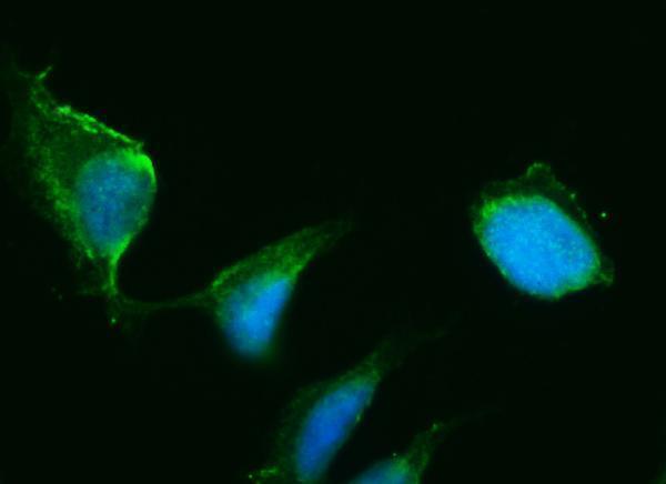
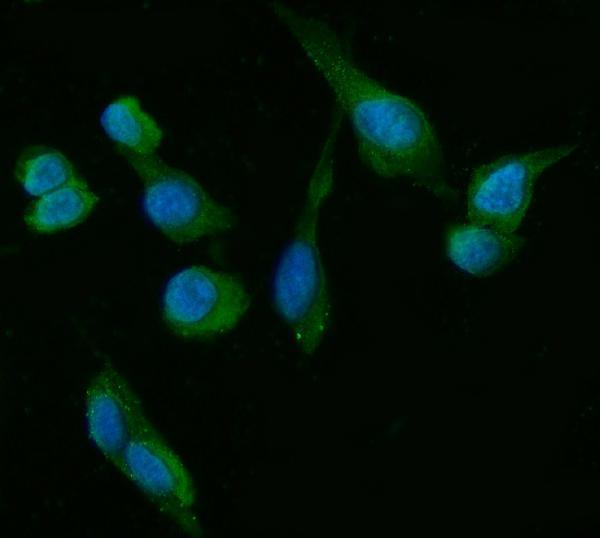
The catalog shows HSD3B1 IHC in human placenta paraffin sections and IF/ICC in human U20S cells (catalog image captions); one IF/ICC antibody also lists mouse and rat reactivity (catalog reactivity).
A02856-2 renders with IHC data from human placenta paraffin sections and lists human reactivity (catalog IHC caption; catalog reactivity). M02856 renders with IF/ICC data from human U20S cells and lists human, mouse, and rat reactivity (catalog IF caption; catalog reactivity).
Which to pick: Choose A02856-2 for paraffin-section IHC: its own caption documents heat retrieval in EDTA at pH 8.0 and chromogenic DAB detection in human placenta; the fixative is unreported (catalog IHC caption). For IF/ICC, A02856-2 has human U20S-cell IF data and a listed concentration of 2 μg/ml, while mouse monoclonal M02856 has its own human U20S-cell IF data at 5 μg/ml (catalog IF captions; catalog host and clone). Choose M02856 when mouse or rat reactivity is needed for IF/ICC, while treating its demonstrated IF image as human-cell evidence (catalog applications, reactivity, and IF caption).