This website uses cookies to ensure you get the best experience on our website.
- Table of Contents
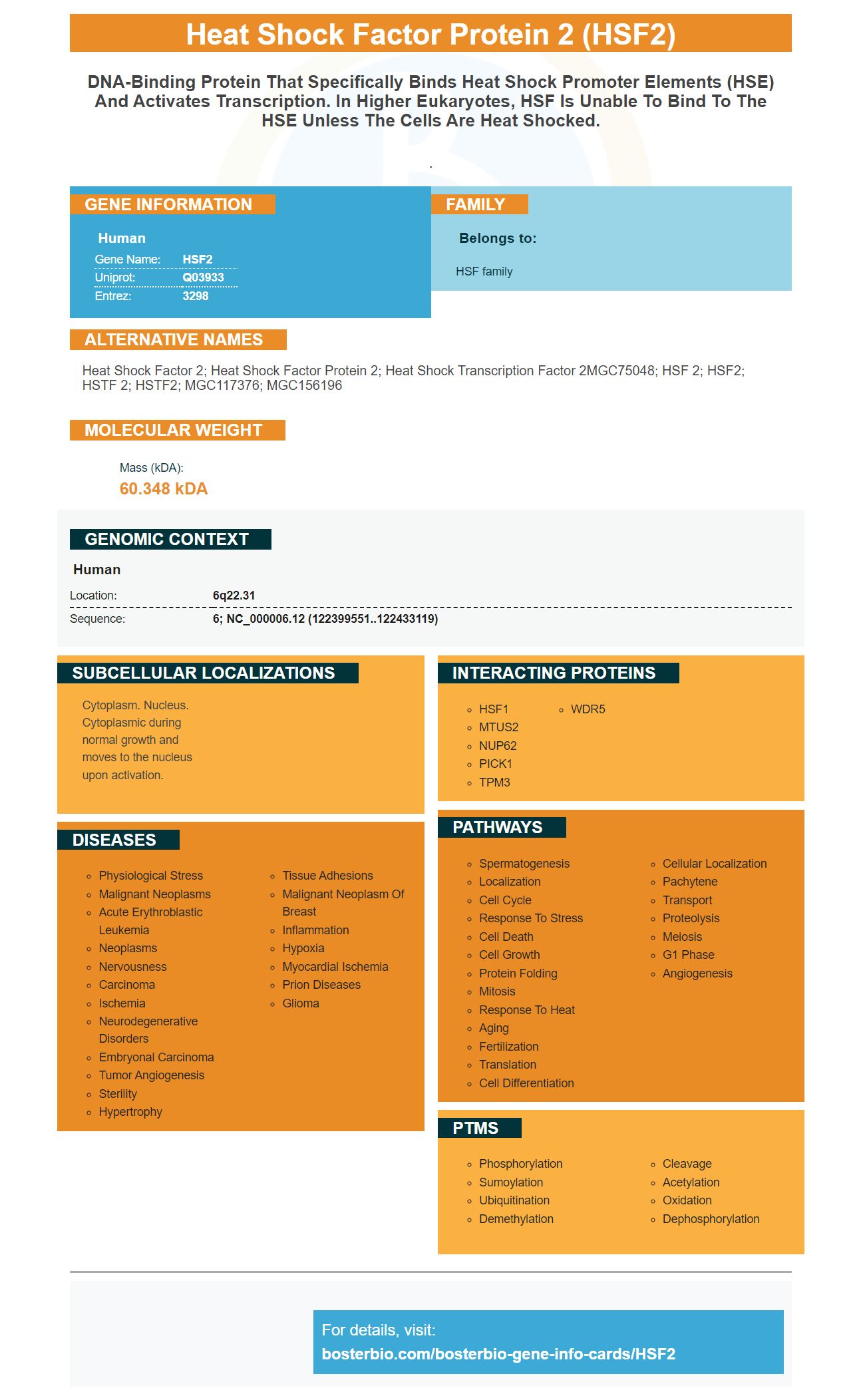
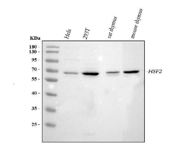
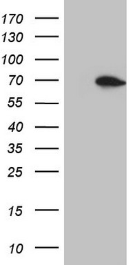
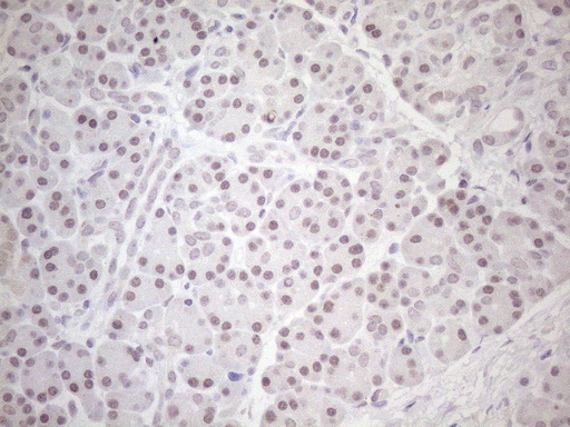
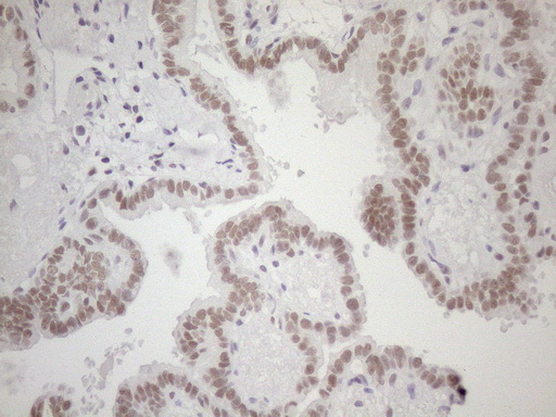
Facts about Heat shock factor protein 2.
.
Human | |
---|---|
Gene Name: | HSF2 |
Uniprot: | Q03933 |
Entrez: | 3298 |
Belongs to: |
---|
HSF family |
heat shock factor 2; heat shock factor protein 2; heat shock transcription factor 2MGC75048; HSF 2; HSF2; HSTF 2; HSTF2; MGC117376; MGC156196
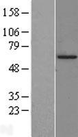
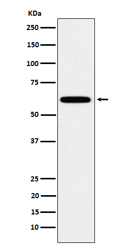
Mass (kDA):
60.348 kDA
Human | |
---|---|
Location: | 6q22.31 |
Sequence: | 6; NC_000006.12 (122399551..122433119) |
Cytoplasm. Nucleus. Cytoplasmic during normal growth and moves to the nucleus upon activation.
This site is for you if you are looking to find high-affinity antibodies that protect against Heat shock factor 2, (HSF2) Boster produces primary antibody that has been used in research for over 25 years. They are approved for use with Western Blotting (Immunohistochemistry) and ELISA. Continue reading to learn more about Boster antibodies and their best uses.
HSF1 triggers the cytoprotective responses in eukaryotes when they are exposed to proteotoxic stimuli. HSF1 forms trimers in the cytosol and translocates to the nucleus where it binds heat shock elements. These proteins interact with many cellular elements and contain a sequence nGAANNTTC. HSF1 binds HSEs, and triggers transcription.
HSF1 interacts directly with HSP70's HR-C domain, allowing for detection of mutant proteins with deletion or truncation mutations. HSF1 binds strongly to mutants that lack either the HRC-C or TADdomains. Although HSF90 binds to other domains of HSF1, its affinity for these domains is low.
HSF1 regulates the heat-shock system. HSF1 regulates the tumor's overall stress levels and the load on HSP-based chaperone machines. This information could help to determine if an HSF90 inhibit will be beneficial for a patient. HSF1 can be used to predict the outcome of patients with cancer.
HSF1 is vital for normal cell survival under lethal stress. Cancer cells can co-opt HSF1 to survive, resulting in dramatic changes in their physiology. HSF1 is a stress-relieving agent that allows cancer cells to deal with stressors. It reprogrammes their metabolic and protein homesostasis networks. Furthermore, HSF1 promoter occupancy was increased immediately after heat shock. This results in an increase of malignant cell proliferation.
Molecular analysis of the ERa-HSF1 interaction revealed that HSF1 and ERa are co-localized at a single-cell level. ERa and HSF1 bind DNA in different places, but their co-localization at the nucleus indicates that they have distinct modes. ChIP-seq data showed that HSF1 (and ERa) are co-located at the nucleus, based on the analysis of their peaks. Immunofluorescence also confirmed their proximity.
It has been shown that HSF1 binds with the human breast-cancer cell ERaprotein protein. HSF1 binds with ERa to increase cell growth when estrogen levels rise. This correlation can be attributed to drug therapies targeting ERa. HSF1 and ERa bind in the same regions. Further truncation of the hsp70 reporter increased its activity.
The interactions between HSF1 and ERa are also involved in the metastasis-associated protein 1 process and chromatin-modulating proteins. HSF1 may influence both liganded and unliganded ERa. Knockout of HSF1 in human breast cancer cells has a different effect on ERa-regulated genes in the presence or absence of estrogen. The hormone status of the tumor cells may influence the effects in real cancer.
Boster Bio's breast cancer cells expressed HSF1 was studied in detail. Researchers discovered that HSF1 positive cancers had poorer outcomes compared to ER-negative ones. The hormone receptor status was taken into account when the study's results were interpreted. The key transcriptional regulator in breast-cancer cells is heat shock factor 1, HSF1 enhances transcription of genes involved in disease progression.
Boster Bio's HSF2 marker is highly specific and sensitive in immunoblotting. It is compatible both with Western blotting and immunoprecipitation. Steven Boster developed it and it is the preferred method for high-affinity primary antibody detection. PicoKine(tm), which is a proprietary platform that allows researchers and scientists to detect antibodies only from one clone, makes it unique.
Many experiments and assays rely on high-affinity prima antibodies. These antibodies bind to antigens to cause a specific reaction. The primary antibody is often used in immunoassays, including Western Blotting, immunohistochemistry, and immunofluorescence, as well as proteomics tests. Another popular technique for measuring the number of cells in a sample is flow cytometry.
For many areas of research, immunolabeling is essential. It is a common tool to detect specific cell or tissue components. Boster Bio HSF2 marker uses a fluorescent dye to label the antibody with an antigen. This tool can be used in order to measure the antigen content of samples and purify the antibodies. In addition, it can be used for QC testing, which helps to ensure the highest quality primary antibodies.
ELISA tests are performed at different phases of antibody development. After an animal is immunized the best-responding animals have been selected and spleen cells taken for hybridoma are fusion. The supernatants of the ELISA titration ELISA are screened to ensure positive responses. The best-responding antibody samples undergo limiting-dilution subcloning until all wells are growing at the same rate. The selected antibodies undergo further application testing.
Boster biomarker, which is highly sensitive in determining HSF2 in human beings, is an option. This antibody can identify a HSF2 molecule in human by analysing its surface chemicals. The HSF1-HSF2 proteins have homodimeric transcription factors that have distinct molecular structures. These proteins can co-purify at an identical elution volume. They are stable in all three steps of tandem purification. Mass spectrometry and crosslinking confirmed that they were the only two non-keratin proteins.
The Human Kinase Library (Catalog #23725) provided the antibodies that were used for detection of HSF2 human protein. The antibody was then incubated with thrombin to generate ARAF kinase-deficient mutants. Amicon Ultra Centricon was used to cleave the 6xHis tag. The protein yield was 2.0 mg/mL.
Co-immunoprecipitation and electrophoretic mobility supershift assays have been used to quantify HSF1-HSF2 interaction in vivo. These methods do not distinguish between direct and indirect interactions. The human HSF2 marker in Boster bio was purified using StrepII-tagged human HSF1 and 6xHis-tagged human HSF2. Tandem differential affinity purification results showed that HSF1 and HSF2 proteins co-elute, indicating that they interact.
The HSF1 and HSF2 DBD structures offer a framework for understanding complex regulation. The unique surfaces of the DBDs of HSFs that wrap around DNA reveal unique surfaces that are unique to each paralog. These surfaces provide the template to interact with regulatory proteins. Further investigation of the HSF1/HSF2 interaction might reveal additional regulatory events. The HSF2-HSF1 interaction in humans may be a new diagnostic tool for cancer diagnosis.
HSF2 is not affected by a proteasome inhibitor and HSF1W2 exhibits enhanced DNA binding. These results indicate that HSF2W2 is a biological function specific to the genomic locus. To determine the exact biological function of HSF2W2, a mechanistic study is required. This research revealed novel aspects of HSF1W2 gene. Hsp70 has been removed from HSF1W2 so that the HSF1W1 genome is indistinguishable.
Human HSF2DBD ligates with both SUMO E1 & SUMO E3 proteins. It was left to incubate at 25 degrees Celsius for 5 minutes. The samples were then placed onto sitting-drop vapor diffuse crystallization trays. They were immunoblotted for HSF2 mRNA levels and protein levels. The HSF2 ligation data is presented in Figure 1.
PMID: 1871106 by Schuetz T.J., et al. Isolation of a cDNA for HSF2: evidence for two heat shock factor genes in humans.
PMID: 8339932 by Sheldon L., et al. Hydrophobic coiled-coil domains regulate the subcellular localization of human heat shock factor 2.