Click image to see more details
-

-
-
-
-
+1
Product Info Summary
| SKU: | PA1607 |
|---|---|
| Size: | 100 μg/vial |
| Reactive Species: | Human, Mouse, Rat |
| Host: | Rabbit |
| Application: | Flow Cytometry, IF, IHC, ICC, WB |
Customers Who Bought This Also Bought

Product info
Product Name
Anti-Heat shock factor protein 2 HSF2 Antibody Picoband®
SKU/Catalog Number
PA1607
BA3206 is an alternative SKU for this antibody, used in previous lots.
Size
100 μg/vial
Form
Lyophilized
Description
Boster Bio Anti-Heat shock factor protein 2 HSF2 Antibody catalog # PA1607. Tested in Flow Cytometry, IF, IHC, ICC, WB applications. This antibody reacts with Human, Mouse, Rat. The brand Picoband indicates this is a premium antibody that guarantees superior quality, high affinity, and strong signals with minimal background in Western blot applications. Only our best-performing antibodies are designated as Picoband, ensuring unmatched performance.
Storage & Handling
Store at -20˚C for one year from date of receipt. After reconstitution, at 4˚C for one month. It can also be aliquotted and stored frozen at -20˚C for six months. Avoid repeated freeze-thaw cycles.
Cite This Product
Anti-Heat shock factor protein 2 HSF2 Antibody Picoband® (Boster Biological Technology, Pleasanton CA, USA, Catalog # PA1607)
Host
Rabbit
Contents
Each vial contains antibody formulated with stabilizing components, 0.9mg NaCl, 0.2mg Na2HPO4, 0.05mg Thimerosal, 0.01mg NaN3.
*This antibody is supplied in a stabilized formulation.
Compatibility with conjugation reactions depends on the chemistry of the conjugation method used.
For conjugation methods that are not compatible with the stabilizing components present in this formulation, a carrier-free antibody format is required.
Clonality
Polyclonal
Isotype
Rabbit IgG
Immunogen
A synthetic peptide corresponding to a sequence in the middle region of human HSF2, identical to the related mouse sequence, and different from the related rat sequence by one amino acid.
Cross-reactivity
No cross-reactivity with other proteins
Reactive Species
PA1607 is reactive to HSF2 in Human, Mouse, Rat
Observed Molecular Weight
60 kDa
Calculated molecular weight
60.3 kDa
Background of HSF2
HSF2 (HEAT-SHOCK TRANSCRIPTION FACTOR 2), also called HEAT-SHOCK FACTOR 2, as well as the related gene HSF1, encodes a protein that binds specifically to the heat-shock element and has homology to HSFs of other species. The International Radiation Hybrid Mapping Consortium maps the HSF2 gene to chromosome 6. Mouse Hsf2 was expressed in all 3 embryonic layers at embryonic day 7.5 and that the head fold was strongly stained at embryonic day 8.5. In adults, Hsf2 expression is detected in spermatocytes and spermatogonia, but not in elongated spermatids, spermatozoa, or Sertoli cells. Hsp70i bookmarking is mediated by a transcription factor called HSF2, which binds this promoter in mitotic cells, recruits protein phosphatase-2A, and interacts with the CAPG subunit of the condensin enzyme to promote efficient dephosphorylation and inactivation of condensin complexes in the vicinity, thereby preventing compaction at this site. And Hsf2-null mice were born at the expected mendelian ratio but had brain abnormalities and meiotic and gametogenic defects in both genders.
Antibody Validation
Boster validates all antibodies on WB, IHC, ICC, Immunofluorescence, and ELISA with known positive control and negative samples to ensure specificity and high affinity, including thorough antibody incubations.
Application & Images
Applications
PA1607 is guaranteed for Flow Cytometry, IF, IHC, ICC, WB Boster Guarantee
Recommend Dilution
| Application | Dilution | Species |
|---|---|---|
| Western blot | 0.1-0.5μg/ml | Human, Mouse, Rat |
| Immunohistochemistry (Paraffin-embedded Section) | 0.5-1μg/ml | Rat |
| Immunocytochemistry/Immunofluorescence | 2μg/ml | Human |
| Flow Cytometry (Fixed) | 1-3μg/1x106 cells | Human |
Tested application
Suggested blocking solution with 5% non-fat milk or BSA; (*)Recommended protein loading: 20-40 µg per lane
Use TE buffer pH 9.0 for antigen retrieval; (*) citrate buffer pH 6.0 is an alternative.
Validation Images & Assay Conditions
Click image to see more details
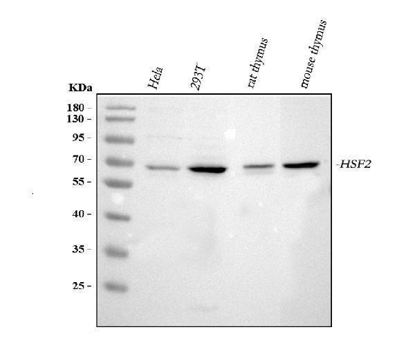
Western blot analysis of HSF2 using anti-HSF2 antibody (PA1607).
Electrophoresis was performed on a 5-20% SDS-PAGE gel at 70V (Stacking gel) / 90V (Resolving gel) for 2-3 hours. The sample well of each lane was loaded with 30 ug of sample under reducing conditions.
Lane 1: human Hela whole cell lysates,
Lane 2: human 293T whole cell lysates,
Lane 3: rat thymus tissue lysates,
Lane 4: mouse thymus tisue lysates.
After electrophoresis, proteins were transferred to a nitrocellulose membrane at 150 mA for 50-90 minutes. Blocked the membrane with 5% non-fat milk/TBS for 1.5 hour at RT. The membrane was incubated with rabbit anti-HSF2 antigen affinity purified polyclonal antibody (Catalog # PA1607) at 0.5 μg/mL overnight at 4°C, then washed with TBS-0.1%Tween 3 times with 5 minutes each and probed with a goat anti-rabbit IgG-HRP secondary antibody at a dilution of 1:5000 for 1.5 hour at RT. The signal is developed using an Enhanced Chemiluminescent detection (ECL) kit (Catalog # EK1002) with Tanon 5200 system. A specific band was detected for HSF2 at approximately 60 kDa. The expected band size for HSF2 is at 60 kDa.

Click image to see more details
Anti-HSF2 antibody, PA1607, IHC(P)
IHC(P): Rat Brain Tissue

Click image to see more details
Anti-HSF2 antibody, PA1607, IHC(P)
IHC(P): Rat Brain Tissue

Click image to see more details
IF analysis of HSF2 using anti-HSF2 antibody (PA1607).
HSF2 was detected in immunocytochemical section of U20S cells. Enzyme antigen retrieval was performed using IHC enzyme antigen retrieval reagent (AR0022) for 15 mins. The cells were blocked with 10% goat serum. And then incubated with 2μg/mL rabbit anti-HSF2 Antibody (PA1607) overnight at 4°C. DyLight®488 Conjugated Goat Anti-Rabbit IgG (BA1127) was used as secondary antibody at 1:100 dilution and incubated for 30 minutes at 37°C. The section was counterstained with DAPI. Visualize using a fluorescence microscope and filter sets appropriate for the label used.

Click image to see more details
Flow Cytometry analysis of 293T cells using anti-HSF2 antibody (PA1607).
Overlay histogram showing 293T cells stained with PA1607 (Blue line). To facilitate intracellular staining, cells were fixed with 4% paraformaldehyde and permeabilized with permeabilization buffer. The cells were blocked with 10% normal goat serum. And then incubated with rabbit anti-HSF2 Antibody (PA1607,1μg/1x106 cells) for 30 min at 20°C. DyLight®488 conjugated goat anti-rabbit IgG (BA1127, 5-10μg/1x106 cells) was used as secondary antibody for 30 minutes at 20°C. Isotype control antibody (Green line) was rabbit IgG (1μg/1x106) used under the same conditions. Unlabelled sample without incubation with primary antibody and secondary antibody (Red line) was used as a blank control.
Specific Publications For Anti-Heat shock factor protein 2 HSF2 Antibody Picoband® (PA1607)
Loading publications
Recommended Resources
Here are featured tools and databases that you might find useful.
- Boster's Pathways Library
- Protein Databases
- Bioscience Research Protocol Resources
- Data Processing & Analysis Software
- Photo Editing Software
- Scientific Literature Resources
- Research Paper Management Tools
- Molecular Biology Software
- Primer Design Tools
- Bioinformatics Tools
- Phylogenetic Tree Analysis
Customer Reviews
Have you used Anti-Heat shock factor protein 2 HSF2 Antibody Picoband®?
Share your experimental results or join a short interview to earn up to $1,000 in product credits or other rewards.
0 Reviews For Anti-Heat shock factor protein 2 HSF2 Antibody Picoband®
Customer Q&As
Have a question?
Find answers in Q&As, reviews.
Can't find your answer?
Submit your question
4 Customer Q&As for Anti-Heat shock factor protein 2 HSF2 Antibody Picoband®
Question
See below the WB image, lot number and protocol we used for female gonad using anti-HSF2 antibody PA1607. Please let me know if you require anything else.
Verified Customer
Verified customer
Asked: 2019-10-01
Answer
Thank you very much for the data. Our lab team are working to resolve this as quickly as possible, and we appreciate your patience and understanding! You have provided everything we needed. Please let me know if there is anything you need in the meantime.
Boster Scientific Support
Answered: 2019-10-01
Question
Is this PA1607 anti-HSF2 antibody reactive to the isotypes of HSF2?
Verified Customer
Verified customer
Asked: 2019-05-27
Answer
The immunogen of PA1607 anti-HSF2 antibody is A synthetic peptide corresponding to a sequence in the middle region of human HSF2(82-102aa KQERDGPVEFQHPYFKQGQDD), identical to the related mouse sequence, and different from the related rat sequence by one amino acid. Could you tell me which isotype you are interested in so I can help see if the immunogen is part of this isotype?
Boster Scientific Support
Answered: 2019-05-27
Question
Thanks for helping with my inquiry over the phone. Here are the WB image, lot number and protocol we used for female gonad using anti-HSF2 antibody PA1607. Let me know if you need anything else.
A. Krishna
Verified customer
Asked: 2016-05-16
Answer
Thank you for the data. You have provided everything we needed. Our lab team are working to resolve your inquiry as quickly as possible, and we appreciate your patience and understanding! Please let me know if there is anything you need in the meantime.
Boster Scientific Support
Answered: 2016-05-16
Question
We are currently using anti-HSF2 antibody PA1607 for mouse tissue, and we are satisfied with the WB results. The species of reactivity given in the datasheet says human, mouse, rat. Is it possible that the antibody can work on goat tissues as well?
N. Mitchell
Verified customer
Asked: 2014-05-26
Answer
The anti-HSF2 antibody (PA1607) has not been tested for cross reactivity specifically with goat tissues, though there is a good chance of cross reactivity. We have an innovator award program that if you test this antibody and show it works in goat you can get your next antibody for free. Please contact me if I can help you with anything.
Boster Scientific Support
Answered: 2014-05-26
