This website uses cookies to ensure you get the best experience on our website.


- Table of Contents
Plan paraffin-section HSF2 IHC using strong spermatogonial staining in testis as a reference (HPA tissue IHC). Assess cytoplasmic and nuclear signal in light of activation-dependent relocation (UniProt), and validate the tissue pattern independently because staining and RNA show low consistency (HPA tissue IHC).

Expected localisation, validated protocols, controls and antibodies — the at-a-glance facts below, then the full design guide.
| Expected localisation | Cytoplasmic at rest; nuclear on activation (UniProt) | |
| Staining pattern | Strong staining in basal spermatogonia; compartment unreported (HPA tissue IHC) | |
| Antigen retrieval | Tris-EDTA pH 9.0 HIER, 95–98 °C, 20 min (rule: nuclear antigen) | |
| Positive control | Testis | |
| Negative control | Adipose tissue+4 more · see all |
| Fixation | Keep fixation consistent across sections (standard IHC practice; not target-specific); target-specific effects are unclear. | |
| Caveat | Staining and RNA show low consistency; verify independently (HPA tissue IHC) | |
| Regulation | Activation redistributes HSF2 to nucleus (UniProt) | |
| Isoform / epitope | 2 isoforms; epitope coverage is unknown (UniProt) |
The catalog antibody protocol is accompanied by published HSF2 IHC methods for human colonic mucosa (PMC3923051) and frozen eye sections (PMC4417422).
| Sample | Tissue sections; selected-image fixative not specified (standard IHC workflow) |
| Fixation | Image fixative and duration unreported (datasheet PA1607); verify before use. |
| Sectioning | 4–5 µm sections on charged slides (standard) |
| Deparaffinisation | Xylene, graded ethanol series to water (standard) |
| Antigen retrieval | Heat-induced epitope retrieval in Tris-EDTA buffer, pH 9.0, 20 min at 95–98 °C (standard rule: nuclear antigen) |
| Peroxidase block | 3% H2O2, 10 min, room temperature (standard) |
| Blocking | 10% normal serum of the secondary host, 30 min, room temperature (standard) |
| Primary antibody | Rabbit anti-HSF2, 0.5-1μg/ml (datasheet PA1607) |
| Primary incubation | Overnight at 4 °C (standard) |
| Detection | HRP-polymer secondary, DAB chromogen 5–10 min (standard) |
| Counterstain | Hematoxylin, blue, dehydrate and mount (standard) |
| Expected result | HSF2-positive staining in spermatogonia cells of testis (HPA tissue IHC: High). HPA tissue profile: Selective expression in basal cells of the seminiferous ducts. No signal in the no-primary control. |
HSF2 can localise to the cytoplasm or nucleus; UniProt describes cytoplasmic localisation during normal growth and movement to the nucleus upon activation (UniProt Q03933). In tissue IHC, HPA reports selective, high staining of spermatogonia at the base of testicular seminiferous ducts (HPA tissue IHC). HPA rates that IHC pattern Approved, while noting low agreement with RNA data and pending external verification (HPA tissue IHC). HSF2 has no transmembrane segment (UniProt Q03933).
| Strong staining in spermatogonia at the base of seminiferous ducts. | This matches HPA's selective, high testis IHC pattern (HPA tissue IHC). Score the identified cells and their location within the duct; staining elsewhere in the section does not substitute for that cell-level match. |
| Predominantly nucleoplasmic staining in identifiable cells. | Nucleoplasmic localisation agrees with HPA ICC-IF images (HPA subcellular). Nuclear localisation is also compatible with activated HSF2 (UniProt Q03933). HPA's tissue IHC summary does not specify a compartment, so compartment alone cannot validate a tissue IHC result. |
| Staining confined to cell membranes or extracellular material. | That distribution is unexpected for HSF2, which is reported in the cytoplasm and nucleus and has no transmembrane segment (UniProt Q03933). Suspect nonspecific staining or detection artefact, and check whether cellular staining also matches the expected testis pattern. |
| Broad staining of cell types reported as negative, or diffuse colour across the section. | HPA reports no detection in several named cell types, including adipocytes in adipose tissue and glandular cells in adrenal gland (HPA tissue IHC). Such staining raises concern for cross-reactivity, endogenous detection activity, or background (standard IHC practice); it does not establish HSF2 expression. |
| No discernible staining in testicular spermatogonia. | This misses the supplied positive tissue benchmark (HPA tissue IHC). First check whether spermatogonia are present and identifiable; then review the IHC detection workflow and controls (standard IHC practice). A negative slide alone cannot distinguish technical failure from a sample-specific result. |
| Tissue and cell selection | Testicular spermatogonia provide the clearest supplied positive reference: HPA calls their staining High and describes selective basal-duct expression (HPA tissue IHC). HPA reports no detection in the listed adipose, adrenal, appendix, marrow, breast, bronchus, caudate and cerebellum cell types; these observations are cell-specific, not whole-tissue absence claims (HPA tissue IHC). |
| Compartment and cell state | UniProt places HSF2 in cytoplasm during normal growth and describes nuclear movement on activation (UniProt Q03933). HPA's ICC-IF summary supports nucleoplasmic localisation (HPA subcellular). Because these sources describe different settings, do not score cytoplasmic staining as artefact solely because an ICC-IF image is nucleoplasmic. |
| Strength of IHC evidence | The supplied IHC antibodies HPA031455 and CAB016067 are each rated Approved (HPA antibodies). HPA also describes low consistency between antibody staining and RNA expression and says external verification is pending (HPA tissue IHC). Treat the reported cell pattern as a useful benchmark, while recording unexpected results rather than assuming every positive cell is confirmed HSF2. |
| Protein form and epitope uncertainty | UniProt lists two HSF2 isoforms, no signal peptide or propeptide, and no transmembrane segment (UniProt Q03933). The payload gives no antibody epitope map or isoform coverage (supplied record). Those entries do not predict retrieval conditions, isoform-specific staining, shedding, or a target-specific fixation effect. |
| Situation | Likely cause | Next action |
|---|---|---|
| Testis section has no signal in identifiable spermatogonia. | The result conflicts with HPA's high spermatogonial IHC staining (HPA tissue IHC); a missing cell population or an unsuccessful IHC run may explain it (standard IHC practice). | Confirm the basal cells are present, inspect the run's positive control, and review retrieval, primary-antibody incubation and detection against the validated workflow (standard IHC practice). Do not infer HSF2 absence from this slide alone. |
| Many unrelated cell types stain as strongly as spermatogonia. | That breadth conflicts with HPA's selective testis pattern and its listed undetected cell types (HPA tissue IHC). Cross-reactivity or endogenous detection activity is possible (standard IHC practice). | Compare staining with the named cell types in the HPA tissue record; run an appropriate no-primary control and assess detection blocking (standard IHC practice). Report the broad pattern as unconfirmed until cellular specificity is resolved. |
| Colour is strongest on membranes or outside cells. | HSF2 is reported in cytoplasm and nucleus, with no transmembrane segment (UniProt Q03933). A membrane-only or extracellular pattern is therefore unexpected and may reflect nonspecific deposition (standard IHC practice). | Check a no-primary control and inspect whether the signal follows cell boundaries or tissue debris (standard IHC practice). Base any positive call on identifiable cells and the supplied testis pattern (HPA tissue IHC). |
| Diffuse colour obscures the nuclei and cell boundaries. | Background from the detection system or inadequate blocking can reduce contrast in chromogenic IHC (standard IHC practice). Diffuse colour cannot establish the selective spermatogonial pattern reported by HPA (HPA tissue IHC). | Examine the no-primary control, review blocking and wash steps, and repeat with conditions from the validated IHC workflow if needed (standard IHC practice). Reassess only where cell identity and localisation remain readable. |
| A cytoplasmic signal appears alongside nuclear staining. | UniProt describes cytoplasmic HSF2 during normal growth and nuclear movement upon activation (UniProt Q03933); HPA ICC-IF supports a nucleoplasmic location (HPA subcellular). The supplied tissue IHC summary does not settle their relative intensity. | Record the compartments separately and judge whether stained cells match the tissue pattern (HPA tissue IHC). Use control staining to investigate diffuse cytoplasmic colour (standard IHC practice); avoid rejecting a result solely for cytoplasmic signal. |
| IF/ICC Q: Does nucleoplasmic fluorescence prove the tissue IHC result is correct? | HPA supports nucleoplasmic localisation in ICC-IF, while its tissue IHC evidence reports selective spermatogonial staining and has an Approved rating with stated verification limits (HPA subcellular; HPA tissue IHC). | No. Compare IF/ICC localisation with the HPA subcellular record, and evaluate paraffin-section IHC against its own cell-level tissue pattern and controls (HPA subcellular; HPA tissue IHC; standard IHC practice). Follow the separate IF/ICC guide for that assay. |




Comprehensive Human Protein Atlas IHC scoring per tissue (reliability: Approved — Low consistency between antibody staining and RNA expression data. Pending external verification.). Rows are taken directly from the HPA tissue chart — click any row's HPA link to view the source.
| Tissue | Cell type | Level | Evidence | Source |
|---|---|---|---|---|
| Testis | Spermatogonia cells | High | Protein (IHC) | HPA → |
| Tissue | Cell type | Level | Evidence | Source |
|---|---|---|---|---|
| Adipose tissue | Adipocytes | Not detected | Protein (IHC) | HPA → |
| Adrenal gland | Glandular cells | Not detected | Protein (IHC) | HPA → |
| Appendix | Glandular cells | Not detected | Protein (IHC) | HPA → |
| Bone marrow | Hematopoietic cells | Not detected | Protein (IHC) | HPA → |
| Breast | Adipocytes | Not detected | Protein (IHC) | HPA → |
Troubleshoot HSF2 staining in paraffin sections by checking retrieval, compartment, cell identity, controls, and scoring before interpreting signal.
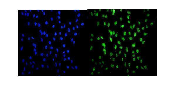
PA1607 has rat brain paraffin-section IHC and U20S-cell IF images; listed reactivity covers human, mouse and rat (PA1607 image captions; catalog reactivity).
PA1607 is the sole card that will render; it lists IHC, IF and ICC applications with human, mouse and rat reactivity (PA1607 catalog). Its own images show rat brain IHC(P) and IF in U20S cells (PA1607 image captions).
Which to pick: Choose PA1607 for tissue IHC: its own image shows rat brain paraffin-section staining, and the fixative is unreported (PA1607 IHC image caption). Choose PA1607 for image-backed IF/ICC; M04692-1 is a human-reactive rabbit monoclonal listing ICC/IF, but has no IF image in this payload (PA1607 IF image caption; M04692-1 catalog). For cross-species work, PA1607 lists human, mouse and rat reactivity, although its tissue IHC image documents rat only (PA1607 catalog reactivity; PA1607 IHC image caption).