HSPA6 / Heat shock 70 kDa protein 6 · IHC design guide

Design Immunohistochemistry for HSPA6

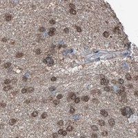
Plan HSPA6 IHC-P using the catalog antibody's 2–5 μg/ml range (datasheet A05402-3). Use the variable cytoplasmic and nuclear tissue staining to guide control selection and scoring (HPA tissue IHC).

Evidence assembled Oct 2026 · For research use; verify linked source records and product datasheet before use
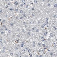
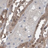
Immunohistochemistry protocol sheet for HSPA6 (IHC for HSPA6): expected localisation Cytoplasmic and nuclear staining (HPA tissue IHC), antibody A05402-3, validated IHC image, and IHC protocol steps
Printable HSPA6 IHC protocol sheet — expected localisation Cytoplasmic and nuclear staining (HPA tissue IHC), antibody A05402-3, controls and protocol steps. Open the full HSPA6 IHC guide →

HSPA6 Immunohistochemistry Experimental Design Guide

Expected localisation, validated protocols, controls and antibodies — the at-a-glance facts below, then the full design guide.

Must know before staining
Expected localisation Cytoplasmic and nuclear staining (HPA tissue IHC)
Staining pattern Variable cytoplasmic and nuclear staining in most tissues (HPA tissue IHC)
Antigen retrieval EDTA pH 8.0 HIER, heat-mediated (datasheet A05402-3)
Positive control ⓘ Caudate+4 more · see all
Negative control ⓘ Bone marrow+1 more · see all
Important caveats
Reasons your staining may differ from the expected pattern.
Fixation Keep fixation consistent across paraffin sections. (standard IHC practice; not target-specific)
Caveat Antibody may detect proteins from more than one gene (HPA tissue IHC)
Regulation Stress response; variable staining (UniProt; HPA tissue IHC)
Isoform / epitope No annotated isoforms or cleavage to shift the epitope (UniProt)
Section 1

Recommended HSPA6 IHC & IF Protocols

The catalog antibody protocol (datasheet: A05402-3) is followed by published IHC methods for gastric cancer specimens (PMC9833989) and lung tissue sections (PMC12918078).

Recommended immunohistochemistry (IHC-P) protocol parameters
SampleParaffin-embedded human esophageal cancer tissue; fixative not specified (datasheet A05402-3)
FixationImage fixative and duration unreported (datasheet A05402-3); verify before use.
Sectioning4–5 µm sections on charged slides (standard)
DeparaffinisationXylene, graded ethanol series to water (standard)
Antigen retrievalHeat retrieval: EDTA pH 8.0 (datasheet A05402-3); 20 min, 95–100 °C (standard)
Peroxidase block3% H2O2, 10 min, room temperature (standard)
Blocking10% goat serum (datasheet A05402-3)
Primary antibodyRabbit anti-HSPA6, 2-5 μg/ml (datasheet A05402-3)
Primary incubationOvernight at 4 °C (datasheet A05402-3)
DetectionStreptavidin-biotin complex (SABC), DAB chromogen (datasheet A05402-3)
CounterstainHematoxylin, blue, dehydrate and mount (standard)
Expected resultHSPA6-positive staining in glial cells of caudate (HPA tissue IHC: High). HPA tissue profile: Cytoplasmic and nuclear expression of varying intensity in most tissues. No signal in the no-primary control.
💡Decision noteStart with heat-mediated EDTA pH 8.0 retrieval (datasheet: A05402-3); compare hot citrate (PMC9833989) or EDTA pH 9.0 (PMC12918078) if staining needs optimization.
Section 2

What Is the Expected HSPA6 Staining Pattern?

In paraffin-section IHC, HSPA6-associated staining is cytoplasmic and nuclear across many tissues (HPA: tissue IHC profile). High staining is reported in glial cells, smooth muscle cells, and late spermatids (HPA: High). HPA rates the tissue profile Approved, with medium RNA–staining consistency, antibodies that may recognize proteins from multiple genes, and external verification pending (HPA: reliability description). UniProt reports no transmembrane segment and no annotated subcellular location (UniProt P17066: topology; subcellular location).

What am I looking at on my slide?
Cytoplasmic and nuclear staining in glial cells, smooth muscle cells, or late spermatids.This fits the reported compartments and High cell-level staining (HPA: tissue IHC profile; High). Compare cells within the section; a positive pattern alone cannot establish HSPA6 specificity because the HPA antibodies may recognize proteins from multiple genes (HPA: reliability description).
Medium staining in bronchial respiratory epithelium or breast myoepithelium.These are reported Medium populations (HPA: Bronchus; Breast). Judge intensity against the chosen counterstain and a known-positive control; staining need not be uniformly High across tissues (HPA: tissue IHC profile; standard IHC practice).
Signal confined to an unexpected compartment, without the reported cytoplasmic or nuclear pattern.Treat it as a possible staining artefact and review morphology, detection controls, and the antibody's validation (HPA: tissue IHC profile; standard IHC practice). UniProt provides no subcellular annotation that would independently validate a new IHC compartment (UniProt P17066: subcellular location).
Strong staining in bone-marrow hematopoietic cells or parathyroid glandular cells.Both populations are reported Not detected (HPA: Bone marrow; Parathyroid gland). Investigate cross-reactivity or endogenous detection activity with appropriate controls before calling the signal HSPA6 (HPA: multi-gene antibody caution; standard IHC practice).
Diffuse color over cells and tissue structures, or no signal in a known-positive control.Diffuse staining can reflect nonspecific antibody binding or detection background; absent control signal makes a negative specimen uninterpretable (standard IHC practice). Check controls and the IHC workflow before drawing a biological conclusion.
💡Expected HSPA6 appearanceA convincing positive shows cytoplasmic and nuclear staining in the relevant cells, potentially High in glia, smooth muscle, or late spermatids (HPA: tissue IHC profile; High); uniform diffuse color or strong staining in HPA Not detected cell populations warrants a false-positive check (HPA: Bone marrow; Parathyroid gland; standard IHC practice).
How each factor affects the staining
Antibody specificity and evidence gradeThree listed antibodies have Approved IHC status, while the tissue profile has medium RNA–staining consistency and awaits external verification (HPA: antibodies; reliability description). Multi-gene targeting limits a single stain's specificity (HPA: reliability description).
Choice of comparison tissueGlial cells, smooth muscle cells, and late spermatids are reported High; bone-marrow hematopoietic and parathyroid glandular cells are Not detected (HPA: tissue IHC). These are useful pattern comparisons, subject to the antibody-specificity caveat (HPA: reliability description).
RNA versus protein patternLung is tissue enhanced by RNA, while alveolar cells are reported Low by IHC (HPA: RNA specificity; Lung). Do not use the RNA label to predict strong alveolar staining (HPA: Lung).
Protein architectureUniProt lists one chain spanning residues 1–643, no signal peptide or propeptide, no transmembrane segment, and no annotated isoforms (UniProt P17066: processing; topology; isoforms). These annotations do not identify an IHC epitope or establish retrieval requirements.
IF/ICC Q: Should its punctate pattern define the IHC result?A: No. IF/ICC reports vesicles, flagellar centriole, and annulus, with additional perinuclear theca and calyx locations; that result carries a multi-gene antibody caution (HPA: subcellular). Use the cytoplasmic and nuclear tissue profile to assess IHC (HPA: tissue IHC profile).
Why is my staining missing, weak or wrong?
SituationLikely causeNext action
No staining in a glial-cell, smooth-muscle, or late-spermatid positive control (HPA: High).An IHC workflow failure is possible; the tissue profile also has medium consistency (HPA: reliability description; standard IHC practice).Check section integrity, antibody and detection reagents, and the catalog antibody's validated IHC conditions. Review antigen retrieval as a general IHC variable; HSPA6-specific fixation or retrieval sensitivity is unreported in the supplied sources.
Diffuse chromogen obscures cell boundaries.Nonspecific antibody binding or detection background may be present (standard IHC practice).Review blocking, antibody dilution, washing, and detection controls; score only interpretable cellular signal (standard IHC practice). Do not infer an HSPA6-specific fixation effect.
Unexpected signal appears in HPA Not detected cell populations (HPA: Bone marrow; Parathyroid gland).Cross-reactivity is plausible given the multi-gene antibody caution; endogenous detection activity is another IHC possibility (HPA: reliability description; standard IHC practice).Run appropriate no-primary and detection controls, assess endogenous activity, and compare with a separately validated antibody when available (standard IHC practice; HPA: multi-gene antibody caution).
Only an unusual compartment stains, with no cytoplasmic or nuclear pattern (HPA: tissue IHC profile).Artefact or nonspecific staining may explain the mismatch; UniProt has no location annotation to resolve it (standard IHC practice; UniProt P17066: subcellular location).Recheck morphology and counterstain, inspect controls, and compare the pattern with the HPA tissue profile before scoring it positive (HPA: tissue IHC profile; standard IHC practice).
Alveolar cells stain weakly despite the lung RNA-enhanced label (HPA: RNA specificity; Lung).Low alveolar-cell IHC staining is itself reported (HPA: Lung).Interpret protein staining against the alveolar-cell IHC entry and an appropriate positive control; do not use tissue-level RNA enrichment as the expected cellular IHC intensity (HPA: Lung; RNA specificity; standard IHC practice).

Sample controls for HSPA6 IHC & IF

🧪Run smooth muscle first; its smooth muscle cells should stain strongly (HPA: High in smooth muscle cells). Use parathyroid gland glandular cells as the negative tissue (HPA: Not detected); on the positive slide, use morphologically distinct non-smooth-muscle cells as background comparators only after confirming they are negative, since their HSPA6 status is unreported (HPA: High in smooth muscle cells).
Positive control tissue: Caudate (Glial cells, HPA High)
Negative control tissue: Bone marrow (HPA Not detected)
ICC-IF cell lines (HPA subcellular resource): HPA ICC-IF images show HSPA6 in CACO-2, PC-3, U2OS, RPTEC/TERT1, hTERT-RPE1 (serum starved), Sperm, with annotated localisation: Vesicles (approved), Flagellar centriole (approved), Annulus (approved) (HPA subcellular).
Technical controls: Include a no-primary secondary-only control, concentration-matched nonimmune rabbit IgG matched to the primary where its isotype is known, and an HSPA6 knockout control or validated peptide competition (caption: rabbit primary; standard IHC specificity practice). For the smooth-muscle slide, quench endogenous peroxidase and check for endogenous biotin before interpreting SABC/DAB staining (caption: SABC/DAB; standard IHC practice).
⚠️Feasibility: A target-specific fixation window or fixation effect is unreported, and the selected A05402-3 paraffin-section caption does not state the fixative (caption: fixative not stated). The reported IHC condition uses heat-mediated EDTA retrieval at pH 8.0 and 2 μg/ml primary overnight at 4°C; the caption does not establish that retrieval is required (caption: EDTA pH 8.0; 2 μg/ml; overnight at 4°C). The supplied evidence does not establish that frozen sections or IF are easier; residual endogenous peroxidase or biotin could confound the reported SABC/DAB detection (caption: SABC/DAB; standard IHC practice).

HPA tissue IHC evidence for HSPA6

Comprehensive Human Protein Atlas IHC scoring per tissue (reliability: Approved — Medium consistency between antibody staining and RNA expression data. Caution, targets protein from more than one gene. Pending external verification.). Rows are taken directly from the HPA tissue chart — click any row's HPA link to view the source.

Positive expression · recommended positive controls

TissueCell typeLevelEvidenceSource
Caudate Glial cells High Protein (IHC) HPA →
Cerebral cortex Glial cells High Protein (IHC) HPA →
Smooth muscle Smooth muscle cells High Protein (IHC) HPA →
Testis Elongated or late spermatids High Protein (IHC) HPA →
Adrenal gland Glandular cells Medium Protein (IHC) HPA →

Undetected expression · recommended negative controls

TissueCell typeLevelEvidenceSource
Bone marrow Hematopoietic cells Not detected Protein (IHC) HPA →
Parathyroid gland Glandular cells Not detected Protein (IHC) HPA →
Section 3

Advanced HSPA6 IHC Tips

Troubleshoot HSPA6 staining in paraffin sections using the catalog antibody’s tissue IHC result, HPA patterns, and compartment-aware controls.

Which retrieval condition should I start with for weak HSPA6 staining?
Start with heat-mediated retrieval in EDTA at pH 8.0 (datasheet A05402-3). The catalog antibody detected HSPA6 in a paraffin-embedded human esophageal cancer section after this retrieval, followed by 2 μg/ml primary antibody overnight at 4°C (datasheet A05402-3). If staining remains weak, adjust heating duration on adjacent sections while keeping buffer and detection conditions constant; include a section processed by the documented method as a reference (datasheet A05402-3). Compare signal in viable cells with background in matched negative controls before increasing retrieval intensity, because stronger heating can compromise morphology and make compartment-level scoring unreliable.
Could fixation explain weak or uneven HSPA6 staining?
Target-specific fixation sensitivity for HSPA6 is unknown: the selected paraffin-section caption does not report a fixative (datasheet A05402-3). Record the actual fixative and fixation duration for each specimen, then compare sections with similar processing, thickness, and retrieval conditions before attributing variation to HSPA6 biology. For a processing check, run the documented EDTA pH 8.0 retrieval and 2 μg/ml overnight primary incubation on a known staining section alongside the problem section (datasheet A05402-3). Assess preservation of cell outlines and nuclear detail, and avoid interpreting damaged or detached tissue as a negative HSPA6 result.
How should I assess cytoplasmic, nuclear, or punctate HSPA6 staining?
Score cytoplasmic and nuclear signal separately because tissue IHC shows both compartments at varying intensity in most tissues (HPA: tissue IHC profile). HSPA6 has no annotated transmembrane segment or signal peptide, and UniProt does not assign a subcellular location (UniProt P17066 topology and subcellular record). Puncta require caution: vesicle, flagellar centriole, annulus, perinuclear theca, and calyx assignments come from subcellular antibody data that may recognize proteins from multiple genes (HPA: subcellular summary and caution). Check whether staining follows intact cell boundaries and persists across adjacent sections; document the compartment and cell type rather than collapsing all brown signal into one score.
What can I conclude if the antibody stains an unexpected compartment?
Do not explain an unexpected IHC pattern by invoking an unlisted splice form: the supplied HSPA6 record lists 0 isoforms and one 1–643 chain (UniProt P17066 processing and isoforms). The record notes lysine methylation at residue 563 in vitro, but gives no antibody epitope or evidence that this modification changes tissue staining (UniProt P17066 modified residues). Compare staining with a no-primary control and an independently validated HSPA6 reagent if available, while recording each compartment separately. Treat antibody specificity as an open question when patterns conflict, because the HPA tissue assessment cautions that its antibody targets proteins from more than one gene (HPA: tissue IHC reliability).
How can IF help check which cells carry the HSPA6 signal?
Use IF as a separate validation experiment, pairing HSPA6 with a glial marker when examining caudate or cerebral cortex, where glial-cell staining is high by tissue IHC (HPA: caudate and cerebral cortex). Choose fluorophores in channels with low measured tissue autofluorescence, and inspect single-stain and unstained controls before calling overlap. Permeabilise sufficiently for access to intracellular epitopes while preserving morphology; HSPA6 has no annotated transmembrane segment or signal peptide, but its antibody epitope is unspecified (UniProt P17066 topology; supplied antibody record). Compare cell identity and compartment with chromogenic sections, while treating punctate subcellular assignments cautiously because the HPA antibodies may recognize proteins from multiple genes (HPA: subcellular caution).
What should I check when DAB background obscures HSPA6?
Run no-primary and secondary-only controls to distinguish nonspecific detection from tissue pigment or endogenous enzyme activity in the chromogenic section. The reported workflow used 10% goat serum blocking, biotinylated goat anti-rabbit secondary for 30 minutes at 37°C, an avidin-biotin complex, and DAB (datasheet A05402-3). Check endogenous peroxidase blocking and, for this biotin-based workflow, endogenous biotin controls as general IHC measures; optimise washes and DAB development using paired sections. Preserve any reproducible cellular signal while reducing diffuse staining, and interpret weak widespread color cautiously because HPA reports cytoplasmic and nuclear staining of varying intensity in most tissues (HPA: tissue IHC profile).
How should I quantify HSPA6 staining across differently processed sections? ⚠ ANSWER MARKED FOR VERIFICATION
Define cell types and compartments before scoring, then record the percentage of positive cells and intensity separately for cytoplasm and nucleus; an H-score can summarize intensity within each category. Use viable, non-necrotic tissue area as the denominator for positive-cell percentage, or report positive-cell density per mm² of annotated tissue. Normalise comparisons to the same cell population, section area, batch controls, retrieval condition, and DAB development time, since HPA reports variable cytoplasmic and nuclear intensity across tissues (HPA: tissue IHC profile). Keep glial cells, smooth muscle cells, and other populations separate where relevant, because their reported staining levels differ (HPA: positive tissue entries).
How do I separate true HSPA6 staining from artefact?
Give greatest weight to reproducible signal in intact, identifiable cells with interpretable cytoplasmic or nuclear staining, consistent with the reported tissue IHC profile (HPA: tissue IHC profile). Check whether candidate positive cells match the sampled tissue: glial cells in caudate and cerebral cortex and smooth muscle cells have high reported staining, whereas bone-marrow hematopoietic cells are reported as not detected (HPA: positive and negative tissue entries). Exclude section edges, folds, necrotic regions, pigment, and staining reproduced in no-primary or peroxidase-control sections. Avoid declaring specificity from a single compartment or image, because the HPA tissue assessment reports medium RNA agreement and warns that its antibody targets proteins from more than one gene (HPA: tissue IHC reliability).
Boster reagents

Best HSPA6 / Heat shock 70 kDa protein 6 IHC Antibodies

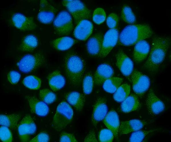
Both antibodies have human tissue IHC images (catalog IHC captions); A05402-3 also has IF data in PC-3 cells (catalog IF caption) and lists Mouse and Rat reactivity (catalog reactivity).

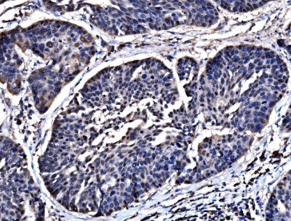
Real IHC data IHC analysis of HSPA6 using anti-HSPA6 antibody (A05402-3). HSPA6 was detected in a paraffin-embedded section of human esophageal cancer tissue. Heat mediated antigen retrieval was performed in EDTA buffer (pH 8.0, epitope retrieval solution). The tissue section was blocked with 10% goat serum. The tissue section was then incubated with 2 μg/ml rabbit anti-HSPA6 Antibody (A05402-3) overnight at 4°C. Biotinylated goat anti-rabbit IgG was used as secondary antibody and incubated for 30 minutes at 37°C. The tissue section was developed using Strepavidin-Biotin-Complex (SABC) (Catalog # SA1022) with DAB as the chromogen.
Anti-HSPA6 Antibody ®
Cat # A05402-3
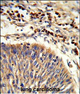
Real IHC data Formalin-fixed and paraffin-embedded human lung carcinoma reacted with HSPA6 Antibody (C-term), which was peroxidase-conjugated to the secondary antibody, followed by DAB staining. This data demonstrates the use of this antibody for immunohistochemistry; clinical relevance has not been evaluated.
Anti-HSPA6 Antibody (C-term)
Cat # A05402-2

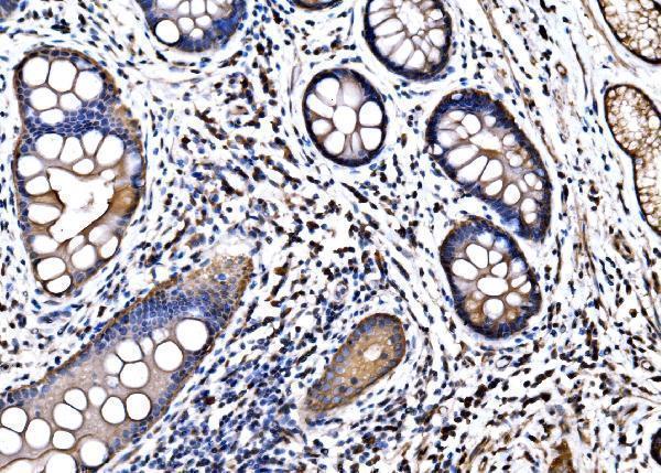
A05402-3 has paraffin-section IHC images from human esophageal cancer, gastric signet ring cell carcinoma, renal pelvis squamous metaplasia, and tonsil, plus IF in PC-3 cells (catalog image captions). A05402-2 has an IHC-P image from formalin-fixed, paraffin-embedded human lung carcinoma (catalog IHC caption).

Which to pick: For tissue IHC, choose A05402-3 for the documented human paraffin-section examples; its captions do not report the fixative (A05402-3 IHC captions). A05402-2 is a rabbit polyclonal option for paraffin-section, paraffin-embedded human tissue (catalog dilution_raw; A05402-2 IHC caption). For IF/ICC or listed Mouse and Rat reactivity, choose A05402-3, which lists both applications and species; its IHC images document human tissue only, and its clonality is unspecified (catalog applications, reactivity, clone; A05402-3 IHC captions). The selected A05402-3 tissue-IHC caption documents paraffin sections, but does not specify the fixative (selected-SKU IHC image A05402-3).

Each figure is that product's own IHC / IF validation image from its datasheet.

References

  1. UniProt Consortium. UniProt entry P17066 (HSP76_HUMAN, Heat shock 70 kDa protein 6).
  2. Human Protein Atlas. HSPA6 tissue IHC expression (reliability: Approved).
  3. Human Protein Atlas. HSPA6 subcellular location (ICC-IF): Mainly localized to vesicles, flagellar centriole and annulus. In addition localized to the perinuclear theca and calyx. Caution: Based on antibodies targeting proteins from multiple genes..
  4. Human Protein Atlas. HSPA6 antibody validation summary (3 antibodies).
  5. HSPA6, a novel prognostic and therapeutic biomarker, associated with Ming classification in gastric cancer. Journal of clinical laboratory analysis 2023 — PMC9833989.
  6. HSPA6 Promotes Ferroptosis in Triple-Negative Breast Cancer by Rewiring Lipid Metabolism to Potentiate Membrane Lipid Peroxidation. International journal of biological sciences 2026 — PMC13137960.
  7. PERP suppresses breast cancer metastasis via the ATF3-HSPA6 signaling pathway. The Journal of international medical research 2025 — PMC12775323.
  8. Exosome-derived Menin from cancer-associated fibroblasts promotes gastric cancer progression by activating the HSPA6/JNK/JunD pathway and inducing EMT. Journal of translational medicine 2025 — PMC12918078.
  9. PubMed PMID:2327978 — UniProt-cited evidence.
  10. PubMed PMID:16710414 — UniProt-cited evidence.
  11. PubMed PMID:15489334 — UniProt-cited evidence.